1,4-DPCA is a novel, potent and competitive inhibitor of prolyl 4-hydroxylase with IC50 value of 3.6 μM. Increased collagen deposition provides physical and biochemical signals to support tumor growth and invasion during breast cancer development. Therefore, inhibition of collagen synthesis and deposition has been considered a strategy to suppress breast cancer progression. Collagen prolyl-4-hydroxylase α subunit 2 (P4HA2), an enzyme hydroxylating proline residues in -X-Pro-Gly- sequences, is a potential therapeutic target for the disorders associated with increased collagen deposition. However, expression and function of P4HA2 in breast cancer progression are not well investigated.
Physicochemical Properties
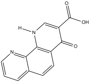
| Molecular Formula | C13H8N2O3 | |
| Molecular Weight | 240.22 | |
| Exact Mass | 240.053 | |
| CAS # | 331830-20-7 | |
| Related CAS # |
|
|
| PubChem CID | 459803 | |
| Appearance | White to off-white solid powder | |
| Density | 1.5±0.1 g/cm3 | |
| Boiling Point | 474.5±45.0 °C at 760 mmHg | |
| Flash Point | 240.8±28.7 °C | |
| Vapour Pressure | 0.0±1.2 mmHg at 25°C | |
| Index of Refraction | 1.727 | |
| LogP | 3.28 | |
| Hydrogen Bond Donor Count | 2 | |
| Hydrogen Bond Acceptor Count | 5 | |
| Rotatable Bond Count | 1 | |
| Heavy Atom Count | 18 | |
| Complexity | 419 | |
| Defined Atom Stereocenter Count | 0 | |
| InChi Key | XZZHOJONZJQARN-UHFFFAOYSA-N | |
| InChi Code | InChI=1S/C13H8N2O3/c16-12-8-4-3-7-2-1-5-14-10(7)11(8)15-6-9(12)13(17)18/h1-6H,(H,15,16)(H,17,18) | |
| Chemical Name |
|
|
| Synonyms |
|
|
| HS Tariff Code | 2934.99.9001 | |
| Storage |
Powder-20°C 3 years 4°C 2 years In solvent -80°C 6 months -20°C 1 month |
|
| Shipping Condition | Room temperature (This product is stable at ambient temperature for a few days during ordinary shipping and time spent in Customs) |
Biological Activity
| Targets | In mouse B6 cells, 1,4-DPCA (24 hours) can treat many elevated target gene expressions, such as the pro-angiogenic target genes Vegfa and Hmox1 and the pro-glycolytic target genes Ldh-a, Pgk1, Pdk1, and 1. ZR-75-1 (20 μM 1,4-DPCA) and T4-2 (10 μM 1,4-DPCA) cells showed a significant reduction in colony size following 4-DPCA therapy. In three-dimensional culture, T4-2 cells treated with DPCA produced localized spheroids. Treatment with 1,4-DPCA significantly decreased MDA-MB-231 (10 μM 1,4-DPCA), ZR-75-1, MDA-MB-157, and T4-2 cells [3]. |
| ln Vitro |
In mouse B6 cells, 1,4-DPCA (24 hours) can treat many elevated target gene expressions, such as the pro-angiogenic target genes Vegfa and Hmox1 and the pro-glycolytic target genes Ldh-a, Pgk1, Pdk1, and 1. ZR-75-1 (20 μM 1,4-DPCA) and T4-2 (10 μM 1,4-DPCA) cells showed a significant reduction in colony size following 4-DPCA therapy. In three-dimensional culture, T4-2 cells treated with DPCA produced localized spheroids. Treatment with 1,4-DPCA significantly decreased MDA-MB-231 (10 μM 1,4-DPCA), ZR-75-1, MDA-MB-157, and T4-2 cells [3]. Treatment of B6 mouse ear fibroblasts with 1,4-DPCA/hydrogel (Gd) for 24 hours increased expression of HIF-1α protein in both cytoplasm and nucleus, as determined by immunohistochemistry and Western blot analysis, but did not affect HIF-2α expression[1] Reverse transcription polymerase chain reaction (RT-PCR) analysis of mRNA from 1,4-DPCA/hydrogel-treated B6 mouse fibroblasts showed increased expression of multiple HIF-1α target genes, including proangiogenic genes Vegfa and Hmox1, and proglycolytic genes Ldh-a, Pgk1, Pdk1, and Glut1[1] Treatment with siRNA against Hif1a (siHif1a_3) blocked the up-regulation of all these target genes induced by 1,4-DPCA/hydrogel[1] B6 mouse ear fibroblasts treated in vitro with 1,4-DPCA/gel for 24 hours stained positive for HIF-1α and a suite of immature cell markers (NANOG, OCT3/4, CD133, PAX7, PREF1, NESTIN, vWF, CD34), displaying staining patterns indistinguishable from MRL mouse ear fibroblasts[1] Quantitative PCR (qPCR) confirmed significant up-regulation of stem cell marker mRNA (e.g., NANOG, OCT3/4) in B6 mouse ear fibroblasts treated with drug/gel compared to untreated cells[1] Treatment of NANOG-positive MRL mouse ear-derived fibroblasts with siHif1a resulted in almost complete suppression of NANOG protein expression[1] |
| ln Vivo |
In intravascular peritoneal poly(lock-co-glycolic acid) (PLGA) discs, treatment with 1,4-DPCA successfully prevented connective tissue ingrowth for a period of 28 days. The pivot disc's intra- and exterior surface-restricted collagen deposition as well as connective tissue ingrowth are successfully inhibited by 1,4-DPCA capture, though not to the same degree as glucocorticoids [2]. Subcutaneous injection of a hydrogel containing 1,4-DPCA (1,4-DPCA/hydrogel) into Swiss Webster mice (a non-regenerative strain) increased stable expression of HIF-1α protein in ear tissue over 5 days, providing a functional measure of drug release in vivo[1] Multiple subcutaneous injections of 1,4-DPCA/hydrogel over a 10-day period (days 0, 5, and 10 post-injury) led to complete regenerative wound healing (ear hole closure) in Swiss Webster mice after ear hole punch injury, with closure ongoing up to day 35[1] Ear hole closure induced by 1,4-DPCA/gel was blocked by simultaneous treatment with siHif1a every other day for 20 days, indicating the healing effect was dependent on HIF-1α expression[1] Treatment with 1,4-DPCA/gel induced several hallmarks of regeneration in Swiss Webster mice: accelerated re-epithelialization (complete by day 2), cellular dedifferentiation (expression of stem cell markers NESTIN, OCT3/4, neurofilament, PAX7), basement membrane breakdown (reduced laminin, increased MMP9), reduced scar formation (reduced collagen cross-linking, reduced expression of Lox14 and Ctgf), and later events including chondrogenesis and new hair follicle formation in the regenerated tissue[1] Western blot analysis showed HIF-1α protein expression was markedly increased in Swiss Webster mice treated with drug/gel on days 7, 14, and 21 post-injury, similar to levels seen in regenerative MRL mice, while HIF-2α expression was barely affected[1] |
| Enzyme Assay |
The compound 1,4-DPCA is reported in the cited references to be a potent inhibitor of prolyl hydroxylases (PHDs), which mediate the hydroxylation and subsequent degradation of HIF-1α protein under normoxic conditions[1] By blocking PHD activity, 1,4-DPCA stabilizes HIF-1α protein[1] |
| Cell Assay |
For in vitro studies, ear-derived fibroblast-like cells from mice were cultured[1] To test drug effects, cells were cultured with normal medium, hydrogel alone (G0), or hydrogel containing 1,4-DPCA (Gd). The hydrogel precursors formed a solid disc in the well plates[1] After 24 hours of treatment, cells were processed for analysis. HIF-1α protein expression was assessed by immunostaining and Western blot analysis[1] For gene expression analysis, mRNA was extracted from treated cells and analyzed by RT-PCR or quantitative PCR (qPCR) for HIF-1α target genes and stem cell markers[1] For siRNA knockdown experiments, cells at 70% confluence were transfected with Hif1a siRNA or scramble siRNA using a transfection reagent according to the manufacturer's protocol. Knockdown efficiency was examined 48 hours post-transfection[1] |
| Animal Protocol |
A locally injectable hydrogel was designed for controlled delivery of 1,4-DPCA over 4 to 10 days. The hydrogel was composed of cross-linked polyethylene glycol molecules (precursors P8Cys and P8NHS) that gel rapidly under physiological conditions[1] Drug-loaded hydrogels were formed by suspending polymer (Pluronic F127)-stabilized 1,4-DPCA microcrystals in an aqueous mixture of the hydrogel precursors, which solidified rapidly to entrap the drug microcrystals[1] Swiss Webster mice (8-10 weeks old) received a 2.1 mm through-and-through ear hole punch injury[1] For single-dose kinetics study: Mice were injected subcutaneously at the back of the neck with 100 µL of hydrogel containing 1,4-DPCA at 2 mg/mL (Gd) or 0 mg/mL (gel alone, G0) several hours after injury. Ear tissue was harvested daily for 5 days for analysis[1] For regenerative response study: Mice were injected subcutaneously at the back of the neck with 100 µL of Gd or G0 on days 0, 5, and 10 after injury (into three adjacent sites). Ear hole closure was monitored by measuring hole diameters over 35 days[1] In some experiments, to test distal effects, mice were injected subcutaneously with drug/gel into the left and right rear flank regions on days 0, 5, and 10[1] For siRNA inhibition in vivo: siHif1a was mixed with an in vivo transfection reagent following the manufacturer's instructions and injected subcutaneously every 48 hours for 20 days beginning on day 0, concurrently with drug/gel treatment[1] Ear hole diameters were measured in the direction of the ear pinna long axis and the perpendicular axis[1] At endpoints, mice were euthanized, and ear tissue was collected for histological analysis (e.g., H&E, Alcian blue, picrosirius red staining), immunohistochemistry, Western blot, and RNA extraction for qPCR[1] |
| References |
[1]. Drug-induced regeneration in adult mice. Sci Transl Med. 2015 Jun 3;7(290):290ra92. [2]. Transient inhibition of connective tissue infiltration and collagen deposition into porous poly(lactic-co-glycolic acid) discs. J Biomed Mater Res A. 2013 Dec;101(12):3599-606. [3]. Prolyl-4-hydroxylase α subunit 2 promotes breast cancer progression and metastasis by regulating collagen deposition. BMC Cancer. 2014 Jan 2;14:1. |
| Additional Infomation |
1,4-DPCA (1,4-dihydrophenonthrolin-4-one-3-carboxylic acid) is a small-molecule inhibitor of prolyl hydroxylases (PHDs)[1] Hypoxia-inducible factor 1α (HIF-1α) is a central regulator identified in the spontaneous regenerative healing of MRL mice. Its protein is degraded under normoxia via PHD-mediated hydroxylation[1] Stabilization of HIF-1α via PHD inhibition by 1,4-DPCA is proposed to mimic a key pro-regenerative mechanism found in regenerative MRL mice, promoting processes like aerobic glycolysis, cell migration, inflammation, and stem cell marker expression[1] Formulating the poorly soluble 1,4-DPCA as microcrystals within a biocompatible hydrogel allowed for sustained local delivery and avoided potential cytotoxicity associated with direct crystal-cell contact[1] The study demonstrates that pharmacological stabilization of HIF-1α with 1,4-DPCA/hydrogel can induce a regenerative healing response in a normally non-regenerating mammal (Swiss Webster mouse) after injury[1] |
Solubility Data
| Solubility (In Vitro) |
|
|||
| Solubility (In Vivo) |
Solubility in Formulation 1: ≥ 0.92 mg/mL (3.83 mM) (saturation unknown) in 10% DMSO + 90% Corn Oil (add these co-solvents sequentially from left to right, and one by one), clear solution. For example, if 1 mL of working solution is to be prepared, you can add 100 μL of 9.2 mg/mL clear DMSO stock solution to 900 μL of corn oil and mix evenly. Solubility in Formulation 2: 10 mg/mL (41.63 mM) in 50% PEG300 50% Saline (add these co-solvents sequentially from left to right, and one by one), suspension solution; with ultrasonication. Preparation of saline: Dissolve 0.9 g of sodium chloride in 100 mL ddH₂ O to obtain a clear solution. (Please use freshly prepared in vivo formulations for optimal results.) |
| Preparing Stock Solutions | 1 mg | 5 mg | 10 mg | |
| 1 mM | 4.1629 mL | 20.8143 mL | 41.6285 mL | |
| 5 mM | 0.8326 mL | 4.1629 mL | 8.3257 mL | |
| 10 mM | 0.4163 mL | 2.0814 mL | 4.1629 mL |