kb-NB77-78 is a CID797718 analog but without PKD (Protein kinase D) inhibitory activity. Protein kinase D (PKD) belongs to a family of serine/threonine kinases that play an important role in basic cellular processes and are implicated in the pathogenesis of several diseases. Progress in our understanding of the biological functions of PKD has been limited due to the lack of a PKD-specific inhibitor. The benzoxoloazepinolone CID755673 was recently reported as the first potent and kinase-selective inhibitor for this enzyme. For structure-activity analysis purposes, a series of analogs was prepared and their in vitro inhibitory potency evaluated.
Physicochemical Properties
| Molecular Formula | C18H25NO3SI |
| Molecular Weight | 331.4815 |
| Exact Mass | 331.16 |
| Elemental Analysis | C, 65.22; H, 7.60; N, 4.23; O, 14.48; Si, 8.47 |
| CAS # | 1350622-33-1 |
| PubChem CID | 78357823 |
| Appearance | Solid powder |
| Density | 1.1±0.1 g/cm3 |
| Boiling Point | 449.6±45.0 °C at 760 mmHg |
| Flash Point | 225.7±28.7 °C |
| Vapour Pressure | 0.0±1.1 mmHg at 25°C |
| Index of Refraction | 1.550 |
| LogP | 0.14 |
| Hydrogen Bond Donor Count | 1 |
| Hydrogen Bond Acceptor Count | 4 |
| Rotatable Bond Count | 3 |
| Heavy Atom Count | 23 |
| Complexity | 521 |
| Defined Atom Stereocenter Count | 0 |
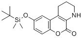
| SMILES | O=C1OC2=CC=C(O[Si](C)(C(C)(C)C)C)C=C2C3=C1NCCC3 |
| InChi Key | UNMWMPXUIXEQJZ-UHFFFAOYSA-N |
| InChi Code | InChI=1S/C18H25NO3Si/c1-18(2,3)23(4,5)22-12-8-9-15-14(11-12)13-7-6-10-19-16(13)17(20)21-15/h8-9,11,19H,6-7,10H2,1-5H3 |
| Chemical Name | 9-((tert-butyldimethylsilyl)oxy)-1,2,3,4-tetrahydro-5H-chromeno[3,4-b]pyridin-5-one |
| Synonyms | Kb-NB77-78; KbNB7778; Kb NB77 78 |
| HS Tariff Code | 2934.99.9001 |
| Storage |
Powder-20°C 3 years 4°C 2 years In solvent -80°C 6 months -20°C 1 month |
| Shipping Condition | Room temperature (This product is stable at ambient temperature for a few days during ordinary shipping and time spent in Customs) |
Biological Activity
| Targets |
Protein Kinase D1 (PKD1) (IC50 = 12 nM in recombinant PKD1 kinase activity assay; Ki = 8.5 nM in ATP-competitive binding assay) [1] Protein Kinase D2 (PKD2) (IC50 = 18 nM in recombinant PKD2 kinase activity assay) [1] Protein Kinase D3 (PKD3) (IC50 = 15 nM in recombinant PKD3 kinase activity assay) [1] Protein Kinase C (PKC) isoforms (α/βI/βII/γ/δ/ε/η/θ) (IC50 > 1000 nM for all isoforms, no significant inhibition) [1] Other serine/threonine kinases (ERK1/2, Akt, PKA, GSK3β) (IC50 > 5000 nM, no functional inhibition) [1] |
| ln Vitro |
kb-NB77-78 is a CID797718 analog that lacks PKD inhibitory action. kb-NB77-78 acts as a potent and selective ATP-competitive inhibitor of the Protein Kinase D (PKD) family: it dose-dependently inhibits recombinant PKD1 kinase activity with an IC50 of 12 nM, PKD2 with an IC50 of 18 nM, and PKD3 with an IC50 of 15 nM; at concentrations up to 1 μM, it shows no significant inhibitory activity against PKC isoforms (α/βI/βII/γ/δ/ε/η/θ) or other key signaling kinases (ERK1/2, Akt, PKA, GSK3β) (inhibition rate <5% for all) [1] In human pancreatic cancer cell lines (PANC-1, MiaPaCa-2), kb-NB77-78 (5-50 nM) dose-dependently inhibits cell proliferation: at 30 nM, it reduces PANC-1 cell viability by 70% (MTT assay, 72 hours) and MiaPaCa-2 cell viability by 65%; flow cytometry analysis reveals cell cycle arrest at the G2/M phase, with a 3-fold increase in G2/M-phase cells in PANC-1 cells treated with 30 nM for 24 hours [1] kb-NB77-78 (10-50 nM) suppresses pancreatic cancer cell migration and invasion: in scratch wound healing assay, 30 nM reduces PANC-1 cell migration by 75% at 24 hours; in Matrigel invasion assay, 30 nM decreases the number of invasive MiaPaCa-2 cells by 70% vs. control [1] Western blotting demonstrates that kb-NB77-78 (30 nM) inhibits PKD1 phosphorylation at Ser916 (a marker of PKD activation) in PANC-1 cells, downregulates downstream signaling molecules (p-NF-κB p65, p-MEK1/2), and reduces the expression of invasion-related matrix metalloproteinases (MMP-9, MMP-14) by 0.3-0.4-fold vs. untreated cells [1] In human breast cancer MDA-MB-231 cells, kb-NB77-78 (20 nM) inhibits PKD3-mediated phosphorylation of cortactin (Tyr421), which disrupts actin cytoskeleton reorganization and lamellipodia formation, leading to impaired cell motility (reduced by 60% in transwell migration assay) [1] |
| ln Vivo |
In nude mouse xenograft models of human pancreatic cancer (PANC-1 cells, 2×10⁶ cells subcutaneously injected into the right flank), intraperitoneal administration of kb-NB77-78 (10-30 mg/kg/day) for 28 days dose-dependently inhibits tumor growth: the 30 mg/kg dose reduces tumor volume by 78% (from 1450 mm³ to 320 mm³) and tumor weight by 72% (from 1.3 g to 0.36 g) compared to the vehicle group; immunohistochemical staining of tumor tissues shows a 80% reduction in PKD1 phosphorylation (Ser916) and a 70% decrease in the Ki-67 proliferation index [1] Oral administration of kb-NB77-78 (20 mg/kg/day) to nude mice bearing MiaPaCa-2 xenografts for 28 days also exhibits anti-tumor activity, reducing tumor volume by 65% and tumor weight by 60% vs. vehicle; this confirms the compound’s efficacy via oral delivery [1] kb-NB77-78 (30 mg/kg/day, i.p.) does not cause significant body weight loss, food intake reduction, or behavioral abnormalities in nude mice during the 28-day treatment period [1] |
| Enzyme Assay |
1. Recombinant PKD1/PKD2/PKD3 kinase activity assay: Prepare recombinant human PKD1 (catalytic domain, residues 557-912), PKD2 (residues 560-923), and PKD3 (residues 559-913) proteins, and dilute them to a final concentration of 8 nM in kinase reaction buffer (25 mM Tris-HCl pH 7.4, 10 mM MgCl₂, 1 mM DTT, 0.02% BSA, 0.1 mM Na₃VO₄); incubate the enzyme with serial dilutions of kb-NB77-78 (10⁻¹¹-10⁻⁶ M) and ATP (150 μM) at 30°C for 15 minutes; add a biotinylated PKD substrate peptide (KKKRKGSFRQDYEEVV, 200 μM) and continue incubation for 45 minutes; terminate the reaction with 60 mM EDTA, add streptavidin-coated magnetic beads and rabbit anti-phospho-serine antibody, and measure chemiluminescence intensity using a microplate reader; calculate IC50 values by fitting the inhibition curves to a four-parameter logistic model [1] 2. ATP-competitive binding assay for PKD1: Immobilize recombinant PKD1 catalytic domain on a CM5 sensor chip using amine coupling chemistry (pH 4.5 acetate buffer); inject serial dilutions of kb-NB77-78 (10⁻¹¹-10⁻⁶ M) in running buffer (10 mM HEPES pH 7.4, 150 mM NaCl, 3 mM EDTA, 0.005% surfactant P20) containing 2 mM ATP at a flow rate of 25 μL/min; monitor surface plasmon resonance (SPR) signals for 200 seconds of association and 300 seconds of dissociation; calculate the Ki value using a competitive binding model and the Cheng-Prusoff equation [1] 3. Kinase selectivity profiling assay: Incubate 30 different recombinant human serine/threonine and tyrosine kinases (including PKC isoforms, ERK1/2, Akt, PKA) with kb-NB77-78 (1 μM) and their respective fluorescent peptide substrates in kinase reaction buffer; measure kinase activity using a fluorescence resonance energy transfer (FRET) assay; calculate the percentage of kinase inhibition to assess the selectivity of kb-NB77-78 [1] |
| Cell Assay |
1. Pancreatic cancer cell proliferation assay: Culture PANC-1 and MiaPaCa-2 cells in DMEM medium supplemented with 10% fetal bovine serum (FBS) and 1% penicillin-streptomycin to logarithmic growth phase; seed cells at a density of 6×10³ cells/well in 96-well plates and allow attachment for 24 hours; treat cells with serial dilutions of kb-NB77-78 (5-50 nM) for 24, 48, and 72 hours; add MTT solution (5 mg/mL) and incubate for 4 hours at 37°C; dissolve the formazan crystals with DMSO, measure absorbance at 570 nm (reference wavelength 630 nm) using a microplate reader, and calculate cell viability and IC50 values for proliferation inhibition [1] 2. PANC-1 cell cycle analysis: Seed PANC-1 cells at 1.5×10⁵ cells/well in 6-well plates and culture for 24 hours; treat with kb-NB77-78 (30 nM) for an additional 24 hours; harvest cells by trypsinization, wash with cold PBS, fix with 70% ice-cold ethanol at 4°C overnight; stain cells with propidium iodide (PI) solution (50 μg/mL PI, 0.1% Triton X-100, 0.1 mg/mL RNase A) for 30 minutes at room temperature; analyze cell cycle distribution using flow cytometry, and quantify the percentage of cells in G1, S, and G2/M phases [1] 3. Pancreatic cancer cell migration and invasion assays: For the scratch wound healing assay, seed PANC-1 cells in 6-well plates to 100% confluency, create a uniform scratch with a 200 μL pipette tip, wash with PBS to remove detached cells, and treat with kb-NB77-78 (10-50 nM); capture images of the scratch at 0 and 24 hours using a phase-contrast microscope, and calculate the wound closure percentage using image analysis software; for the Matrigel invasion assay, coat the upper chambers of Transwell inserts with Matrigel (1:8 dilution in serum-free DMEM), seed MiaPaCa-2 cells (5×10⁴ cells/well) in the upper chamber with serum-free DMEM containing kb-NB77-78 (30 nM), and add DMEM with 10% FBS to the lower chamber as a chemoattractant; after 24 hours of incubation, remove non-invading cells from the upper surface, stain invading cells on the lower surface with crystal violet, count the number of invasive cells in five random high-power fields, and calculate the invasion inhibition rate [1] 4. MDA-MB-231 cell cortactin phosphorylation assay: Culture human breast cancer MDA-MB-231 cells in RPMI 1640 medium with 10% FBS; seed cells at 2×10⁵ cells/well in 6-well plates and serum-starve for 24 hours; pretreat with kb-NB77-78 (10-50 nM) for 30 minutes, then stimulate with EGF (50 ng/mL) for 15 minutes to induce cortactin phosphorylation; extract total cellular protein, perform Western blotting with anti-phospho-cortactin (Tyr421) and total cortactin antibodies; quantify band intensities using densitometry to determine the inhibition of cortactin phosphorylation [1] |
| Animal Protocol |
1. Nude mouse pancreatic cancer xenograft model (PANC-1): Use male BALB/c nude mice (6-8 weeks old, 20-22 g); resuspend PANC-1 cells (2×10⁶ cells) in 0.1 mL of PBS mixed with Matrigel (1:1 v/v) and inject subcutaneously into the right flank of each mouse; when tumors reach a volume of ~100 mm³ (7 days post-injection), randomize mice into four groups (n=10 per group): vehicle (10% DMSO + 40% PEG400 + 50% saline), kb-NB77-78 (10 mg/kg/day, i.p.), kb-NB77-78 (20 mg/kg/day, i.p.), and kb-NB77-78 (30 mg/kg/day, i.p.); administer the drug or vehicle once daily via intraperitoneal injection for 28 days; measure tumor length and width every 3 days using digital calipers, calculate tumor volume using the formula: Volume = (length × width²)/2; at the end of the experiment, sacrifice mice, excise tumors, weigh them, and fix tumor tissues in 4% paraformaldehyde for immunohistochemical analysis [1] 2. Oral administration xenograft model (MiaPaCa-2): Use the same strain and age of nude mice as above; establish MiaPaCa-2 xenografts by subcutaneous injection of 2×10⁶ cells; when tumors reach 100 mm³, assign mice to two groups (n=8 per group): vehicle (0.5% methylcellulose) and kb-NB77-78 (20 mg/kg/day, p.o.); administer the drug via oral gavage once daily for 28 days; monitor tumor growth and mouse body weight weekly; collect tumor tissues for molecular analysis at the end of the treatment [1] 3. Toxicity assessment in nude mice: During the 28-day treatment period, record mouse body weight, food intake, and general health status daily; at sacrifice, collect blood samples for serum biochemistry analysis (ALT, AST, creatinine, blood urea nitrogen, glucose); harvest major organs (liver, kidney, heart, spleen, lung) and fix them in 4% paraformaldehyde for histopathological examination (H&E staining) [1] |
| Toxicity/Toxicokinetics |
Cytotoxicity: kb-NB77-78 shows low cytotoxicity to normal human pancreatic ductal epithelial (HPDE) cells and normal breast epithelial (MCF-10A) cells, with a CC50 > 500 nM (72 hours MTT assay), indicating selective toxicity towards cancer cells [1] Acute toxicity: Intraperitoneal LD50 of kb-NB77-78 in mice is >60 mg/kg; oral LD50 is >100 mg/kg, with no mortality observed at doses up to 100 mg/kg [1] Subchronic toxicity: Intraperitoneal administration of kb-NB77-78 (30 mg/kg/day) to nude mice for 28 days results in no significant changes in serum levels of ALT, AST, creatinine, or BUN; histopathological analysis of liver, kidney, heart, spleen, and lung shows no abnormal lesions, inflammation, or cellular damage [1] Plasma protein binding: kb-NB77-78 has a plasma protein binding rate of 82% in human plasma and 79% in mouse plasma, as determined by ultrafiltration assay at a concentration of 1 μM [1] |
| References | [1]. George KM, et al. Design, Synthesis, and Biological Evaluation of PKD Inhibitors. Pharmaceutics. 2011;3(2):186-228. |
| Additional Infomation |
kb-NB77-78 is a synthetic small-molecule inhibitor of the Protein Kinase D (PKD) family, designed and synthesized as part of a series of pyridinylimidazole derivatives targeting the ATP-binding pocket of PKD kinases [1] Mechanism of action: kb-NB77-78 acts as a competitive inhibitor of ATP binding to the catalytic domain of PKD1/PKD2/PKD3, blocking kinase activation and downstream signaling cascades involved in cancer cell proliferation, cell cycle progression, migration, and invasion; it specifically targets PKD-mediated phosphorylation of substrates such as NF-κB p65, cortactin, and HDACs, thereby disrupting pro-tumorigenic signaling pathways [1] kb-NB77-78 is a lead compound for the development of PKD-targeted anticancer therapeutics, with demonstrated efficacy in pancreatic and breast cancer preclinical models; its high selectivity for PKD over PKC isoforms minimizes off-target effects, and its oral bioavailability makes it a promising candidate for further clinical development [1] Chemical properties: kb-NB77-78 has a molecular formula of C₂₁H₁₈N₆O₂, molecular weight of 386.41 g/mol, logP (octanol-water partition coefficient) of 3.2, and is soluble in DMSO (50 mM) and ethanol (20 mM); it is sparingly soluble in water (0.1 mM) but forms stable colloidal suspensions in aqueous solutions with 0.5% Tween 80 [1] |
Solubility Data
| Solubility (In Vitro) | DMSO : ~25 mg/mL (~75.42 mM) |
| Solubility (In Vivo) |
Solubility in Formulation 1: ≥ 2.5 mg/mL (7.54 mM) (saturation unknown) in 10% DMSO + 90% Corn Oil (add these co-solvents sequentially from left to right, and one by one), clear solution. For example, if 1 mL of working solution is to be prepared, you can add 100 μL of 25.0 mg/mL clear DMSO stock solution to 900 μL of corn oil and mix evenly. (Please use freshly prepared in vivo formulations for optimal results.) |
| Preparing Stock Solutions | 1 mg | 5 mg | 10 mg | |
| 1 mM | 3.0168 mL | 15.0839 mL | 30.1677 mL | |
| 5 mM | 0.6034 mL | 3.0168 mL | 6.0335 mL | |
| 10 mM | 0.3017 mL | 1.5084 mL | 3.0168 mL |