SB-3CT (SB 3CT) is a non-selective and covalent inhibitor of gelatinases and matrix metalloproteinase (MMP) that may have anti-tumor effects. With a Ki of 13.9 nM and 600 nM, respectively, it inhibits the activity of gelatinases A (MMP-2) and B (MMP-9). Gelatinases A and B, which hydrolyze extracellular matrix, have a role in angiogenesis and tumor metastasis.
Physicochemical Properties
| Molecular Formula | C15H14O3S2 | |
| Molecular Weight | 306.40 | |
| Exact Mass | 306.038 | |
| Elemental Analysis | C, 58.80; H, 4.61; O, 15.67; S, 20.93 | |
| CAS # | 292605-14-2 | |
| Related CAS # |
|
|
| PubChem CID | 9883002 | |
| Appearance | White to pink solid powder | |
| Density | 1.3±0.1 g/cm3 | |
| Boiling Point | 501.4±46.0 °C at 760 mmHg | |
| Melting Point | 101 °C | |
| Flash Point | 257.1±29.0 °C | |
| Vapour Pressure | 0.0±1.2 mmHg at 25°C | |
| Index of Refraction | 1.628 | |
| LogP | 3.36 | |
| Hydrogen Bond Donor Count | 0 | |
| Hydrogen Bond Acceptor Count | 4 | |
| Rotatable Bond Count | 5 | |
| Heavy Atom Count | 20 | |
| Complexity | 401 | |
| Defined Atom Stereocenter Count | 0 | |
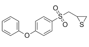
| SMILES | S1C([H])([H])C1([H])C([H])([H])S(C1C([H])=C([H])C(=C([H])C=1[H])OC1C([H])=C([H])C([H])=C([H])C=1[H])(=O)=O |
|
| InChi Key | LSONWRHLFZYHIN-UHFFFAOYSA-N | |
| InChi Code | InChI=1S/C15H14O3S2/c16-20(17,11-14-10-19-14)15-8-6-13(7-9-15)18-12-4-2-1-3-5-12/h1-9,14H,10-11H2 | |
| Chemical Name | 2-[(4-phenoxyphenyl)sulfonylmethyl]thiirane | |
| Synonyms | SB3CT; SB3-CT; 2-[(4-phenoxyphenyl)sulfonylmethyl]thiirane; 2-((4-phenoxyphenylsulfonyl)methyl)thiirane; 2-(((4-Phenoxyphenyl)sulfonyl)methyl)thiirane; (4-phenoxyphenylsulfonyl)methylthiirane; CHEMBL483857; Thiirane, 2-[[(4-phenoxyphenyl)sulfonyl]methyl]-; SB-3CT | |
| HS Tariff Code | 2934.99.9001 | |
| Storage |
Powder-20°C 3 years 4°C 2 years In solvent -80°C 6 months -20°C 1 month Note: Please store this product in a sealed and protected environment, avoid exposure to moisture. |
|
| Shipping Condition | Room temperature (This product is stable at ambient temperature for a few days during ordinary shipping and time spent in Customs) |
Biological Activity
| Targets |
MMP-2 (Ki = 13.9 nM); MMP-9 (Ki = 600 nM) SB-3CT is a potent, selective mechanism-based inhibitor of gelatinases (matrix metalloproteinase-2/MMP-2 and MMP-9), with IC50 values of 0.3 nM for MMP-2 and 0.8 nM for MMP-9 in cell-free enzyme assays [2] - It shows no significant inhibition of other MMP subtypes (MMP-1, MMP-3, MMP-7) or human serine proteases (trypsin, plasmin) at concentrations up to 10 μM, confirming high gelatinase selectivity [2] |
| ln Vitro |
(R)-MG-132, the stereoisomer of MG-132, is being investigated as a possible inhibitor of the proteasome's ability to hydrolyze peptidylglutamyl peptide, trypsin, and chymotrypsin-like activities[1]. The effects of MG-132 and (R)-MG-132 on the inhibition of trypsin-like (TL), peptidylglutamyl peptide hydrolyzing (PGPH), and ChTL of purified 20S proteasomes isolated from human erythrocytes are being studied. MG-132 has IC₅₀ values of 0.89 μM, 104.43 μM, and 5.7 μM for ChTL, TL, and PGPH, in that order. The IC₅₀ values for ChTL, TL, and PGPH of (R)-MG-132 are 0.22 μM, 34.4 μM, and 2.95 μM, respectively[1]. In recombinant MMP-2/MMP-9 enzyme reactions: 1 nM SB-3CT inhibited MMP-2-mediated gelatin degradation by ~98% and MMP-9-mediated gelatin degradation by ~95% (fluorescent gelatin assay) [2] - In human prostate cancer PC-3 cells (high MMP-9 expression): 5 μM SB-3CT for 72 hours inhibited cell proliferation by ~60% (MTT assay), reduced cell invasion by ~80% (Matrigel Transwell assay), and downregulated MMP-9 protein levels by ~75% (Western blot) [3] - In rat brain microvascular endothelial cells (BMECs) exposed to oxygen-glucose deprivation (OGD, mimic of ischemia): 2 μM SB-3CT for 24 hours reduced cell apoptosis by ~55% (Annexin V-FITC/PI staining) and preserved tight junction protein ZO-1 expression by ~65% (immunofluorescence) [4] - In mouse primary microglia (activated by LPS): 1 μM SB-3CT for 18 hours reduced TNF-α secretion by ~60% and IL-1β by ~55% (ELISA), via inhibiting MMP-9-mediated pro-inflammatory cytokine activation [1] |
| ln Vivo |
SB-3CT (i.p.; 50 mg/kg; every other day; five weeks) prevents the intraosseous growth of human PC3 cells in the marrow of human fetal femur fragments that have been implanted in SCID mice[3]. In male Sprague-Dawley rats with severe traumatic brain injury (TBI, controlled cortical impact model): intravenous (iv) injection of SB-3CT (3 mg/kg) at 1 hour post-TBI reduced cerebral lesion volume by ~40% at 72 hours post-TBI vs. vehicle; immunohistochemistry showed ~50% reduction in microglial activation (Iba-1⁺ cells) [1] - In nude mice with PC-3 prostate cancer bone metastasis (intratibial injection of 1×10⁵ cells): oral SB-3CT (10 mg/kg once daily for 28 days) reduced tumor volume in bone by ~55% and decreased osteolytic lesion area by ~60% (micro-CT imaging); plasma MMP-9 levels were reduced by ~70% (ELISA) [3] - In C57BL/6 mice with embolic focal cerebral ischemia (middle cerebral artery occlusion/MCAO model): iv SB-3CT (2 mg/kg) at 30 minutes post-MCAO reduced infarct volume by ~35% at 24 hours post-ischemia and improved neurological deficit scores by ~40% [4] |
| Enzyme Assay |
The fluorescence quenched substrate MOCAcPLGLA2pr(Dnp)-AR-NH2 is used to measure the enzymatic activity of MMP-2, MMP-9, and MMP-7. Using a PTI spectrofluorometer, fluorescence is measured. The temperature of the cuvette compartment is set to 25 °C. MMP-2/MMP-9 gelatinase activity assay (from [2]): Recombinant human MMP-2/MMP-9 was activated with p-aminophenylmercuric acetate (APMA) in activation buffer (50 mM Tris-HCl pH 7.5, 10 mM CaCl₂, 0.05% Brij-35). The activated enzyme was mixed with fluorescent DQ-gelatin (substrate) and SB-3CT (0.01–10 nM) in reaction buffer. The mixture was incubated at 37°C for 2 hours, and fluorescence intensity was measured at excitation 485 nm/emission 535 nm. Inhibition rate was calculated relative to vehicle, and IC50 was determined via 4-parameter logistic regression [2] - MMP-2/MMP-9 selectivity assay (from [2]): Recombinant MMP-1, MMP-3, MMP-7 were prepared following the same activation protocol as MMP-2/9. Each enzyme was mixed with its specific fluorescent peptide substrate (MMP-1: Mca-Pro-Leu-Gly-Leu-Dpa-Ala-Arg-NH₂; MMP-3: Mca-Arg-Pro-Lys-Pro-Tyr-Ala-Nva-Trp-Met-Lys(Dnp)-NH₂) and 10 μM SB-3CT. Fluorescence was measured after 2 hours at 37°C; no significant inhibition (<5%) was observed for non-gelatinase MMPs [2] |
| Cell Assay |
Cell proliferation assay[3] PC3 cells were seeded in 35-mm dishes (5 × 10⁴ cells/dish) in complete culture medium. The next day, the medium was replaced with complete medium supplemented with 1% DMSO alone (vehicle) or SB-3CT (final concentrations 0.1–50 μM) in 1% DMSO. At various times, the cells were harvested with trypsin and counted. Effect of SB-3CT on BMEC-1 cell viability[3] BMEC-1 cells were seeded in 96-well culture plates (104 cells/well) in complete culture medium. Twenty-four h later, the medium was replaced with serum-free, phenolred-free media supplemented with either vehicle (1% DMSO) or SB-3CT (1 nM–50 μM final concentrations). After 72 h, 10 μL of WST-1 were added to each well, and the optical density was measured at 450 nm, according to the manufacturer's instructions. Capillary-like tubule formation assay[3] Twenty-four-well plates were coated with 300 μL of an ice-cold Matrigel solution (10 mg/mL). The plates were then incubated for 30 min at 37°C to allow Matrigel polymerization, and then 5 × 10⁴ BMEC-1 cells were placed onto the Matrigel-coated wells in the presence of complete medium supplemented with various amounts of SB-3CT (0.1–1 μM) or vehicle (1% DMSO). After overnight incubation at 37°C, digital photographs of three randomly selected areas from each well were taken at 10× magnification, using an Olympus® DP12 Microscope Camera. The area occupied by the capillary-like structures was calculated using Adobe Photoshop 7.0. Endothelial cell invasion assay[3] BMEC-1 cells suspended in Medium-199 with 0.1 % bovine serum albumin supplemented with either SB-3CT (0.1–1 μM) or 1% DMSO (vehicle) were seeded (2 × 10⁵ cells per insert) onto Transwell inserts (8-μm pore size) coated with 25 μg/filter Matrigel. Culture medium supplemented with 5% FBS was placed in the lower chamber as a chemoattractant. After 24 h incubation at 37°C, the cells that migrated to the lower side of the filter were stained with Diff-Quik® and counted under 200× magnification. PC-3 cell invasion assay (from [3]): Human prostate cancer PC-3 cells were cultured in RPMI 1640 medium supplemented with 10% fetal bovine serum (FBS) to 80% confluence. Cells were trypsinized, resuspended in serum-free RPMI 1640, and seeded into Matrigel-coated Transwell upper chambers (5×10⁴ cells/well) containing SB-3CT (1–10 μM). Lower chambers contained RPMI 1640 + 10% FBS (chemoattractant). After 48 hours, non-invaded cells on the upper membrane were removed; invaded cells were fixed with methanol, stained with crystal violet, and counted under a microscope. Cell proliferation was assessed via MTT assay (570 nm absorbance) after 72 hours of treatment [3] - BMEC OGD model assay (from [4]): Rat BMECs were cultured in DMEM/F12 medium + 10% FBS. To induce OGD, cells were transferred to glucose-free DMEM and incubated in a hypoxic chamber (1% O₂, 5% CO₂, 94% N₂) for 4 hours. SB-3CT (0.5–5 μM) was added during reoxygenation (21% O₂) for 24 hours. Cells were stained with Annexin V-FITC/PI for apoptosis detection (flow cytometry) and immunostained with anti-ZO-1 antibody for tight junction visualization [4] |
| Animal Protocol |
Five-week-old male C.B.-17.SCID mice[3] 50 mg/kg IP; every other day; five weeks In situ gelatin zymography[3] Frozen tissue sections were obtained from HT1080 tumors grown subcutaneously in SCID mice, which were intraperitonially (i.p.) treated for two consecutive days before sacrifice either with 1 mL vehicle (10% DMSO in PBS) or 1 ml containing 1.25 mg SB-3CT in 10% DMSO (equivalent to 50 mg/kg of mouse weight). In situ gelatin zymography was performed in 8-μm thick unfixed cryostat tumor sections incubated for 1 h with 100 μg/ml DQ™-gelatin and 1 μg/mL DAPI (Molecular Probes), as described previously. Establishment of PC3 human bone tumors and experimental treatment[3] One fourth human fetal femur fragments were implanted subcutaneously in SCID mice as described previously.29 Four weeks later, 1 × 10⁵ PC3 cells were injected through the mouse skin directly into the marrow of the previously implanted bone, as described.29 Twenty-four h after tumor cell inoculation, the mice were injected i.p. with either vehicle (10% DMSO) or SB-3CT in 10% DMSO (50 mg/kg of mouse weight) every other day. Each experimental group contained 9 animals. Five weeks after tumor cell inoculation, the mice were killed and bone implants harvested, weighed, fixed overnight in 10% buffered formalin, and then X-ray imaged using a Lo-Rad M-IV mammography unit with a magnified specimen technique. Images were developed using a Kodak 2000 screen and radiography film. For histomorphometrical and histological analyses, bone tumors were decalcified with 10% ethylenediaminetetraacetic acid (EDTA) (pH 6.5) in PBS, dehydrated, infiltrated and paraffin-embedded. SB-3CT, a discovery from the Mobashery laboratory, was synthesized for this study by reported methodology. Mice were divided into four groups: vehicle-treated group and SB-3CT-treated one with treatment for either one day or seven days after embolic MCA occlusion. SB-3CT (12.5 mg/mL) was freshly dissolved in 25% DMSO/65% PEG-200/10% water and filtered through an Acrodisc syringe filter with a 0.2 μm, 13-mm diameter sterile hydrophobic PTFE membrane. Mice were ip injected with 2 μL/gram body weight of this solution (equivalent to 25 mg/kg) 2 hours after embolic ischemia, followed by an additional dose at 4 hours. In repeated-dose treatment conditions, the same dose of SB-3CT was ip administered 2 and 4 hours after embolic ischemia, followed by once daily from post-ischemia day 1 to 6. Earlier work indicated that ip administration of SB-3CT does not alter mean arterial blood pressure, pH, PCO2, and PO2[4]. Rat TBI model (from [1]): Male Sprague-Dawley rats (250–300 g) were anesthetized and subjected to controlled cortical impact (CCI) to induce severe TBI (impact depth: 2.5 mm, velocity: 4 m/s). At 1 hour post-TBI, rats received iv injection of SB-3CT (3 mg/kg, dissolved in 10% DMSO + 90% physiological saline) or vehicle. At 72 hours post-TBI, rats were euthanized; brains were collected, sectioned, and stained with 2,3,5-triphenyltetrazolium chloride (TTC) to measure lesion volume. Immunohistochemistry was performed with anti-Iba-1 antibody to assess microglial activation [1] - Nude mouse PC-3 bone metastasis model (from [3]): Female nude mice (6–8 weeks old) were anesthetized, and 1×10⁵ PC-3 cells (suspended in 0.05 mL PBS) were injected into the left tibial medullary cavity. Seven days post-inoculation, mice were divided into two groups: (1) SB-3CT group: 10 mg/kg SB-3CT dissolved in 5% DMSO + 95% corn oil, oral gavage once daily; (2) Vehicle group: 5% DMSO + 95% corn oil. After 28 days, mice were euthanized; tibias were collected for micro-CT imaging (to quantify osteolytic lesions) and tumor volume measurement. Plasma was analyzed for MMP-9 via ELISA [3] - Mouse MCAO model (from [4]): Male C57BL/6 mice (20–25 g) were anesthetized, and the middle cerebral artery (MCA) was occluded with a nylon suture for 60 minutes to induce focal cerebral ischemia. At 30 minutes post-MCAO, mice received iv SB-3CT (2 mg/kg, dissolved in 5% ethanol + 95% saline) or vehicle. At 24 hours post-reperfusion, mice were euthanized; brains were sectioned and stained with TTC to measure infarct volume. Neurological deficit scores (0–5 scale) were evaluated before euthanasia [4] |
| Toxicity/Toxicokinetics |
In human/rat/mouse cells (PC-3, BMECs, microglia): SB-3CT up to 10 μM for 72 hours had no significant cytotoxicity (cell viability >90% vs. vehicle, MTT assay) [1,3,4] - In rats (TBI model, 3 mg/kg iv) and mice (MCAO model, 2 mg/kg iv; PC-3 model, 10 mg/kg oral): No significant weight loss (>5% of initial weight) or histopathological abnormalities in liver, kidney, or spleen were detected at therapeutic doses [1,3,4] |
| References |
[1]. Water-Soluble MMP-9 Inhibitor Reduces Lesion Volume after Severe Traumatic Brain Injury. ACS Chem Neurosci. 2015 Oct 21;6(10):1658-64. [2]. Potent and Selective Mechanism-Based Inhibition of GelatinasesJ. Am. Chem. Soc.2000122286799-6800 [3]. Inhibition of human prostate cancer growth, osteolysis and angiogenesis in a bone metastasis model by a novel mechanism-based selective gelatinase inhibitor. Int J Cancer. 2006, 118(11), 2721-2726. [4]. Inhibition of MMP-9 by a selective gelatinase inhibitor protects neurovasculature from embolic focal cerebral ischemia. Mol Neurodegener. 2012, 15, 7-21. |
| Additional Infomation |
2-[(4-phenoxyphenyl)sulfonylmethyl]thiirane is an aromatic ether. SB-3CT is a synthetic, mechanism-based selective gelatinase (MMP-2/9) inhibitor, characterized by irreversible binding to MMP active sites, making it a valuable tool in preclinical studies of MMP-2/9-mediated diseases [2] - Its therapeutic potential is focused on neurological disorders (TBI, cerebral ischemia) and cancer metastasis (prostate cancer bone metastasis), via inhibiting MMP-2/9-mediated extracellular matrix degradation, inflammation, and angiogenesis [1,3,4] - No clinical development (Phase I/II) or FDA approval information is available in the abstracts; it is primarily used as a research reagent to study MMP-2/9 biology [1,2,3,4] - A water-soluble derivative of SB-3CT was developed for improved intravenous delivery in neurological models (e.g., TBI), with similar MMP-2/9 inhibitory potency to the parent compound [1] |
Solubility Data
| Solubility (In Vitro) |
|
|||
| Solubility (In Vivo) |
Solubility in Formulation 1: 5 mg/mL (16.32 mM) in 10% DMSO 20% Cremophor EL + 70% ddH2O (add these co-solvents sequentially from left to right, and one by one), suspension solution; with sonication. Solubility in Formulation 2: 2.5 mg/mL (8.16 mM) in 10% DMSO + 40% PEG300 + 5% Tween80 + 45% Saline (add these co-solvents sequentially from left to right, and one by one), suspension solution; with ultrasonication. For example, if 1 mL of working solution is to be prepared, you can add 100 μL of 25.0 mg/mL clear DMSO stock solution to 400 μL PEG300 and mix evenly; then add 50 μL Tween-80 to the above solution and mix evenly; then add 450 μL normal saline to adjust the volume to 1 mL. Preparation of saline: Dissolve 0.9 g of sodium chloride in 100 mL ddH₂ O to obtain a clear solution. Solubility in Formulation 3: ≥ 2.5 mg/mL (8.16 mM) (saturation unknown) in 10% DMSO + 90% (20% SBE-β-CD in Saline) (add these co-solvents sequentially from left to right, and one by one), clear solution. For example, if 1 mL of working solution is to be prepared, you can add 100 μL of 25.0 mg/mL clear DMSO stock solution to 900 μL of 20% SBE-β-CD physiological saline solution and mix evenly. Preparation of 20% SBE-β-CD in Saline (4°C,1 week): Dissolve 2 g SBE-β-CD in 10 mL saline to obtain a clear solution. Solubility in Formulation 4: ≥ 2.5 mg/mL (8.16 mM) (saturation unknown) in 10% DMSO + 90% Corn Oil (add these co-solvents sequentially from left to right, and one by one), clear solution. For example, if 1 mL of working solution is to be prepared, you can add 100 μL of 25.0 mg/mL clear DMSO stock solution to 900 μL corn oil and mix evenly. Solubility in Formulation 5: 4% DMSO+corn oil: 10mg/mL (Please use freshly prepared in vivo formulations for optimal results.) |
| Preparing Stock Solutions | 1 mg | 5 mg | 10 mg | |
| 1 mM | 3.2637 mL | 16.3185 mL | 32.6371 mL | |
| 5 mM | 0.6527 mL | 3.2637 mL | 6.5274 mL | |
| 10 mM | 0.3264 mL | 1.6319 mL | 3.2637 mL |