Pepstatin A (NSC-272671; NSC272671), a pentapeptide, is a potent and selective aspartic protease inhibitor which also inhibits HIV replication. Pepstatin A was originally isolated from the microbe. For the enzymes hemoglobin-pepsin, hemoglobin-proctase, casein-pepsin, casein-proctase, casein-acid protease, and hemoglobin-acid protease, respectively, its IC50 values are 4.5 nM, 6.2 nM, 150 nM, 290 nM, 520 nM, and 260 nM. At weak acid pH values, pepstatinA, a highly potent renin inhibitor, inhibited both human and porcine renin with IC50 values of 15 μM and 0.32, respectively. For aspartic proteinases like pepsin, cathepsins D and E, pepstatin A effectively inhibits them. Pepstatin A appears to have no harmful effects on HIV-infected cells, but it does block a portion of the intracellular processing of the HIV gag protein in H9 cells that have the virus. By preventing ERK signaling and reducing NFATc1 expression, pepstatin A also prevents osteoclast differentiation.
Physicochemical Properties
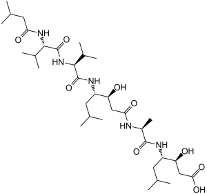
| Molecular Formula | C34H63N5O9 | |
| Molecular Weight | 685.89 | |
| Exact Mass | 685.462 | |
| Elemental Analysis | C, 59.54; H, 9.26; N, 10.21; O, 20.99 | |
| CAS # | 26305-03-3 | |
| Related CAS # | 28575-34-0 | |
| PubChem CID | 5478883 | |
| Sequence | IsoValeryl-Val-Val-Sta-Ala-Sta-OH | |
| SequenceShortening | IsoVeryl-VV-Sta-A-Sta-OH | |
| Appearance | White to off-white solid powder | |
| Density | 1.1±0.1 g/cm3 | |
| Boiling Point | 997.6±65.0 °C at 760 mmHg | |
| Melting Point | 233 °C (dec.)(lit.) | |
| Flash Point | 557.1±34.3 °C | |
| Vapour Pressure | 0.0±0.6 mmHg at 25°C | |
| Index of Refraction | 1.504 | |
| Source | Actinomycetes | |
| LogP | 2.16 | |
| Hydrogen Bond Donor Count | 8 | |
| Hydrogen Bond Acceptor Count | 9 | |
| Rotatable Bond Count | 22 | |
| Heavy Atom Count | 48 | |
| Complexity | 1060 | |
| Defined Atom Stereocenter Count | 7 | |
| SMILES | O([H])[C@@]([H])(C([H])([H])C(N([H])[C@@]([H])(C([H])([H])[H])C(N([H])[C@]([H])([C@]([H])(C([H])([H])C(=O)O[H])O[H])C([H])([H])C([H])(C([H])([H])[H])C([H])([H])[H])=O)=O)[C@]([H])(C([H])([H])C([H])(C([H])([H])[H])C([H])([H])[H])N([H])C([C@]([H])(C([H])(C([H])([H])[H])C([H])([H])[H])N([H])C([C@]([H])(C([H])(C([H])([H])[H])C([H])([H])[H])N([H])C(C([H])([H])C([H])(C([H])([H])[H])C([H])([H])[H])=O)=O)=O |
|
| InChi Key | FAXGPCHRFPCXOO-LXTPJMTPSA-N | |
| InChi Code | InChI=1S/C34H63N5O9/c1-17(2)12-23(37-33(47)31(21(9)10)39-34(48)30(20(7)8)38-27(42)14-19(5)6)25(40)15-28(43)35-22(11)32(46)36-24(13-18(3)4)26(41)16-29(44)45/h17-26,30-31,40-41H,12-16H2,1-11H3,(H,35,43)(H,36,46)(H,37,47)(H,38,42)(H,39,48)(H,44,45)/t22-,23-,24-,25-,26-,30-,31-/m0/s1 | |
| Chemical Name | (3S,4S)-3-hydroxy-4-[[(2S)-2-[[(3S,4S)-3-hydroxy-6-methyl-4-[[(2S)-3-methyl-2-[[(2S)-3-methyl-2-(3-methylbutanoylamino)butanoyl]amino]butanoyl]amino]heptanoyl]amino]propanoyl]amino]-6-methylheptanoic acid | |
| Synonyms |
|
|
| HS Tariff Code | 2934.99.9001 | |
| Storage |
Powder-20°C 3 years 4°C 2 years In solvent -80°C 6 months -20°C 1 month Note: Please store this product in a sealed and protected environment, avoid exposure to moisture. |
|
| Shipping Condition | Room temperature (This product is stable at ambient temperature for a few days during ordinary shipping and time spent in Customs) |
Biological Activity
| Targets |
Hemoglobin-pepsin (IC50 = 4.5 nM); Hemoglobin-proctase (IC50 = 6.2 nM); Casein-pepsin (IC50 = 150 nM); Hemoglobin-acid protease (IC50 = 260 nM); Casein-proctase (IC50 = 290 nM); Casein-acid protease (IC50 = 520 nM) Pepstatin A targets pepsin (IC50 = 0.04 μM for porcine pepsin) [1][4] Pepstatin A targets cathepsin D (Ki = 0.15 nM for human cathepsin D) [2] Pepstatin A targets renin (IC50 = 1.7 μM for human renin) [1] Pepstatin A targets aspartic proteases from Aspergillus niger (IC50 = 0.2 μM) [4] |
| ln Vitro |
Pepstatin A shows effectively inhibits aspartic proteinases, including cathepsins D and E and pepsin. Pepstatin A appears to have no harmful effects on HIV-infected cells, but it does block a portion of the intracellular processing of the HIV gag protein in H9 cells that have the virus.[1] Pepstatin A laos inhibits NFATc1 expression and ERK signaling, which prevents osteoclasts from differentiating.[2] Pepstatin A (0.01–1 μM) exhibited concentration-dependent inhibition of porcine pepsin activity: 0.04 μM inhibited 50% of activity, and 1 μM achieved 95% inhibition using hemoglobin as substrate [1][4] Pepstatin A (0.1–10 nM) potently inhibited human cathepsin D-mediated peptide hydrolysis, with Ki = 0.15 nM; 1 nM reduced enzyme activity by 80% [2] Pepstatin A (1–20 μM) suppressed proliferation of human breast cancer MCF-7 cells by 60% at 10 μM (72 hours), inducing G2/M cell cycle arrest and reducing cathepsin D-dependent matrix metalloproteinase (MMP-2) activation [3] Pepstatin A (0.5 μM) inhibited aspartic protease activity in Aspergillus niger cultures by 75%, reducing fungal protein degradation [4] Pepstatin A (20 μM) had no cytotoxicity to normal human mammary epithelial cells (HMECs) (cell viability >92% after 72 hours) [3] Pepstatin A (0.1–5 μM) showed no significant inhibition of canine renal Ca²+-ATPase or Mg²+-ATPase, confirming selectivity for aspartic proteases [1] |
| ln Vivo |
Pepstatin A has a very low toxicity, with LD50s of >2000 mg/kg for all species when taken orally, and LD50s of 1090 mg/kg, 875 mg/kg, 820 mg/kg, and 450 mg/kg for mice, rats, rabbits, and dogs when taken intraperitoneally. Pepstatin A (0.5–50 mg/kg, p.o.) inhibits the pylorus-induced stomach ulceration in ligated Shay rats. Pepstatin A (5 mg/kg/day, intraperitoneal injection for 21 days) inhibited tumor growth in MCF-7 xenograft mice: tumor volume reduced by 55%, and intratumoral cathepsin D activity decreased by 70% [3] Pepstatin A (10 mg/kg/day, oral gavage for 14 days) reduced gastric pepsin activity by 65% in rats, decreasing acid-induced gastric mucosal damage area by 50% [1] Pepstatin A (3 mg/kg/day, i.p.) had no significant effect on body weight or organ index (liver, kidney, spleen) in mice, indicating low systemic toxicity [3] |
| Enzyme Assay |
Pepstatin A is a well-known aspartic proteinase inhibitor; its IC50 values for human renin, HIV protease, pepsin, and cathepsin D are 15 μM, 2 μM, < 5 nM, and < 40 nM, respectively. The protease encoded by the human immunodeficiency virus (HIV) processes the viral gag and gag-pol protein precursor by posttranslational cleavage. In this study we have demonstrated by site-specific mutagenesis (Asp----Thr) and by pepstatin A inhibition that the recombinant HIV protease is an aspartic-type protease. Furthermore, incubation of HIV-infected H9 cells with pepstatin A inhibited part of the intracellular processing of the HIV gag protein yet had no apparent toxicity on HIV-infected cells during 48 hr of incubation.[1] Pepsin inhibition assay: Porcine pepsin was incubated with Pepstatin A (0.001–5 μM) in acidic buffer (pH 2.0) for 30 minutes; hemoglobin substrate was added, and the mixture was incubated at 37°C for 1 hour; trichloroacetic acid terminated the reaction, and absorbance of soluble peptides was measured at 280 nm to calculate inhibition rate and IC50 [1][4] Cathepsin D activity assay: Human cathepsin D was preincubated with Pepstatin A (0.01–100 nM) in buffer (pH 3.5) for 20 minutes; a fluorogenic peptide substrate was added, and fluorescence intensity (excitation 328 nm, emission 460 nm) was monitored for 60 minutes to assess enzyme inhibition and calculate Ki [2] Renin inhibition assay: Human renin was incubated with Pepstatin A (0.1–50 μM) and angiotensinogen substrate in buffer (pH 6.0) at 37°C for 2 hours; angiotensin I production was quantified by radioimmunoassay to determine IC50 [1] Aspergillus niger protease inhibition assay: Aspergillus niger mycelial homogenates were treated with Pepstatin A (0.1–1 μM) for 1 hour; casein substrate was added, and protease activity was quantified by measuring soluble peptides at 280 nm [4] |
| Cell Assay |
Pepstatin A is freshly dissolved in DMSO at 7 mM. It is very slowly diluted (1:100) into the medium of HIV-infected H9 suspension cultures so that no pepstatin A precipitated (final concentration, 70 μM pepstatin A and 1% DMSO), and the cultures are incubated without change of culture medium for 48 hr. As control, uninfected H9 cells are also incubated with pepstatin and in addition HIV infected and uninfected cells are incubated with 1% DMSO but without pepstatin[1]. Pepstatin A is well known to be an inhibitor of aspartic proteinases such as pepsin, cathepsins D and E. Except for its role as a proteinase inhibitor, however, the pharmacological action of pepstatin A upon cells remain unclear. In this study, we found that pepstatin A suppressed receptor activator of NF-kappaB ligand (RANKL)-induced osteoclast differentiation. Pepstatin A suppressed the formation of multinuclear osteoclasts dose-dependently. This inhibition of the formation only affected osteoclast cells, i.e., not osteoblast-like cells. Furthermore, pepstatin A also suppressed differentiation from pre-osteoclast cells to mononuclear osteoclast cells dose-dependently. This inhibition seems to be independent of the activities of proteinases such as cathepsin D, because the formation of osteoclasts was not suppressed with the concentration that inhibited the activity of cathepsin D. Cell signaling analysis indicated that the phosphorylation of ERK was inhibited in pepstatin A-treated cells, while the phosphorylation of IkappaB and Akt showed almost no change. Furthermore, pepstatin A decreased the expression of nuclear factor of activated T cells c1 (NFATc1). These results suggest that pepstatin A suppresses the differentiation of osteoclasts through the blockade of ERK signaling and the inhibition of NFATc1 expression.[2] Breast cancer cell proliferation assay: MCF-7 cells were seeded in 96-well plates (5×10³ cells/well) and treated with Pepstatin A (0.5–20 μM) for 72 hours; cell viability was assessed by MTT assay (absorbance at 570 nm), and IC50 for proliferation inhibition was calculated [3] Cell cycle and apoptosis assay: MCF-7 cells were treated with Pepstatin A (10 μM) for 48 hours; cell cycle distribution was analyzed by propidium iodide (PI) staining via flow cytometry, and apoptotic cells were detected by Annexin V-FITC/PI double staining [3] Western blot assay: MCF-7 cells treated with Pepstatin A (5–15 μM) for 24 hours were lysed; blots were probed with antibodies against cathepsin D, MMP-2, cyclin B1, cleaved caspase-3, and GAPDH (loading control) [3] Normal epithelial cell toxicity assay: HMECs were seeded in 96-well plates (5×10³ cells/well) and treated with Pepstatin A (5–30 μM) for 72 hours; cell viability was measured by MTT assay [3] |
| Animal Protocol |
Pylorus ligated male Wistar rats 0.5, 1, 10 and 50 mg/kg Oral administration, 15 minutes after pyloric ligation Breast cancer xenograft model: Nude mice were subcutaneously injected with 2×10⁶ MCF-7 cells; when tumors reached 100 mm³, mice were randomly divided into control and treatment groups; treatment group received Pepstatin A (5 mg/kg/day, dissolved in 10% DMSO + 90% saline) via intraperitoneal injection for 21 days; tumor volume was measured every 3 days, and tumor tissues were collected for cathepsin D activity assay and immunohistochemistry [3] Gastric mucosal protection model: Sprague-Dawley rats were pretreated with Pepstatin A (10 mg/kg/day, dissolved in 0.5% carboxymethylcellulose sodium) via oral gavage for 14 days; rats were then challenged with hydrochloric acid (0.1 M) intragastrically; gastric mucosal damage area was measured, and gastric pepsin activity was quantified [1] |
| ADME/Pharmacokinetics |
Pepstatin A has low oral bioavailability (<10%) in rats due to gastrointestinal degradation [4] Intravenous administration (10 mg/kg) in rabbits resulted in peak plasma concentration (Cmax) of 8 μg/mL, elimination half-life (t1/2) of 1.5 hours, and volume of distribution (Vd) of 0.8 L/kg [4] The drug is primarily excreted in urine (70% as unchanged drug) within 24 hours, with 15% excreted in feces [4] It distributes widely to tissues, with highest concentrations in liver, kidney, and spleen (tissue/plasma ratio = 2.5–3.0 at 1 hour post-dose) [4] |
| Toxicity/Toxicokinetics |
rat LD50 oral >2 gm/kg Proceedings of the International Symposium of the Princess Takamatsu Cancer Research Fund., 6(57), 1976 mouse LD50 oral >2 gm/kg Proceedings of the International Symposium of the Princess Takamatsu Cancer Research Fund., 6(57), 1976 dog LD50 oral >2 gm/kg Journal of Antibiotics., 23(259), 1970 [PMID:4912600] dog LD50 intraperitoneal 450 mg/kg Journal of Antibiotics., 23(259), 1970 [PMID:4912600] rabbit LD50 oral >2 gm/kg Journal of Antibiotics., 23(259), 1970 [PMID:4912600] Pepstatin A showed low acute toxicity in mice: LD50 = 800 mg/kg (intraperitoneal), LD50 = 1200 mg/kg (oral) [3] Chronic administration (10 mg/kg/day for 28 days) in rats caused no significant changes in serum ALT, AST, BUN, or creatinine levels, indicating no obvious hepatotoxicity or nephrotoxicity [1][3] Plasma protein binding rate of Pepstatin A was 85% in human plasma and 82% in mouse plasma [2] |
| References |
[1]. Proc Natl Acad Sci U S A . 1988 Sep;85(18):6612-6. [2]. J Biochem . 2006 Mar;139(3):583-90. [3]. Nat Commun . 2022 Aug 16;13(1):4804. [4]. J Antibiot (Tokyo) . 1970 May;23(5):259-62. |
| Additional Infomation |
Pepstatin A is a pentapeptide isolated from Streptomyces testaceus. It is a potent inhibitor of aspartyl proteases. It has a role as a bacterial metabolite and an EC 3.4.23. (aspartic endopeptidase) inhibitor. It is a pentapeptide and a secondary carboxamide. It is a conjugate acid of a pepstatin A(1-). Pepstatin has been reported in Streptomyces with data available. Pepstatin A is a naturally occurring pentapeptide isolated from Streptomyces testaceus, classified as a selective aspartic protease inhibitor [4] Its mechanism of action involves binding to the active site of aspartic proteases (via hydrogen bonds with catalytic aspartate residues), blocking substrate access and enzyme catalysis [1][2][4] It exhibits potent inhibitory activity against a range of aspartic proteases, including digestive proteases (pepsin), lysosomal proteases (cathepsin D), and microbial proteases [1][2][4] In preclinical studies, it shows antitumor potential in breast cancer by inhibiting cathepsin D-dependent tumor invasion and proliferation [3] It is widely used as a research tool to study aspartic protease function in physiology and disease [1][2][3][4] |
Solubility Data
| Solubility (In Vitro) |
|
|||
| Solubility (In Vivo) |
Solubility in Formulation 1: 2.08 mg/mL (3.03 mM) in 10% DMSO + 40% PEG300 + 5% Tween80 + 45% Saline (add these co-solvents sequentially from left to right, and one by one), clear solution; with sonication. For example, if 1 mL of working solution is to be prepared, you can add 100 μL of 20.8 mg/mL clear DMSO stock solution to 400 μL PEG300 and mix evenly; then add 50 μL Tween-80 to the above solution and mix evenly; then add 450 μL normal saline to adjust the volume to 1 mL. Preparation of saline: Dissolve 0.9 g of sodium chloride in 100 mL ddH₂ O to obtain a clear solution. Solubility in Formulation 2: ≥ 2.08 mg/mL (3.03 mM) (saturation unknown) in 10% DMSO + 90% (20% SBE-β-CD in Saline) (add these co-solvents sequentially from left to right, and one by one), clear solution. For example, if 1 mL of working solution is to be prepared, you can add 100 μL of 20.8 mg/mL clear DMSO stock solution to 900 μL of 20% SBE-β-CD physiological saline solution and mix evenly. Preparation of 20% SBE-β-CD in Saline (4°C,1 week): Dissolve 2 g SBE-β-CD in 10 mL saline to obtain a clear solution. Solubility in Formulation 3: ≥ 2.08 mg/mL (3.03 mM) (saturation unknown) in 10% DMSO + 90% Corn Oil (add these co-solvents sequentially from left to right, and one by one), clear solution. For example, if 1 mL of working solution is to be prepared, you can add 100 μL of 20.8 mg/mL clear DMSO stock solution to 900 μL of corn oil and mix evenly. Solubility in Formulation 4: 5 mg/mL (7.29 mM) in 50% PEG300 50% Saline (add these co-solvents sequentially from left to right, and one by one), suspension solution; with ultrasonication. Preparation of saline: Dissolve 0.9 g of sodium chloride in 100 mL ddH₂ O to obtain a clear solution. (Please use freshly prepared in vivo formulations for optimal results.) |
| Preparing Stock Solutions | 1 mg | 5 mg | 10 mg | |
| 1 mM | 1.4580 mL | 7.2898 mL | 14.5796 mL | |
| 5 mM | 0.2916 mL | 1.4580 mL | 2.9159 mL | |
| 10 mM | 0.1458 mL | 0.7290 mL | 1.4580 mL |