Parthenolide, formerly known as NSC-157035, is a novel sesquiterpene lactone that was isolated from the herb feverfew with the potential to treat cancer. It works as an NF-κB activation inhibitor. In HeLa cells, parthenolide promotes apoptosis and growth inhibition via autophagy by inhibiting the PI3K/Akt signaling pathway, as well as mitochondrial membrane depolarization and ROS production.
Physicochemical Properties
| Molecular Formula | C15H20O3 |
| Molecular Weight | 248.322 |
| Exact Mass | 248.141 |
| Elemental Analysis | C, 72.55; H, 8.12; O, 19.33 |
| CAS # | 20554-84-1 |
| Related CAS # | 20554-84-1 |
| PubChem CID | 6473881 |
| Appearance | White to off-white solid powder |
| Density | 1.1±0.1 g/cm3 |
| Boiling Point | 394.1±42.0 °C at 760 mmHg |
| Melting Point | 115-116ºC(lit.) |
| Flash Point | 166.3±22.5 °C |
| Vapour Pressure | 0.0±0.9 mmHg at 25°C |
| Index of Refraction | 1.533 |
| LogP | 2.42 |
| Hydrogen Bond Donor Count | 0 |
| Hydrogen Bond Acceptor Count | 3 |
| Rotatable Bond Count | 0 |
| Heavy Atom Count | 18 |
| Complexity | 437 |
| Defined Atom Stereocenter Count | 4 |
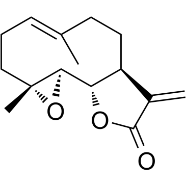
| SMILES | O1[C@@]2([H])[C@]3([H])[C@]([H])(C(=C([H])[H])C(=O)O3)C([H])([H])C([H])([H])C(C([H])([H])[H])=C([H])C([H])([H])C([H])([H])[C@@]12C([H])([H])[H] |c:25| |
| InChi Key | KTEXNACQROZXEV-PVLRGYAZSA-N |
| InChi Code | InChI=1S/C15H20O3/c1-9-5-4-8-15(3)13(18-15)12-11(7-6-9)10(2)14(16)17-12/h5,11-13H,2,4,6-8H2,1,3H3/b9-5+/t11-,12-,13+,15+/m0/s1 |
| Chemical Name | (1S,2R,4R,7E,11S)-4,8-dimethyl-12-methylidene-3,14-dioxatricyclo[9.3.0.02,4]tetradec-7-en-13-one |
| Synonyms | Parthenolide; NSC 157035; NSC157035; NSC-157035 |
| HS Tariff Code | 2934.99.9001 |
| Storage |
Powder-20°C 3 years 4°C 2 years In solvent -80°C 6 months -20°C 1 month |
| Shipping Condition | Room temperature (This product is stable at ambient temperature for a few days during ordinary shipping and time spent in Customs) |
Biological Activity
| Targets |
HDAC1 ; NF-κB; MDM2; p53 Parthenolide is an inhibitor of the transcription factor nuclear factor-kappaB (NF-κB). Its analog, dimethylaminoparthenolide (DMAPT), also inhibits NF-κB. Parthenolide and its analog DMAPT reduce the levels of histone deacetylase 1 (HDAC-1) and DNA methyltransferase 1 (DNMT1) independently of NF-κB inhibition. |
| ln Vitro |
Parthenolide (PTL), a sesquiterpene lactone isolated from feverfew shoots, inhibits the activity of the enzyme ubiquitin-specific peptidase 7 (USP7) and deubiquitinates and stabilizes β-catenin, the primary transcriptional factor of the Wnt signaling pathway, indicating that PTL is a promising anticancer agent that targets aberrant USP7/Wnt signaling.[4] Parthenolide and its water-soluble analog DMAPT exhibit anti-tumor activity in multiple cancers. DMAPT treatment (10 µM, 12 hours) increases histone H3 lysine 36 trimethylation (H3K36me3) by 1.6 to 2.5-fold and histone H4 lysine 20 trimethylation (H4K20me3) by 2.8 to 16-fold in bladder cancer cell lines (UMUC-3 and RT-4). This epigenetic modulation is mediated through the upregulation of histone methyltransferases NSD1 (KMT3B), SETD2 (KMT3A), and KMT5C (SUV4-20H2). DMAPT-mediated NF-κB inhibition results in elevated NSD1 and SETD2 expression, while KMT5C induction is NF-κB-independent. DMAPT treatment reduces levels of HDAC-1, EZH2, C-terminal binding protein 1 (CtBP1), and poly (ADP-ribose) polymerase 1 (PARP-1) in a cell type-dependent manner. It also increases expression of the cell cycle inhibitor p21 and the pro-apoptotic protein BIM. NSD1 is essential for DMAPT-induced BIM expression. DMAPT induces apoptosis in wild-type mouse embryonic fibroblasts (MEFs) but not in p65-deficient (p65 -/-) MEFs, indicating the role of NF-κB inhibition in its apoptotic effect. |
| ln Vivo |
In a subcutaneous xenograft mouse model of gastric cancer (GC), parthenolide (PN), a P65 inhibitor, significantly increases tumor volumes that can be inhibited by gastrin.[5] Parthenolide administration reduces liver damage and fibrosis in liver-specific Phb1 knockout (Phb1 KO) mice subjected to bile duct ligation (BDL), an experimental model of obstructive cholestasis. Intraperitoneal injection of parthenolide (3 mg/kg) 24 hours and 1 hour before BDL, or twice a week for two weeks, improved survival rates in Phb1 KO mice after BDL. Treated mice showed reduced liver injury markers: decreased necrotic areas, lower TUNEL-positive cells, reduced serum ALT (from 8431 ± 957 U/L to 4225 ± 210 U/L) and AST (from 4805 ± 300 U/L to 2242 ± 438 U/L) activities. Fibrosis markers (αSMA protein levels) and proinflammatory cytokines (TNFα, IL-6, TNFR2 mRNA levels) were also reduced. Parthenolide treatment lowered hepatic HDAC4 mRNA and protein levels, restored FXR and CYP7A1 expression levels, and increased hepatic 26S proteasome activity, promoting ubiquitin-dependent degradation of proteins including HDAC4. In Phb1 KO mice with spontaneous liver fibrosis, parthenolide treatment for 15 days attenuated the fibrotic phenotype: reduced serum AST activity (from 268 ± 68 U/L to 63 ± 16 U/L), decreased protein levels of Smad2/3 and F4/80, lowered mRNA levels of profibrogenic markers (TGFβ, αSMA), reduced protein levels of the inflammatory cytokine TNFα, increased FXR levels, decreased CYP7A1 levels, and reduced global ubiquitin levels. Parthenolide treatment specifically decreased HDAC4 protein levels (with no change in its mRNA), restored proteasome activity, and reduced ubiquitinated HDAC4 levels. |
| Cell Assay |
HEK293W cells are seeded in 96-well plates with three repeats and treated with PTL for 24 hours to confirm that PTL is a Wnt signaling inhibitor. HCT116 and SW480 cells are seeded in 96-well plates and transfected with Renilla reporter plasmid (8 ng/well) and SuperTOPFlash (80 ng/well), two luciferase reporter plasmids that are responsive to Wnt/-catenin signaling. Cells are transfected for 3 hours, given different PTL concentrations for 24 hours, and then lysed. Using the Dual-Luciferase Reporter Assay kit, the activities of both Firefly and Renilla luciferases are determined. For epigenetic marker analysis, cells (e.g., UMUC-3, RT-4, MDA-MB-231, MEFs) were treated with 10 µM DMAPT for specified durations (e.g., 6h, 12h, 24h). Proteins were extracted for western blotting. Histones were extracted using a protocol involving cell lysis with Triton Extraction Buffer, centrifugation to collect nuclei, and acid extraction with 0.2 M HCl. Western blotting was performed using specific antibodies against histone modifications (H3K36me3, H4K20me3, etc.) and epigenetic regulators (NSD1, SETD2, KMT5C, HDAC-1, EZH2, PARP-1, BIM, p21). For gene expression analysis, total RNA was extracted, and first-strand cDNA was synthesized using random hexamers and reverse transcriptase. Quantitative reverse transcription-PCR (qRT-PCR) was performed using SYBR green chemistry, with β-actin as the internal control. Specific primers were used to measure mRNA levels of target genes (NSD1, SETD2, KMT5C, CXCL1). For siRNA experiments, cells were transfected with 25 nM of siRNA targeting NSD1 or SETD2 using a lipofectamine reagent. After 3 days, cells were treated with DMAPT for 24 hours and then harvested for protein or RNA analysis to assess the effects on BIM, p21 expression, and cell growth. For apoptosis assay, cells were treated with DMAPT for 24 hours. Both floating and adherent cells were collected, stained with Alexa Fluor-488-conjugated Annexin V and propidium iodide, and analyzed by flow cytometry to quantify apoptotic (Annexin V-positive) cells. For electrophoretic mobility shift assay (EMSA), cells were harvested with or without TNF-α treatment (5 ng/ml, 15 min). Nuclear extracts were prepared and incubated with labeled DNA probes for NF-κB or SP-1. For supershift assays, specific antibodies against NF-κB subunits (p65, p50, c-Rel) were added. |
| Animal Protocol |
Mice: They use Phb1 knockout mice. We treat males who are 8 to 12 weeks old. A dose of 3 mg/kg of parthenolide is intraperitoneally injected 24 hours and 1 hour prior to bile duct ligation (BDL) or twice weekly over a period of two weeks. For further analysis, liver specimens are quickly frozen. For the bile duct ligation (BDL) model, 8-12 week old male Phb1 KO mice and wild-type controls were used. Parthenolide was administered via intraperitoneal injection at a dose of 3 mg/kg. Two dosing regimens were used: 1) Pre-BDL treatment: parthenolide was injected 24 hours and 1 hour before performing the BDL surgery. 2) Post-BDL treatment: parthenolide was injected twice a week for two weeks following BDL. Control animals received vehicle. Liver specimens were collected at specified time points (e.g., 3, 7, or 14 days post-BDL) and snap-frozen for subsequent biochemical, histological, and molecular analyses (e.g., serum ALT/AST measurement, H&E, Sirius Red, TUNEL, immunohistochemistry for αSMA, CK19, F4/80, Western blot, qPCR). For the study of spontaneous fibrosis in Phb1 KO mice, animals were treated with parthenolide (3 mg/kg, i.p.) for 15 days, and liver tissues were analyzed similarly. |
| References |
[1]. NF-κB-dependent and -independent epigenetic modulation using the novel anti-cancer agent DMAPT. Cell Death Dis. 2015 Jan 22;6:e1608. [2]. Parthenolide induces apoptosis via TNFRSF10B and PMAIP1 pathways in human lung cancer cells. J Exp Clin Cancer Res. 2014 Jan 6;33:3. [3]. Histone deacetylase 4 promotes cholestatic liver injury in the absence of prohibitin-1. Hepatology. 2015 Oct;62(4):1237-48. |
| Additional Infomation |
(1Ar,7aS,10aS,10bS)-1a,5-dimethyl-8-methylidene-2,3,6,7,7a,8,10a,10b-octahydrooxireno[9,10]cyclodeca[1,2-b]furan-9(1aH)-one is a germacranolide. Parthenolide has been used in trials studying the diagnostic of Allergic Contact Dermatitis. (1aR,7aS,10aS,10bS)-1a,5-dimethyl-8-methylidene-2,3,6,7,7a,8,10a,10b-octahydrooxireno[9,10]cyclodeca[1,2-b]furan-9(1aH)-one has been reported in Cyathocline purpurea, Magnolia compressa, and other organisms with data available. See also: Parthenolide (annotation moved to). Parthenolide is a sesquiterpene lactone. Its analog, dimethylaminoparthenolide (DMAPT), is a clinical-grade, water-soluble compound developed as a potent NF-κB inhibitor. This study identifies DMAPT as an epigenetic modulator that functions in both NF-κB-dependent and independent manners. By inhibiting NF-κB, DMAPT reverses the repression of tumor suppressors NSD1 and SETD2, leading to increased H3K36me3. Independently of NF-κB, it induces KMT5C and increases H4K20me3. These actions can reverse cancer-specific epigenetic abnormalities. NSD1 and SETD2 are frequently mutated or deleted in cancers like bladder and breast cancer. Lower expression of these genes correlates with poor prognosis in breast cancer. DMAPT's ability to restore their expression and modify histone marks suggests its potential as a therapeutic agent to reverse epigenetic dysregulation in cancer. |
Solubility Data
| Solubility (In Vitro) |
DMSO: ~50 mg/mL (~201.4 mM) Ethanol: ~50 mg/mL (~201.4 mM) |
| Solubility (In Vivo) |
Solubility in Formulation 1: ≥ 2.5 mg/mL (10.07 mM) (saturation unknown) in 10% DMSO + 90% Corn Oil (add these co-solvents sequentially from left to right, and one by one), clear solution. For example, if 1 mL of working solution is to be prepared, you can add 100 μL of 25.0 mg/mL clear DMSO stock solution to 900 μL of corn oil and mix evenly. Solubility in Formulation 2: ≥ 2.08 mg/mL (8.38 mM) (saturation unknown) in 10% DMSO + 40% PEG300 + 5% Tween80 + 45% Saline (add these co-solvents sequentially from left to right, and one by one), clear solution. For example, if 1 mL of working solution is to be prepared, you can add 100 μL of 20.8 mg/mL clear DMSO stock solution to 400 μL PEG300 and mix evenly; then add 50 μL Tween-80 to the above solution and mix evenly; then add 450 μL normal saline to adjust the volume to 1 mL. Preparation of saline: Dissolve 0.9 g of sodium chloride in 100 mL ddH₂ O to obtain a clear solution. Solubility in Formulation 3: 2% DMSO+corn oil: 10mg/mL (Please use freshly prepared in vivo formulations for optimal results.) |
| Preparing Stock Solutions | 1 mg | 5 mg | 10 mg | |
| 1 mM | 4.0271 mL | 20.1353 mL | 40.2706 mL | |
| 5 mM | 0.8054 mL | 4.0271 mL | 8.0541 mL | |
| 10 mM | 0.4027 mL | 2.0135 mL | 4.0271 mL |