Berbamine is a naturally occuring isoquinoline alkaloid found in traditional Chinese medicine Barberry with anti-tumor, immunomodulatory and cardiovascular effects. Calcium channel blockers include berbamine. It can be used to treat or prevent cardiac arrhythmias, and it may have an impact on the action potential's polarization-repolarization phase, excitability or refractoriness, impulse conduction, or membrane responsiveness within cardiac fibers.
Physicochemical Properties
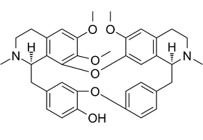
| Molecular Formula | C₃₇H₄₀N₂O₆ |
| Molecular Weight | 608.72 |
| Exact Mass | 608.288 |
| CAS # | 478-61-5 |
| Related CAS # | Berbamine dihydrochloride;6078-17-7 |
| PubChem CID | 275182 |
| Appearance | White to yellow solid |
| Density | 1.2±0.1 g/cm3 |
| Boiling Point | 707.0±60.0 °C at 760 mmHg |
| Melting Point | 225°C |
| Flash Point | 381.4±32.9 °C |
| Vapour Pressure | 0.0±2.3 mmHg at 25°C |
| Index of Refraction | 1.602 |
| LogP | 3.89 |
| Hydrogen Bond Donor Count | 1 |
| Hydrogen Bond Acceptor Count | 8 |
| Rotatable Bond Count | 3 |
| Heavy Atom Count | 45 |
| Complexity | 963 |
| Defined Atom Stereocenter Count | 2 |
| SMILES | O1C2=C(C([H])=C3C([H])([H])C([H])([H])N(C([H])([H])[H])[C@@]([H])(C([H])([H])C4C([H])=C([H])C(=C([H])C=4[H])OC4=C(C([H])=C([H])C(=C4[H])C([H])([H])[C@]4([H])C5=C1C(=C(C([H])=C5C([H])([H])C([H])([H])N4C([H])([H])[H])OC([H])([H])[H])OC([H])([H])[H])O[H])C3=C2[H])OC([H])([H])[H] |
| InChi Key | DFOCUWZXJBAUSQ-URLMMPGGSA-N |
| InChi Code | InChI=1S/C37H40N2O6/c1-38-14-12-24-19-32(41-3)33-21-27(24)28(38)16-22-6-9-26(10-7-22)44-31-18-23(8-11-30(31)40)17-29-35-25(13-15-39(29)2)20-34(42-4)36(43-5)37(35)45-33/h6-11,18-21,28-29,40H,12-17H2,1-5H3/t28-,29+/m0/s1 |
| Chemical Name | (1S,14R)-20,21,25-trimethoxy-15,30-dimethyl-7,23-dioxa-15,30-diazaheptacyclo[22.6.2.23,6.18,12.114,18.027,31.022,33]hexatriaconta-3(36),4,6(35),8,10,12(34),18,20,22(33),24,26,31-dodecaen-9-ol |
| Synonyms | Berbamine HCl; BERBAMINE; (+)-Berbamine; d-Berbamine; 478-61-5; Berbenine; V5KM4XJ0WM; CHEBI:3063; CHEMBL504323; UNII-V5KM4XJ0WM; V5KM4XJ0WM; CCRIS 6538; Berbamine hydrochloride |
| HS Tariff Code | 2934.99.9001 |
| Storage |
Powder-20°C 3 years 4°C 2 years In solvent -80°C 6 months -20°C 1 month |
| Shipping Condition | Room temperature (This product is stable at ambient temperature for a few days during ordinary shipping and time spent in Customs) |
Biological Activity
| Targets |
Bcr-Abl; NF-κB; CaMKII - `Berbamine` targets Nuclear Factor kappaB (NF-κB) pathway; it inhibits NF-κB activation with an IC50 of ~10 μM in human myeloma RPMI 8226 cells, and inhibits the viability of RPMI 8226, U266, and MM.1S myeloma cells with IC50 values of 8.5 μM, 9.2 μM, and 10.1 μM, respectively [1] - `Berbamine` targets Ca²⁺/calmodulin-dependent protein kinase II (CaMKII); it inhibits CaMKIIα activity with an IC50 of 6.8 μM, and inhibits the viability of human liver cancer HepG2, Huh7, and PLC/PRF/5 cells with IC50 values of 7.3 μM, 6.9 μM, and 8.1 μM, respectively [2] |
| ln Vitro |
KM3 cell growth is inhibited by beribamine (8.17 μg/mL, 24 h) at a 50% rate [1]. Normal cell growth is induced by beribamine (185.20 μg/mL, 48 h), and the cell inhibition rate reaches 50% [1]. The growth of KM3 cells is inhibited by berabamine (8 μg/mL, 24 h), and 14.32% of the cells are stained[1]. IKKα expression and p65 nuclear translocation are inhibited by berabamine (8 μg/mL, 24 hours) [1]. - In RPMI 8226 myeloma cells, treatment with `Berbamine` (5-20 μM) for 48 hours reduced cell viability in a dose-dependent manner: 5 μM caused 28% viability reduction, 10 μM caused 56% reduction, and 20 μM caused 89% reduction (measured by MTT assay). Additionally, 10 μM `Berbamine` increased apoptotic rate from 3.2% (control) to 35.7% (measured by flow cytometry with Annexin V/PI staining) and downregulated NF-κB p65, phospho-p65, and IκBα proteins (measured by Western blot) [1] - In HepG2 liver cancer cells, `Berbamine` (4-16 μM) for 72 hours inhibited cell proliferation dose-dependently: 4 μM reduced proliferation by 22%, 8 μM by 51%, and 16 μM by 83% (CCK-8 assay). In liver cancer-initiating cells (LCICs), 10 μM `Berbamine` reduced sphere formation efficiency from 12.3% (control) to 3.1% and downregulated CaMKIIα, phospho-CaMKIIα, and c-Myc proteins (Western blot and PCR) [2] |
| ln Vivo |
Berbamine (100 mg/kg, barrier) causes a 70% weight loss and time-dependently suppresses the formation of Huh7 xenograft tumors [2]. Furthermore, berbamine inhibited the in vivo tumorigenicity of liver cancer cells in NOD/SCID mice and downregulated the self-renewal abilities of liver cancer-initiating cells. Chemical inhibition or short hairpin RNA-mediated knockdown of CAMKII recapitulated the effects of berbamine, whereas overexpression of CAMKII promoted cancer cell proliferation and increased the resistance of liver cancer cells to berbamine treatments. Western blot analyses of human liver cancer specimens showed that CAMKII was hyperphosphorylated in liver tumors compared with the paired peritumor tissues, which supports a role of CAMKII in promoting human liver cancer progression and the potential clinical use of berbamine for liver cancer therapies. Our data suggest that berbamine and its derivatives are promising agents to suppress liver cancer growth by targeting CAMKII. [2] - In nude mice bearing RPMI 8226 myeloma xenografts (n=6 per group), intraperitoneal injection of `Berbamine` (20 mg/kg/day or 40 mg/kg/day) for 21 days reduced tumor volume by 42% (20 mg/kg) and 68% (40 mg/kg) compared to the vehicle group. Tumor weight in the 40 mg/kg group was 0.32 ± 0.05 g, significantly lower than the vehicle group’s 0.98 ± 0.11 g; no significant weight loss was observed [1] - In nude mice bearing HepG2 liver cancer xenografts (n=6 per group), oral gavage of `Berbamine` (30 mg/kg/day or 60 mg/kg/day) for 28 days reduced tumor volume by 38% (30 mg/kg) and 71% (60 mg/kg) vs. vehicle. The 60 mg/kg group also showed reduced LCIC frequency in tumors (from 1/500 to 1/2000) and downregulated CaMKIIα expression (immunohistochemistry) [2] |
| Enzyme Assay |
Berbamine inhibits the growth of liver cancer cells and cancer-initiating cells by targeting Ca²⁺/calmodulin-dependent protein kinase II. The human CAMKIIγ coding sequence with a kozak site was cloned into the retroviral vectors pMSCV-puro (Addgene 24828) and pRetroX-Tight-puro. A MOI of 3–5 was used for retroviral transduction of the liver cancer cells. The retroviral experiments were performed following the manual of Retro-X™ Tet-On® Advanced Inducible Expression System. A lentiviral vector pLKO.1-TRC (Addgene 10878) was used for the knockdown of CAMK2γ. The following targets in the coding sequences were selected for the design of shRNAs: GGATATGTCGACTTCTGAAAC, GGAGCCTATGATTTCCCATCA, GCCACAAACCACTGTGGTACA, GCATCCATGATGCATCGTCAGGA. A MOI of 3 was applied for the infection of the target cells. Puromycin was used to select the cells after lentiviral infection. The stable cells were used for the following animal experiments. Both retroviruses and lentiviruses were packaged in Hek293T cells and titrated with HT1080 cells.[2] - CaMKIIα activity assay (for `Berbamine` targeting): The reaction system contained 50 mM Tris-HCl (pH 7.5), 10 mM MgCl₂, 1 mM CaCl₂, 2 μM calmodulin, 0.2 mM ATP, 0.1 mg/mL histone H2B (substrate), and 0.1-20 μM `Berbamine`. The mixture was incubated at 30°C for 30 minutes, and the amount of phosphorylated histone H2B was measured by ELISA (detection at 450 nm). The assay showed that `Berbamine` inhibited CaMKIIα activity in a dose-dependent manner, with IC50=6.8 μM [2] |
| Cell Assay |
Cell viability assay [1] Cell Types: KM3 cells Tested Concentrations: 1−32 μg/mL Incubation Duration: 24, 48 or 72 h Experimental Results: Inhibited the growth of KM3 cells in a dose- and time-dependent manner. Apoptosis analysis[1] Cell Types: KM3 Cell Tested Concentrations: 4 μg/mL Incubation Duration: 6, 12 or 24 hrs (hours) Experimental Results: Treatment with 8 μg/mL induces apoptosis in a time-dependent manner. Western Blot Analysis[1] Cell Types: KM3 Cell Tested Concentrations: 8 μg/mL Incubation Duration: 0, 6, 12 or 24 h Experimental Results: Inhibition of p65 nuclear translocation and IKKα expression. - Myeloma cell viability and apoptosis assay (RPMI 8226 cells): Cells were seeded in 96-well plates (5×10³ cells/well) and treated with `Berbamine` (0-20 μM) for 48 hours. For viability, MTT solution (5 mg/mL) was added, incubated for 4 hours, then DMSO was added to dissolve formazan, and absorbance was measured at 570 nm. For apoptosis, cells were treated with 10 μM `Berbamine` for 48 hours, stained with Annexin V-FITC and PI, and analyzed by flow cytometry [1] - Liver cancer cell proliferation and LCIC sphere assay: HepG2 cells were seeded (3×10³ cells/well) and treated with `Berbamine` (0-16 μM) for 72 hours; CCK-8 reagent was added, and absorbance was measured at 450 nm. For LCIC spheres, single HepG2 cells were seeded in serum-free medium (with growth factors) and treated with 10 μM `Berbamine`; spheres (>50 μm) were counted after 7 days [2] |
| Animal Protocol |
Animal/Disease Models: Huh7 xenograft NOD/SCID mouse model [2] Doses: 100mg/kg Route of Administration: po (oral gavage), twice a day for 5 days. Experimental Results: Inhibited tumor growth and diminished tumor weight by 70%. Berbamine (BBM) was dissolved in pure sterile water for animal experiments. 5 × 10⁶ Huh7 cells in 50% Matrigel (BD bioscience, San Jose, CA) dissolved in PBS were inoculated in a NOD/SCID mouse. 5 × 10⁶ SK-Hep-1 cells were applied for each xenograft without Matrigel. 100 mg/kg of BBM was orally treated to mice with a regimen of twice a day for 5 consecutive days after the tumors reached a size of 2 mm in diameter. After 2 days withdraw, the regimen was repeated once. All the procedures followed the National Institutes of Health guidelines for the care and use of laboratory animals.[2] - Myeloma xenograft model (RPMI 8226): 5×10⁶ RPMI 8226 cells were subcutaneously injected into the right flank of nude mice (6-8 weeks old). When tumors reached ~100 mm³, mice were divided into 3 groups: vehicle (0.9% saline with 0.1% DMSO), `Berbamine` 20 mg/kg, and `Berbamine` 40 mg/kg. `Berbamine` was administered via intraperitoneal injection once daily for 21 days. Tumor volume (calculated as length×width²/2) and mouse body weight were measured every 3 days; tumors were excised and weighed at the end of the experiment [1] - Liver cancer xenograft model (HepG2): 1×10⁷ HepG2 cells were subcutaneously injected into nude mice. When tumors reached ~150 mm³, mice were grouped into vehicle (0.5% carboxymethyl cellulose), `Berbamine` 30 mg/kg, and `Berbamine` 60 mg/kg. `Berbamine` was given by oral gavage once daily for 28 days. Tumor volume and weight were measured, and tumor tissues were collected for immunohistochemistry (CaMKIIα staining) [2] |
| Toxicity/Toxicokinetics |
rat LDLo intraperitoneal 500 mg/kg National Academy of Sciences, National Research Council, Chemical-Biological Coordination Center, Review., 5(26), 1953 mouse LD50 oral 1700 mg/kg Zhongcaoyao. Chinese Traditional and Herbal Medicine., 14(45), 1983 mouse LD50 intraperitoneal 75 mg/kg Chemical and Pharmaceutical Bulletin., 24(2413), 1976 [PMID:1017086] mouse LD50 intravenous 17430 ug/kg Zhongcaoyao. Chinese Traditional and Herbal Medicine., 14(45), 1983 - In nude mice treated with `Berbamine` (up to 40 mg/kg, ip, 21 days) or (up to 60 mg/kg, po, 28 days), no significant body weight loss (weight change <5%) or mortality was observed. Serum levels of alanine transaminase (ALT), aspartate transaminase (AST), creatinine, and urea nitrogen were within normal ranges, indicating no obvious (hepatic or renal) toxicity [1][2] |
| References |
[1]. Berbamine, a novel nuclear factor kappaB inhibitor, inhibits growth and induces apoptosis in human myeloma cells. Acta Pharmacol Sin. 2009 Dec;30(12):1659-65. [2]. Berbamine inhibits the growth of liver cancer cells and cancer-initiating cells by targeting Ca2+/calmodulin-dependent protein kinase II. Mol Cancer Ther. 2013 Oct;12(10):2067-77. |
| Additional Infomation |
Berbamine is a member of isoquinolines and a bisbenzylisoquinoline alkaloid. Berbamine has been reported in Berberis silva-taroucana, Berberis ferdinandi-coburgii, and other organisms with data available. Aim: We sought to investigate the effect of berbamine on the growth of human multiple myeloma cell line KM3 and elucidate the mechanism of its action. Methods: MTT assay was used to determine the inhibitory effect of berbamine alone or combined with chemotherapeutic drugs. Flow cytometry was performed to characterize cell cycle profile in response to berbamine treatment. Western blot was used to measure the protein levels of p65, IkappaB Kinase alpha (IKKalpha), TNFAIP3 (A20), IkappaBalpha, p-IkappaBalpha, cyclinD1, Bcl-2, BAX, Bcl-x(L), Bid, and survivin. Results: Berbamine inhibits the proliferation of KM3 cells in a dose- and time-dependent manner. Combination of berbamine with dexamethasone (Dex), doxorubicin (Dox) or arsenic trioxide (ATO) resulted in enhanced inhibition of cell growth. Flow cytometric analysis revealed that KM3 cells were arrested at G(1) phase and apoptotic cells increased from 0.54% to 51.83% for 36 h. Morphological changes of cells undergoing apoptosis were observed under light microscope. Berbamine treatment led to increased expression of A20, down-regulation of IKKalpha, p-IkappaBalpha, and followed by inhibition of p65 nuclear localization. As a result, NF-kappaB downstream targets such as cyclinD1, Bcl-x(L), Bid and survivin were down-regulated. Conclusion: Berbamine inhibits the growth of KM3 cells by inducing G(1) arrest as well as apoptosis. Berbamine blocks NF-kappaB signaling pathway through up-regulating A20, down-regulating IKKalpha, p-IkappaBalpha, and then inhibiting p65 nuclear translocation, and resulting in decreased expression of the downstream targets of NF-kappaB. Our results suggest that berbamine is a novel inhibitor of NF-kappaB activity with remarkable anti-myeloma efficacy.[1] Liver cancer is the third leading cause of cancer deaths worldwide but no effective treatment toward liver cancer is available so far. Therefore, there is an unmet medical need to identify novel therapies to efficiently treat liver cancer and improve the prognosis of this disease. Here, we report that berbamine and one of its derivatives, bbd24, potently suppressed liver cancer cell proliferation and induced cancer cell death by targeting Ca(2+)/calmodulin-dependent protein kinase II (CAMKII). Furthermore, berbamine inhibited the in vivo tumorigenicity of liver cancer cells in NOD/SCID mice and downregulated the self-renewal abilities of liver cancer-initiating cells. Chemical inhibition or short hairpin RNA-mediated knockdown of CAMKII recapitulated the effects of berbamine, whereas overexpression of CAMKII promoted cancer cell proliferation and increased the resistance of liver cancer cells to berbamine treatments. Western blot analyses of human liver cancer specimens showed that CAMKII was hyperphosphorylated in liver tumors compared with the paired peritumor tissues, which supports a role of CAMKII in promoting human liver cancer progression and the potential clinical use of berbamine for liver cancer therapies. Our data suggest that berbamine and its derivatives are promising agents to suppress liver cancer growth by targeting CAMKII. [2] - `Berbamine` is a natural alkaloid isolated from plants of the Berberidaceae family (e.g., Berberis amurensis) [1] - The anti-myeloma mechanism of `Berbamine` involves inhibiting NF-κB activation by blocking the phosphorylation and nuclear translocation of NF-κB p65, thereby suppressing the expression of NF-κB-targeted anti-apoptotic genes (e.g., Bcl-2, XIAP) [1] - The anti-liver cancer mechanism of `Berbamine` includes inhibiting CaMKIIα activity to reduce the phosphorylation of c-Myc, which in turn suppresses the proliferation of liver cancer cells and the self-renewal of LCICs [2] |
Solubility Data
| Solubility (In Vitro) | DMSO: ~100 mg/mL (~164.3 mM) |
| Solubility (In Vivo) |
Solubility in Formulation 1: ≥ 2.5 mg/mL (4.11 mM) (saturation unknown) in 10% DMSO + 40% PEG300 + 5% Tween80 + 45% Saline (add these co-solvents sequentially from left to right, and one by one), clear solution. For example, if 1 mL of working solution is to be prepared, you can add 100 μL of 25.0 mg/mL clear DMSO stock solution to 400 μL PEG300 and mix evenly; then add 50 μL Tween-80 to the above solution and mix evenly; then add 450 μL normal saline to adjust the volume to 1 mL. Preparation of saline: Dissolve 0.9 g of sodium chloride in 100 mL ddH₂ O to obtain a clear solution. Solubility in Formulation 2: ≥ 2.5 mg/mL (4.11 mM) (saturation unknown) in 10% DMSO + 90% Corn Oil (add these co-solvents sequentially from left to right, and one by one), clear solution. For example, if 1 mL of working solution is to be prepared, you can add 100 μL of 25.0 mg/mL clear DMSO stock solution to 900 μL of corn oil and mix evenly. (Please use freshly prepared in vivo formulations for optimal results.) |
| Preparing Stock Solutions | 1 mg | 5 mg | 10 mg | |
| 1 mM | 1.6428 mL | 8.2140 mL | 16.4279 mL | |
| 5 mM | 0.3286 mL | 1.6428 mL | 3.2856 mL | |
| 10 mM | 0.1643 mL | 0.8214 mL | 1.6428 mL |