Ki16198 (Ki 16198; Ki-16198), the methyl ester of Ki16425, is a potent LPA (Lysophosphatidic acid) receptor antagonist with important biological activity. It suppresses the production of inositol phosphate induced by LPA1 and LPA3, with Ki values of 0.34 μM and 0.93 μM, respectively.
Physicochemical Properties
| Molecular Formula | C24H25CLN2O5S | |
| Molecular Weight | 488.98 | |
| Exact Mass | 488.117 | |
| Elemental Analysis | C, 58.95; H, 5.15; Cl, 7.25; N, 5.73; O, 16.36; S, 6.56 | |
| CAS # | 355025-13-7 | |
| Related CAS # |
|
|
| PubChem CID | 9913405 | |
| Appearance | White to off-white solid powder | |
| Density | 1.3±0.1 g/cm3 | |
| Boiling Point | 594.2±50.0 °C at 760 mmHg | |
| Flash Point | 313.2±30.1 °C | |
| Vapour Pressure | 0.0±1.7 mmHg at 25°C | |
| Index of Refraction | 1.604 | |
| LogP | 5.19 | |
| Hydrogen Bond Donor Count | 1 | |
| Hydrogen Bond Acceptor Count | 7 | |
| Rotatable Bond Count | 11 | |
| Heavy Atom Count | 33 | |
| Complexity | 634 | |
| Defined Atom Stereocenter Count | 0 | |
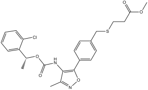
| SMILES | O=C(OC)CCSCC1=CC=C(C2=C(NC(OC(C3=CC=CC=C3Cl)C)=O)C(C)=NO2)C=C1 |
|
| InChi Key | HHVJBROTJWPHHX-UHFFFAOYSA-N | |
| InChi Code | InChI=1S/C24H25ClN2O5S/c1-15-22(26-24(29)31-16(2)19-6-4-5-7-20(19)25)23(32-27-15)18-10-8-17(9-11-18)14-33-13-12-21(28)30-3/h4-11,16H,12-14H2,1-3H3,(H,26,29) | |
| Chemical Name | methyl 3-[[4-[4-[1-(2-chlorophenyl)ethoxycarbonylamino]-3-methyl-1,2-oxazol-5-yl]phenyl]methylsulfanyl]propanoate | |
| Synonyms |
|
|
| HS Tariff Code | 2934.99.9001 | |
| Storage |
Powder-20°C 3 years 4°C 2 years In solvent -80°C 6 months -20°C 1 month |
|
| Shipping Condition | Room temperature (This product is stable at ambient temperature for a few days during ordinary shipping and time spent in Customs) |
Biological Activity
| Targets | LPA1 receptor ( Ki = 0.34 μM ); LPA1 receptor ( Ki = 0.34 μM ) | ||
| ln Vitro |
|
||
| ln Vivo |
|
||
| Enzyme Assay | On collagen-coated 12-well dishes, RH7777 cells expressing LPA1, LPA2, LPA3, LPA4, or LPA5 are cultured in the growth medium. After that, the medium is changed to TCM199 containing 2 μCi/mL [3H]inositol and 0.1% (w/v) BSA (fraction V). The cells are then incubated for 30 minutes with the indicated concentrations of Ki16425 or Ki16198 with or without 1 μM LPA in the presence of 10 mM LiCl in the same medium at a final volume of 0.5 mL. Twenty-four hours later, the cells are rinsed three times with HEPES-buffered medium, which consisted of 20 mM Hepes (pH 7.4), 134 mM NaCl, 4.7 mM KCl, 1.2 mM KH2PO4, 1.2 mM MgSO4, 2.5 mM NaHCO3, 5 mM glucose, and 0.1% (w/v) BSA. The cells are frozen and 1 N HCl (0.1 mL) is added to stop the reaction. The [3H]inositol phosphate fractions are separated using the supernatant (0.5 mL of acid extract) of the thawed cells. The data are normalized to 105 dpm of the total radioactivity that is integrated into the inositol lipids in cells. The total radioactivity of the trichloroacetic acid (5%)-insoluble fraction is determined. | ||
| Cell Assay | On 12-well plates, YAPC-PD or Panc-1 cells are seeded at a density of 1 × 10⁴ cells per milliliter. A change to RPMI1640 containing 0.1% BSA is made to the medium sixteen hours prior to the experiments. The cells are then stimulated in the same medium, with or without Ki16198, for a duration of 24 hours. The ability of the cells to reduce MTT (3-(4,5-Dimethyl-2-thiazoyl)-2,5-diphenyltetrazolium bromide) is used to measure the proliferation activity. | ||
| Animal Protocol |
|
||
| References |
[1]. Cancer Sci . 2012 Jun;103(6):1099-104. [2]. Biochim Biophys Acta . 2008 May;1783(5):748-59. [3]. Cardiovasc Res . 2011 Oct 1;92(1):149-58. |
||
| Additional Infomation |
Pancreatic cancer is highly metastatic and has a poor prognosis. However, there is no established treatment for pancreatic cancer. Lysophosphatidic acid (LPA) has been shown to be present in effluents of cancers and involved in migration and proliferation in a variety of cancer cells, including pancreatic cancer cells, in vitro. In the current study, we examined whether an orally active LPA antagonist is effective for pancreatic cancer tumorigenesis and metastasis in vivo. Oral administration of Ki16198, which is effective for LPA(1) and LPA(3), into YAPC-PD pancreatic cancer cell-inoculated nude mice significantly inhibited tumor weight and remarkably attenuated invasion and metastasis to lung, liver, and brain, in association with inhibition of matrix metalloproteinase (MMP) accumulation in ascites in vivo. Ki16198 inhibited LPA-induced migration and invasion in several pancreatic cancer cells in vitro, which was associated with the inhibition of LPA-induced MMP production. In conclusion, Ki16198 is a promising orally active LPA antagonist for inhibiting the invasion and metastasis of pancreatic cancer cells. The inhibitory effects of the antagonist on invasion and metastasis in vivo may be partially explained by the inhibition of motility activity and MMP production in cancer cells.[1] Lysophosphatidic acid (LPA) is an extracellular signaling lipid that regulates cell proliferation, survival, and motility of normal and cancer cells. These effects are produced through G protein-coupled LPA receptors, LPA(1) to LPA(5). We generated an LPA(1) mutant lacking the SerValVal sequence of the C-terminal PDZ-binding domain to examine the role of this domain in intracellular signaling and other cellular functions. B103 neuroblastoma cells expressing the mutant LPA(1) showed rapid cell proliferation and tended to form colonies under serum-free conditions. The enhanced cell proliferation of the mutant cells was inhibited by exogenous expression of the plasmids inhibiting G proteins including G(betagamma), G(alphai) and G(alphaq) or G(alpha12/13), or treatment with pertussis toxin, phosphoinositide 3-kinase (PI3K) inhibitors or a Rho inhibitor. We confirmed that the PI3K-Akt and Rho pathways were intrinsically activated in mutant cells by detecting increases in phosphorylated Akt in western blot analyses or by directly measuring Rho activity. Interestingly, expression of the mutant LPA(1) in non-tumor mouse fibroblasts induced colony formation in a clonogenic soft agar assay, indicating that oncogenic pathways were activated. Taken together, these observations suggest that the mutant LPA(1) constitutively activates the G protein signaling leading to PI3K-Akt and Rho pathways, resulting in enhanced cell proliferation.[2] Aims: We investigated the mechanisms of action of lysophosphatidic acid (LPA) to regulate vascular endothelial (VE)-cadherin dynamics and cell-cell contact. Methods and results: While a low concentration of LPA stimulated VE-cadherin internalization and subsequent cell-cell dissociation, a high concentration of LPA masked the disruptive actions on VE-cadherin and protected the barrier function in human vascular endothelial cells. Knockdown experiments of major LPA receptor subtypes, i.e. LPA(1) and p2y5 (also termed LPA(6)), with their specific small interfering RNAs, showed that LPA(1) and LPA(6) mediate the LPA-induced disruptive and protective actions on barrier integrity, respectively. LPA(6)-mediated tube formation, reflecting stabilization of barrier integrity, was confirmed by in vitro angiogenesis assay. The LPA(1)-mediated disruptive actions were inhibited by pertussis toxin, dominant-negative Rac1, and inhibitors for c-Jun NH(2)-terminal kinase (JNK) and p38 mitogen-activated protein kinase (p38MAPK), but not by dominant-negative RhoA. In contrast, the LPA(6)-mediated protective actions were associated with activation of Src and Rap1 and attenuated by abrogation of their activities. Further characterization showed that Rap1 is located downstream of Src and dependent on C3G, a Rap1 guanine nucleotide exchange factor. Finally, an LPA antagonist significantly inhibited lactic acid-induced limb lesions in vivo, which may be attributed to dysfunction of endothelial cells. Conclusion: LPA induced disruption and protection of VE-cadherin integrity through LPA(1)-G(i) protein-Rac1-JNK/p38MAPK and LPA(6)-G(12/13) protein-Src-C3G-Rap1 pathways, respectively.[3] |
Solubility Data
| Solubility (In Vitro) |
|
|||
| Solubility (In Vivo) |
Solubility in Formulation 1: 2.5 mg/mL (5.11 mM) in 10% DMSO + 40% PEG300 + 5% Tween80 + 45% Saline (add these co-solvents sequentially from left to right, and one by one), suspension solution; with sonication. For example, if 1 mL of working solution is to be prepared, you can add 100 μL of 25.0 mg/mL clear DMSO stock solution to 400 μL PEG300 and mix evenly; then add 50 μL Tween-80 to the above solution and mix evenly; then add 450 μL normal saline to adjust the volume to 1 mL. Preparation of saline: Dissolve 0.9 g of sodium chloride in 100 mL ddH₂ O to obtain a clear solution. Solubility in Formulation 2: 2.5 mg/mL (5.11 mM) in 10% DMSO + 90% (20% SBE-β-CD in Saline) (add these co-solvents sequentially from left to right, and one by one), suspension solution; with ultrasonication. For example, if 1 mL of working solution is to be prepared, you can add 100 μL of 25.0 mg/mL clear DMSO stock solution to 900 μL of 20% SBE-β-CD physiological saline solution and mix evenly. Preparation of 20% SBE-β-CD in Saline (4°C,1 week): Dissolve 2 g SBE-β-CD in 10 mL saline to obtain a clear solution. Solubility in Formulation 3: ≥ 2.5 mg/mL (5.11 mM) (saturation unknown) in 10% DMSO + 90% Corn Oil (add these co-solvents sequentially from left to right, and one by one), clear solution. For example, if 1 mL of working solution is to be prepared, you can add 100 μL of 25.0 mg/mL clear DMSO stock solution to 900 μL of corn oil and mix evenly. Solubility in Formulation 4: 1% DMSO +30% polyethylene glycol+1% Tween 80 : 30 mg/mL (Please use freshly prepared in vivo formulations for optimal results.) |
| Preparing Stock Solutions | 1 mg | 5 mg | 10 mg | |
| 1 mM | 2.0451 mL | 10.2254 mL | 20.4507 mL | |
| 5 mM | 0.4090 mL | 2.0451 mL | 4.0901 mL | |
| 10 mM | 0.2045 mL | 1.0225 mL | 2.0451 mL |