Physicochemical Properties
| Molecular Formula | C19H19CLN4O |
| Molecular Weight | 354.833362817764 |
| Exact Mass | 354.12 |
| Elemental Analysis | C, 64.31; H, 5.40; Cl, 9.99; N, 15.79; O, 4.51 |
| CAS # | 732973-87-4 |
| PubChem CID | 2802665 |
| Appearance | Off-white to light yellow solid powder |
| LogP | 3.5 |
| Hydrogen Bond Donor Count | 2 |
| Hydrogen Bond Acceptor Count | 2 |
| Rotatable Bond Count | 2 |
| Heavy Atom Count | 25 |
| Complexity | 460 |
| Defined Atom Stereocenter Count | 0 |
| InChi Key | VVVZKBOSNCHQEJ-UHFFFAOYSA-N |
| InChi Code | InChI=1S/C19H19ClN4O/c20-14-4-6-15(7-5-14)22-19(25)24-12-10-23(11-13-24)18-3-1-2-17-16(18)8-9-21-17/h1-9,21H,10-13H2,(H,22,25) |
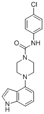
| Chemical Name | N-(4-chlorophenyl)-4-(1H-indol-4-yl)piperazine-1-carboxamide |
| Synonyms | GOT1 inhibitor 2c; VUN73874; VUN-73874; VUN 73874; |
| HS Tariff Code | 2934.99.9001 |
| Storage |
Powder-20°C 3 years 4°C 2 years In solvent -80°C 6 months -20°C 1 month |
| Shipping Condition | Room temperature (This product is stable at ambient temperature for a few days during ordinary shipping and time spent in Customs) |
Biological Activity
| Targets |
Aspartate Aminotransferase 1 (GOT1) (IC50 = 0.8 μM); Aspartate Aminotransferase 2 (GOT2) (IC50 > 10 μM) [1] |
| ln Vitro |
To preserve redox homeostasis and continue to proliferate, PDAC tumors depend on metabolic pathways involving aspartate aminotransferase 1 (glutamate-oxaloacetate aminotransferase 1; GOT1). Research on cancer may find new directions with the use of small molecule inhibitors that target this metabolic pathway. GOT1 inhibitor-1 demonstrated an inhibiting effect on GOT1 activity in the MDH-coupled GOT1 enzyme assay, with an IC50 value of 8.2 uM[1]. GOT1 enzyme inhibition: GOT1 inhibitor 2c potently inhibited recombinant human GOT1 activity with IC50 = 0.8 μM, showing high selectivity over GOT2 (IC50 > 10 μM) (colorimetric assay for oxaloacetate production) [1] - Antiproliferative activity against PDAC cells: GOT1 inhibitor 2c dose-dependently inhibited proliferation of pancreatic ductal adenocarcinoma (PDAC) cell lines: IC50 = 3.2 μM (PANC-1), 4.5 μM (MIA PaCa-2), and 5.1 μM (BxPC-3) after 72 h incubation (MTT assay); it exhibited low toxicity to normal human pancreatic epithelial cells (HPDE6-C7) with IC50 > 20 μM [1] - Disruption of redox balance in PDAC cells: GOT1 inhibitor 2c (5 μM, 24 h) reduced NADH/NAD+ ratio by ~60% and increased intracellular reactive oxygen species (ROS) levels by ~85% in PANC-1 cells (DCFH-DA staining and NADH/NAD+ assay kit) [1] - Induction of apoptosis in PDAC cells: GOT1 inhibitor 2c (5 μM, 48 h) induced apoptosis in PANC-1 cells, with apoptotic rate increasing from 4.1% (control) to 27.3% (Annexin V-FITC/PI double staining, flow cytometry) [1] - Downregulation of anti-apoptotic proteins: GOT1 inhibitor 2c (5 μM, 24 h) reduced Bcl-2 protein level by ~55% and increased Bax protein level by ~1.8-fold in PANC-1 cells (western blot) [1] |
| Enzyme Assay |
Recombinant human GOT1 and GOT2 proteins were separately resuspended in assay buffer. Serial dilutions of GOT1 inhibitor 2c (0.01-20 μM) were mixed with each enzyme, followed by addition of substrates L-aspartate and α-ketoglutarate. The reaction was incubated at 37°C for 30 minutes, and the production of oxaloacetate was quantified by a colorimetric method using 2,4-dinitrophenylhydrazine. IC50 values were calculated by nonlinear regression of dose-response inhibition curves [1] |
| Cell Assay |
Cell proliferation assay: PDAC cells (PANC-1, MIA PaCa-2, BxPC-3) and normal HPDE6-C7 cells were seeded in 96-well plates (5×10³ cells/well) and cultured overnight. GOT1 inhibitor 2c (0.1-20 μM) was added, and cells were incubated for 72 h. MTT reagent was added, incubated for 4 h, formazan crystals were dissolved in DMSO, and absorbance was measured at 570 nm to calculate IC50 values [1] - Redox balance assay: PANC-1 cells were seeded in 24-well plates (2×10⁵ cells/well) and treated with GOT1 inhibitor 2c (5 μM) for 24 h. Intracellular ROS levels were detected by DCFH-DA staining and flow cytometry. NADH/NAD+ ratio was measured using a commercial assay kit according to the manufacturer’s protocol [1] - Apoptosis and protein expression assay: PANC-1 cells were seeded in 6-well plates (3×10⁵ cells/well) and treated with GOT1 inhibitor 2c (5 μM) for 24-48 h. For apoptosis detection, cells were stained with Annexin V-FITC and PI, then analyzed by flow cytometry. For protein analysis, cells were lysed, proteins were separated by SDS-PAGE, transferred to PVDF membranes, and probed with antibodies against Bcl-2, Bax, and β-actin (loading control) [1] |
| References |
[1]. Discovery and optimization of aspartate aminotransferase 1 inhibitors to target redox balance in pancreatic ductal adenocarcinoma. Bioorg Med Chem Lett. 2018 Sep 1;28(16):2675-2678. |
| Additional Infomation |
GOT1 inhibitor 2c is a small-molecule selective inhibitor of aspartate aminotransferase 1 (GOT1) [1] - Its antitumor mechanism involves inhibiting GOT1-mediated aspartate metabolism, disrupting redox balance (reducing NADH/NAD+ ratio, increasing ROS production) in PDAC cells, and inducing apoptosis through regulating Bcl-2/Bax signaling [1] - The compound shows high selectivity for GOT1 over GOT2 (selectivity index > 12.5) and low toxicity to normal pancreatic epithelial cells, indicating a favorable therapeutic window [1] - It is proposed as a potential therapeutic agent for pancreatic ductal adenocarcinoma (PDAC), targeting the redox dependency of PDAC cells on GOT1 activity [1] |
Solubility Data
| Solubility (In Vitro) | DMSO : ~125 mg/mL (~352.28 mM) |
| Solubility (In Vivo) |
Solubility in Formulation 1: ≥ 2.08 mg/mL (5.86 mM) (saturation unknown) in 10% DMSO + 40% PEG300 + 5% Tween80 + 45% Saline (add these co-solvents sequentially from left to right, and one by one), clear solution. For example, if 1 mL of working solution is to be prepared, you can add 100 μL of 20.8 mg/mL clear DMSO stock solution to 400 μL PEG300 and mix evenly; then add 50 μL Tween-80 to the above solution and mix evenly; then add 450 μL normal saline to adjust the volume to 1 mL. Preparation of saline: Dissolve 0.9 g of sodium chloride in 100 mL ddH₂ O to obtain a clear solution. Solubility in Formulation 2: 2.08 mg/mL (5.86 mM) in 10% DMSO + 90% (20% SBE-β-CD in Saline) (add these co-solvents sequentially from left to right, and one by one), suspension solution; with ultrasonication. For example, if 1 mL of working solution is to be prepared, you can add 100 μL of 20.8 mg/mL clear DMSO stock solution to 900 μL of 20% SBE-β-CD physiological saline solution and mix evenly. Preparation of 20% SBE-β-CD in Saline (4°C,1 week): Dissolve 2 g SBE-β-CD in 10 mL saline to obtain a clear solution. Solubility in Formulation 3: ≥ 2.08 mg/mL (5.86 mM) (saturation unknown) in 10% DMSO + 90% Corn Oil (add these co-solvents sequentially from left to right, and one by one), clear solution. For example, if 1 mL of working solution is to be prepared, you can add 100 μL of 20.8 mg/mL clear DMSO stock solution to 900 μL of corn oil and mix evenly. (Please use freshly prepared in vivo formulations for optimal results.) |
| Preparing Stock Solutions | 1 mg | 5 mg | 10 mg | |
| 1 mM | 2.8183 mL | 14.0913 mL | 28.1825 mL | |
| 5 mM | 0.5637 mL | 2.8183 mL | 5.6365 mL | |
| 10 mM | 0.2818 mL | 1.4091 mL | 2.8183 mL |