GANT61 (GANT 61; nsc 136476; GANT-61; nsc-136476) is a novel and potent small-molecule inhibitor for GLI1 (glioma-associated oncogene 1) and GLI2-induced transcription with potential anticancer activity. In GLI1-expressing HEK293T cells, it inhibits the hedgehog pathway with an IC50 of 5 μM and exhibits selectivity against other pathways like TNF and glucocorticoid receptor gene transactivation. The way that GANT61 works is by stopping DNA binding. Its ability to cause dose-dependent cell death in Ewing's sarcoma family tumor (ESFT) cell lines has been shown.
Physicochemical Properties
| Molecular Formula | C27H35N5 | |
| Molecular Weight | 429.6 | |
| Exact Mass | 429.289 | |
| Elemental Analysis | C, 75.49; H, 8.21; N, 16.30 | |
| CAS # | 500579-04-4 | |
| Related CAS # |
|
|
| PubChem CID | 421610 | |
| Appearance | White to off-white solid powder | |
| Density | 1.1±0.1 g/cm3 | |
| Boiling Point | 549.0±50.0 °C at 760 mmHg | |
| Flash Point | 285.8±30.1 °C | |
| Vapour Pressure | 0.0±1.5 mmHg at 25°C | |
| Index of Refraction | 1.632 | |
| LogP | 3.53 | |
| Hydrogen Bond Donor Count | 0 | |
| Hydrogen Bond Acceptor Count | 5 | |
| Rotatable Bond Count | 7 | |
| Heavy Atom Count | 32 | |
| Complexity | 508 | |
| Defined Atom Stereocenter Count | 0 | |
| SMILES | N1(C([H])([H])C2=C([H])C([H])=C([H])C([H])=C2N(C([H])([H])[H])C([H])([H])[H])C([H])([H])C([H])([H])C([H])([H])N(C([H])([H])C2=C([H])C([H])=C([H])C([H])=C2N(C([H])([H])[H])C([H])([H])[H])C1([H])C1C([H])=C([H])N=C([H])C=1[H] |
|
| InChi Key | KVQOGDQTWWCZFX-UHFFFAOYSA-N | |
| InChi Code | InChI=1S/C27H35N5/c1-29(2)25-12-7-5-10-23(25)20-31-18-9-19-32(27(31)22-14-16-28-17-15-22)21-24-11-6-8-13-26(24)30(3)4/h5-8,10-17,27H,9,18-21H2,1-4H3 | |
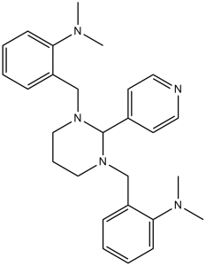
| Chemical Name | 2-[[3-[[2-(dimethylamino)phenyl]methyl]-2-pyridin-4-yl-1,3-diazinan-1-yl]methyl]-N,N-dimethylaniline | |
| Synonyms |
|
|
| HS Tariff Code | 2934.99.9001 | |
| Storage |
Powder-20°C 3 years 4°C 2 years In solvent -80°C 6 months -20°C 1 month |
|
| Shipping Condition | Room temperature (This product is stable at ambient temperature for a few days during ordinary shipping and time spent in Customs) |
Biological Activity
| Targets | GLI1 ( IC50 = 5 μM ) | |
| ln Vitro |
|
|
| ln Vivo |
|
|
| Enzyme Assay |
ChIP analysis[4] HT29 cells were treated with GANT61 (20 µM) for 1 hr or 24 hr and ChIP analysis was conducted using Gli1 or Gli2 antibodies and Abcam ChIP kit, according to the manufacturer’s protocol. Details are provided in Supplementary Materials and Methods. Luciferase reporter assays[4] The Gli-luciferase reporter construct has been previously described. The NF-κB-luciferase plasmid p5XIP10κB was previously reported. The AP1-Luciferase contains a basic promoter element (TATA box) joined to tandem repeats of the AP1 binding element. Transient transfection for 24 hr with luciferase reporters was performed 24 hr after GANT61 (20 µM) treatment, as described. RT-PCR[4] HT29 cell were treated with GANT61 (20 µM) for 1 hr followed by RNA isolation for up to a further 4 hr. Following conversion into cDNA, samples were used for qPCR as described previously. On 10-cm plates (day 0), HEK293 cells are transfected with the GLI1 expression plasmid along with the reporter plasmids 12×GliBSLuc and R-Luc. After twenty-four hours, cells are seeded at a density of 15,000 per well in white, transparent 96-well plates. Compounds are added to cells at a final concentration of 10 μM in DMSO (0.5% final DMSO concentration) after allowing the cells to attach (day 1.5). After growing the cells for a further twenty-four hours, they are lysed and the Dual Luciferase kit is used for analysis. |
|
| Cell Assay |
Clonogenic assays[3] The cells were plated at a density of 1,500 (HT29, HCT8, HCT116), and 3,000 (SW480, GC3/c1, VRC5/c1) cells/well in 6-well plates. Following overnight attachment, cells were treated, in triplicate, with varied concentrations of GANT61 (0–20 μM) for 72 hr. Drug was removed and replaced with fresh media containing dThd (20 μM) for a period equivalent to 7 cell doublings (7 days for HT29, SW480, GC3/c1 and VRC5/c1; 5 days for HCT8 and HCT116). Cells were washed with 1X Dulbecco’s PBS (without Ca++ or Mg++) and allowed to dry overnight. The following day, cells were stained with crystal violet, and colonies analyzed using an Alpha Innotech imager. Cell cycle distribution, Bivariate Flow Cytometric Analysis and BrdU incorporation[4] For cell cycle distribution and Bivariate flow cytometry, cells were analyzed as previously described. For analysis of BrdU incorporation, cells were plated (50,000 cells/well) in a 6-well format, and treated with GANT61 (20µM) or cyclopamine (20µM) for up to 48 hr. Cells were pulsed with BrdU (10 µM) for 30–45 min and analyzed by flow cytometry for distribution within the cell cycle as per the manufacturer protocol. Confocal microscopy[4] Cells were plated (50,000/well) on coverslips in 6-well plates. The cells were treated with GANT61 (20µM) or cyclopamine (20µM) for up to 48 hr and processed for microscopy. Details of microscopy are described in the Supplementary Materials and Methods. BrdU Incorporation Assay. On white 96-well plates with clear bottoms, subconfluent cells are grown in reduced FBS (2.5%) for 48 hours while being exposed to 5 μM test compound (or DMSO). The cells are then fixed, labeled with BrdU for two hours, and examined. |
|
| Animal Protocol |
|
|
| References |
[1]. Proc Natl Acad Sci U S A . 2007 May 15;104(20):8455-60. [2]. Oncogene . 2010 Sep 2;29(35):4885-95. [3]. Cancer Res . 2011 Feb 1;71(3):1092-102. [4]. Cancer Res . 2011 Sep 1;71(17):5904-14. [5]. Leuk Res . 2012 Jun;36(6):742-8. [6]. Int J Cancer . 2013 Apr 1;132(7):1516-24. |
|
| Additional Infomation |
GANT61 is an aminal that is hexahydropyrimidine which is substituted on each nitrogen by a 2-(dimethylamino)benzyl group, and at the aminal carbon by a pyridin-4-yl group. A Hedgehog signaling pathway and Gli protein inhibitor. It has a role as a Hedgehog signaling pathway inhibitor, a glioma-associated oncogene inhibitor, an antineoplastic agent and an apoptosis inducer. It is a tertiary amino compound, a member of pyridines, a substituted aniline and an aminal. The developmentally important Hedgehog (Hh) signaling pathway has recently been implicated in several forms of solid cancer. Current drug development programs focus on targeting the protooncogene Smoothened, a key transmembrane pathway member. These drug candidates, albeit promising, do not address the scenario in which pathway activation occurs downstream of Smoothened, as observed in cases of medulloblastoma, glioma, pericytoma, breast cancer, and prostate cancer. A cellular screen for small-molecule antagonists of GLI-mediated transcription, which constitutes the final step in the Hh pathway, revealed two molecules that are able to selectively inhibit GLI-mediated gene transactivation. We provide genetic evidence of downstream pathway blockade by these compounds and demonstrate the ineffectiveness of upstream antagonists such as cyclopamine in such situations. Mechanistically, both inhibitors act in the nucleus to block GLI function, and one of them interferes with GLI1 DNA binding in living cells. Importantly, the discovered compounds efficiently inhibited in vitro tumor cell proliferation in a GLI-dependent manner and successfully blocked cell growth in an in vivo xenograft model using human prostate cancer cells harboring downstream activation of the Hh pathway.[1] The Hedgehog (Hh) pathway regulates cell proliferation and survival and contributes to tumorigenesis. We investigated the expression and function of this pathway in B-cell chronic lymphocytic leukemia (CLL) cells and in healthy B lymphocytes. Profiling of cognate Hh pathway members revealed reduced expression of two key Hh signaling effectors, Smoothened (SMOH) and GLI, in CLL cells, whereas transcription levels of other investigated members resembled normal B-lymphocyte levels. Examining the functional role of SMOH and GLI in cell survival, we found that CLL cells were hardly sensitive toward specific SMOH inhibition, but showed an unspecific decline in cell viability in response to high concentrations of the SMOH antagonist cyclopamine. In contrast, treatment with the novel GLI antagonist GANT61 reduced expression of the target gene Patched and preferentially decreased the viability of malignant cells. Specific RNA interference knockdown experiments in a CLL-derived cell line confirmed the autonomous role of GLI in malignant cell survival. GANT61-induced apoptosis in primary leukemic cells was partly attenuated by protective stromal cells, but not soluble sonic hedgehog ligand. In summary, our data show a downregulation of the classical Hh pathway in CLL and suggest an intrinsic SMOH-independent role of GLI in the ex vivo survival of CLL cells.[2] Aberrant activation of Hedgehog (HH) signaling is implicated in many human cancers. Classical HH signaling is characterized by Smoothened (Smo)-dependent activation of Gli1 and Gli2, which transcriptionally regulate target genes. A small molecule inhibitor of Gli1 and Gli2, GANT61, was used to block HH signaling in human colon carcinoma cell lines that express HH signaling components. GANT61 administration induced robust cytotoxicity in 5 of 6 cell lines and moderate cytotoxicity in the remaining 1 cell line. In comparison, the classical Smo inhibitor, cyclopamine, induced modest cytotoxicity. Further, GANT61 treatment abolished the clonogenicity of all six human colon carcinoma cell lines. Analysis of the molecular mechanisms of GANT61-induced cytotoxicity in HT29 cells showed increased Fas expression and decreased expression of PDGFRα, which also regulates Fas. Furthermore, DR5 expression was increased whereas Bcl-2 (direct target of Gli2) was downregulated following GANT61 treatment. Suppression of Gli1 by shRNA mimicked the changes in gene expression observed in GANT61-treated cells. Overexpression of dominant-negative FADD (to abrogate Fas/DR5-mediated death receptor signaling) and/or Bcl-2 (to block mitochondria-mediated apoptosis) partially rescued GANT61-induced cytotoxicity in HT29 cells. Thus, activated GLI genes repress DR5 and Fas expressions while upregulating Bcl-2 and PDGFRα expressions to inhibit Fas and facilitate cell survival. Collectively, these results highlight the importance of Gli activation downstream of Smo as a therapeutic target in models of human colon carcinoma.[3] Canonical Hedgehog (HH) signaling is characterized by Smoothened (Smo)-dependent activation of the transcription factors Gli1 and Gli2, which regulate HH target genes. In human colon carcinoma cells, treatment with the Gli small-molecule inhibitor GANT61 induces extensive cell death in contrast to the Smo inhibitor cyclopamine. Here we elucidate cellular events upstream of cell death elicited by GANT61, which reveal the basis for its unique cytotoxic activity in colon carcinoma cells. Unlike cyclopamine, GANT61 induced transient cellular accumulation at G(1)-S (24 hours) and in early S-phase (32 hours), with elevated p21(Cip1), cyclin E, and cyclin A in HT29 cells. GANT61 induced DNA damage within 24 hours, with the appearance of p-ATM and p-Chk2. Pharmacologic inhibition of Gli1 and Gli2 by GANT61 or genetic inhibition by transient transfection of the Gli3 repressor (Gli3R) downregulated Gli1 and Gli2 expression and induced γH2AX, PARP cleavage, caspase-3 activation, and cell death. GANT61 induced γH2AX nuclear foci, while transient transfection of Gli3R showed expression of Gli3R and γH2AX foci within the same nuclei in HT29, SW480, and HCT116. GANT61 specifically targeted Gli1 and Gli2 substantiated by specific inhibition of (i) direct binding of Gli1 and Gli2 to the promoters of target genes HIP1 and BCL-2, (ii) Gli-luciferase activity, and (iii) transcriptional activation of BCL-2. Taken together, these findings establish that inhibition of HH signaling at the level of the GLI genes downstream of Smo is critical in the induction of DNA damage in early S-phase, leading to cell death in human colon carcinoma cells.[4] |
Solubility Data
| Solubility (In Vitro) |
|
|||
| Solubility (In Vivo) |
Solubility in Formulation 1: ≥ 5 mg/mL (11.64 mM) (saturation unknown) in 10% EtOH + 40% PEG300 + 5% Tween80 + 45% Saline (add these co-solvents sequentially from left to right, and one by one), clear solution. For example, if 1 mL of working solution is to be prepared, you can add 100 μL of 50.0 mg/mL clear EtOH stock solution to 400 μL PEG300 and mix evenly; then add 50 μL Tween-80 to the above solution and mix evenly; then add 450 μL normal saline to adjust the volume to 1 mL. Preparation of saline: Dissolve 0.9 g of sodium chloride in 100 mL ddH₂ O to obtain a clear solution. Solubility in Formulation 2: 5 mg/mL (11.64 mM) in 10% EtOH + 90% (20% SBE-β-CD in Saline) (add these co-solvents sequentially from left to right, and one by one), suspension solution; with ultrasonication. For example, if 1 mL of working solution is to be prepared, you can add 100 μL of 50.0 mg/mL clear EtOH stock solution to 900 μL of 20% SBE-β-CD physiological saline solution and mix evenly. Preparation of 20% SBE-β-CD in Saline (4°C,1 week): Dissolve 2 g SBE-β-CD in 10 mL saline to obtain a clear solution. Solubility in Formulation 3: ≥ 5 mg/mL (11.64 mM) (saturation unknown) in 10% EtOH + 90% Corn Oil (add these co-solvents sequentially from left to right, and one by one), clear solution. For example, if 1 mL of working solution is to be prepared, you can add 100 μL of 50.0 mg/mL clear EtOH stock solution to 900 μL of corn oil and mix well. Solubility in Formulation 4: ≥ 2.5 mg/mL (5.82 mM) (saturation unknown) in 10% DMSO + 40% PEG300 + 5% Tween80 + 45% Saline (add these co-solvents sequentially from left to right, and one by one), clear solution. For example, if 1 mL of working solution is to be prepared, you can add 100 μL of 25.0 mg/mL clear DMSO stock solution to 400 μL PEG300 and mix evenly; then add 50 μL Tween-80 to the above solution and mix evenly; then add 450 μL normal saline to adjust the volume to 1 mL. Preparation of saline: Dissolve 0.9 g of sodium chloride in 100 mL ddH₂ O to obtain a clear solution. Solubility in Formulation 5: 2.5 mg/mL (5.82 mM) in 10% DMSO + 90% (20% SBE-β-CD in Saline) (add these co-solvents sequentially from left to right, and one by one), suspension solution; with ultrasonication. For example, if 1 mL of working solution is to be prepared, you can add 100 μL of 25.0 mg/mL clear DMSO stock solution to 900 μL of 20% SBE-β-CD physiological saline solution and mix evenly. Preparation of 20% SBE-β-CD in Saline (4°C,1 week): Dissolve 2 g SBE-β-CD in 10 mL saline to obtain a clear solution. Solubility in Formulation 6: ≥ 2.5 mg/mL (5.82 mM) (saturation unknown) in 10% DMSO + 90% Corn Oil (add these co-solvents sequentially from left to right, and one by one), clear solution. For example, if 1 mL of working solution is to be prepared, you can add 100 μL of 25.0 mg/mL clear DMSO stock solution to 900 μL of corn oil and mix evenly. Solubility in Formulation 7: ≥ 2.5 mg/mL (5.82 mM) (saturation unknown) in 5% DMSO + 40% PEG300 + 5% Tween80 + 50% Saline (add these co-solvents sequentially from left to right, and one by one), clear solution. Preparation of saline: Dissolve 0.9 g of sodium chloride in 100 mL ddH₂ O to obtain a clear solution. Solubility in Formulation 8: 2.5 mg/mL (5.82 mM) in 5% DMSO + 95% (20% SBE-β-CD in Saline) (add these co-solvents sequentially from left to right, and one by one), suspension solution; with ultrasonication. Preparation of 20% SBE-β-CD in Saline (4°C,1 week): Dissolve 2 g SBE-β-CD in 10 mL saline to obtain a clear solution. Solubility in Formulation 9: 5% DMSO+95% Corn oil: 30 mg/mL Solubility in Formulation 10: 8 mg/mL (18.62 mM) in Cremophor EL (add these co-solvents sequentially from left to right, and one by one), clear solution; with ultrasonication. (Please use freshly prepared in vivo formulations for optimal results.) |
| Preparing Stock Solutions | 1 mg | 5 mg | 10 mg | |
| 1 mM | 2.3277 mL | 11.6387 mL | 23.2775 mL | |
| 5 mM | 0.4655 mL | 2.3277 mL | 4.6555 mL | |
| 10 mM | 0.2328 mL | 1.1639 mL | 2.3277 mL |