Physicochemical Properties
| Molecular Formula | C40H56N10O10 |
| Molecular Weight | 836.93 |
| Exact Mass | 836.418 |
| CAS # | 2370952-98-8 |
| PubChem CID | 138454802 |
| Appearance | White to off-white solid powder |
| Density | 1.43±0.1 g/cm3(Predicted) |
| Boiling Point | 1143.9±65.0 °C(Predicted) |
| LogP | -7.2 |
| Hydrogen Bond Donor Count | 4 |
| Hydrogen Bond Acceptor Count | 17 |
| Rotatable Bond Count | 16 |
| Heavy Atom Count | 60 |
| Complexity | 1490 |
| Defined Atom Stereocenter Count | 1 |
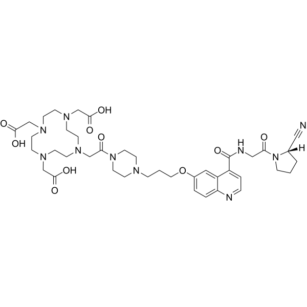
| SMILES | C1C[C@H](N(C1)C(=O)CNC(=O)C2=C3C=C(C=CC3=NC=C2)OCCCN4CCN(CC4)C(=O)CN5CCN(CCN(CCN(CC5)CC(=O)O)CC(=O)O)CC(=O)O)C#N |
| InChi Key | CRWOFMLXEOYIEP-PMERELPUSA-N |
| InChi Code | InChI=1S/C40H56N10O10/c41-24-30-3-1-9-50(30)35(51)25-43-40(59)32-6-7-42-34-5-4-31(23-33(32)34)60-22-2-8-44-18-20-49(21-19-44)36(52)26-45-10-12-46(27-37(53)54)14-16-48(29-39(57)58)17-15-47(13-11-45)28-38(55)56/h4-7,23,30H,1-3,8-22,25-29H2,(H,43,59)(H,53,54)(H,55,56)(H,57,58)/t30-/m0/s1 |
| Chemical Name | 2-[4,7-bis(carboxymethyl)-10-[2-[4-[3-[4-[[2-[(2S)-2-cyanopyrrolidin-1-yl]-2-oxoethyl]carbamoyl]quinolin-6-yl]oxypropyl]piperazin-1-yl]-2-oxoethyl]-1,4,7,10-tetrazacyclododec-1-yl]acetic acid |
| Synonyms | FAPI-2; 2370952-98-8; UNII-54MD7EU3MC; 54MD7EU3MC; FAPI-02; SCHEMBL21257060; |
| HS Tariff Code | 2934.99.9001 |
| Storage |
Powder-20°C 3 years 4°C 2 years In solvent -80°C 6 months -20°C 1 month |
| Shipping Condition | Room temperature (This product is stable at ambient temperature for a few days during ordinary shipping and time spent in Customs) |
Biological Activity
| Targets | FAP (fibroblast activation protein) |
| ln Vitro | Of 15 synthesized FAPIs, FAPI-04 was identified as the most promising tracer for clinical application. Compared with the previously published ligand, FAPI-02, FAPI-04 showed excellent stability in human serum, higher affinity for FAP as opposed to CD26, and slower excretion in vitro. https://pubmed.ncbi.nlm.nih.gov/29626119/ |
| ln Vivo |
In vivo, a higher SUV was reached in tumor-bearing animals, leading to larger areas under the curve as calculated from biodistribution experiments. Finally, PET/CT scans with 68Ga-FAPI-04 in 2 patients with metastasized breast cancer revealed high tracer uptake in metastases and a reduction in pain symptoms after therapy with a considerably low dose of 90Y-FAPI-04. Conclusion: FAPI-04 represents a promising tracer for both diagnostic imaging and, possibly, targeted therapy of malignant tumors with a high content of activated fibroblasts, such as breast cancer.https://pubmed.ncbi.nlm.nih.gov/29626119/ Results: Similar to literature values for 18F-FDG, 68Ga-DOTATATE, and 68Ga-PSMA-11, an examination with 200 MBq of 68Ga-FAPI-2 or 68Ga-FAPI-4 corresponds to an equivalent dose of approximately 3-4 mSv. After a fast clearance via the kidneys, the normal organs showed a low tracer uptake with only minimal changes between 10 min and 3 h after injection. In 68Ga-FAPI-2, the tumor uptake from 1 to 3 h after injection decreased by 75%, whereas the tumor retention was prolonged with 68Ga-FAPI-4 (25% washout). Regarding tumor-to-background ratios, at 1 h after injection both 68Ga-FAPI tracers performed equally. In comparison to 18F-FDG, the tumor uptake was almost equal (average SUVmax, 7.41 for 18F-FDG and 7.37 for 68Ga-FAPI-2; not statistically significant); the background uptake in brain (11.01 vs. 0.32), liver (2.77 vs. 1.69), and oral/pharyngeal mucosa (4.88 vs. 2.57) was significantly lower with 68Ga-FAPI. Other organs did not relevantly differ between 18F-FDG and 68Ga-FAPI. Conclusion: FAPI PET/CT is a new diagnostic method in imaging cancer patients. In contrast to 18F-FDG, no diet or fasting in preparation for the examination is necessary, and image acquisition can potentially be started a few minutes after tracer application. Tumor-to-background contrast ratios were equal to or even better than those of 18F-FDG.[1] |
| Cell Assay | FAPIs based on a quinoline structure were synthesized and characterized with respect to binding, internalization, and efflux in cells expressing human and murine FAP as well as CD26.https://pubmed.ncbi.nlm.nih.gov/29626119/ |
| Animal Protocol |
Preclinical pharmacokinetics were determined in tumor-bearing animals with biodistribution experiments and small-animal PET. Finally, a proof-of-concept approach toward imaging and therapy was chosen for 2 patients with metastasized breast cancer. https://pubmed.ncbi.nlm.nih.gov/29626119/ Methods: A preliminary dosimetry estimate for 68Ga-FAPI-2 and 68Ga-FAPI-4 was based on 2 patients examined at 0.2, 1, and 3 h after tracer injection using the QDOSE dosimetry software suit. Further PET/CT scans of tumor patients were acquired 1 h after injection of either 68Ga-FAPI-2 (n = 25) or 68Ga-FAPI-4 (n = 25); for 6 patients an intraindividual related 18F-FDG scan (also acquired 1 h after injection) was available. For the normal tissue of 16 organs, a 2-cm spheric volume of interest was placed in the parenchyma; for tumor lesions, a threshold-segmented volume of interest was used to quantify SUVmean and SUVmax[1] |
| References |
[1]. 68Ga-FAPI PET/CT: Biodistribution and Preliminary Dosimetry Estimate of 2 DOTA-Containing FAP-Targeting Agents in Patients with Various Cancers. J Nucl Med. 2019 Mar;60(3):386-392. [2]. Fibroblast Activation Protein (FAP) targeting homodimeric FAP inhibitor radiotheranostics: a step to improve tumor uptake and retention time. Am J Nucl Med Mol Imaging. 2021 Dec 15;11(6):476-491. [3]. Clinical summary of fibroblast activation protein inhibitor-based radiopharmaceuticals: cancer and beyond. Eur J Nucl Med Mol Imaging. 2022 Jul;49(8):2844-2868. doi: 10.1007/s00259-022-05706-y. Epub 2022 Jan 31. Erratum in: Eur J Nucl Med Mol Imaging. 2022 Jul;49(8):3007. |
| Additional Infomation |
Several radiopharmaceuticals targeting fibroblast activation protein (FAP) based on the highly potent FAP inhibitor UAMC1110 are currently under investigation. Pre-clinical as well as clinical research exhibited the potential of these imaging agents. However, the monomeric small molecules seemed to have a short retention time in the tumor in combination with fast renal clearance. Therefore, our strategy was to develop homodimeric systems having two FAP inhibitors to improve residence time and tumor accumulation. The homodimers with two squaramide coupled FAP inhibitor conjugates DOTA.(SA.FAPi)2 and DOTAGA.(SA.FAPi)2 were synthesized and radiochemically evaluated with gallium-68. [68Ga]Ga-DOTAGA.(SA.FAPi)2 was tested for its in vitro stability, lipophilicity and affinity properties. In addition, human PET/CT scans were performed for [68Ga]Ga-DOTAGA.(SA.FAPi)2 with a head-to-head comparison with [68Ga]Ga-DOTA.SA.FAPi and [18F]FDG. Labeling with gallium-68 demonstrated high radiochemical yields. Inhibition measurements revealed excellent affinity and selectivity with low nanomolar IC50 values for FAP. In PET/CT human studies, significantly higher tumor uptake as well as longer tumor retention could be observed for [68Ga]Ga-DOTAGA.(SA.FAPi)2 compared to [68Ga]Ga-DOTA.SA.FAPi. Therefore, the introduction of the dimer led to an advance in human PET imaging indicated by increased tumor accumulation and prolonged retention times in vivo and thus, the use of dimeric structures could be the next step towards prolonged uptake of FAP inhibitors resulting in radiotherapeutic analogs of FAP inhibitors.[2] Fibroblast activation protein (FAP) is a type II membrane-bound glycoprotein which is overexpressed in cancer-associated fibroblasts and activated fibroblasts at wound healing/inflammatory sites. Since the first clinical application of quinoline-based FAP ligands in 2018, FAP inhibitor (FAPI)-based PET imaging and radiotherapy have been investigated for a wide variety of diseases, both cancerous and non-cancerous. As a consequence, promising strides have been made in particular to improve the understanding of FAPI-based PET imaging and the potential value of FAPI-based tumor radiotherapy. Herein, we present a comprehensive review of radiolabeled FAPI, including their clinical translation, in order to clarify the current and potential future role of this class of molecules in nuclear medicine. In particular, this review underlines the value of FAPI radiopharmaceuticals in the diagnosis or therapy of tumors or benign conditions. However, limitations in present studies have hampered a precise evaluation of FAPI radiopharmaceuticals. Despite this, it will likely be worthwhile to further explore the clinical value of FAPI in diagnosis and therapy through better-designed and larger-population clinical trials in the future.[3] |
Solubility Data
| Solubility (In Vitro) | DMSO: 100 mg/mL (119.48 mM) |
| Solubility (In Vivo) |
Solubility in Formulation 1: ≥ 2.5 mg/mL (2.99 mM) (saturation unknown) in 10% DMSO + 40% PEG300 + 5% Tween80 + 45% Saline (add these co-solvents sequentially from left to right, and one by one), clear solution. For example, if 1 mL of working solution is to be prepared, you can add 100 μL of 25.0 mg/mL clear DMSO stock solution to 400 μL PEG300 and mix evenly; then add 50 μL Tween-80 to the above solution and mix evenly; then add 450 μL normal saline to adjust the volume to 1 mL. Preparation of saline: Dissolve 0.9 g of sodium chloride in 100 mL ddH₂ O to obtain a clear solution. Solubility in Formulation 2: ≥ 2.5 mg/mL (2.99 mM) (saturation unknown) in 10% DMSO + 90% (20% SBE-β-CD in Saline) (add these co-solvents sequentially from left to right, and one by one), clear solution. For example, if 1 mL of working solution is to be prepared, you can add 100 μL of 25.0 mg/mL clear DMSO stock solution to 900 μL of 20% SBE-β-CD physiological saline solution and mix evenly. Preparation of 20% SBE-β-CD in Saline (4°C,1 week): Dissolve 2 g SBE-β-CD in 10 mL saline to obtain a clear solution. Solubility in Formulation 3: ≥ 2.5 mg/mL (2.99 mM) (saturation unknown) in 10% DMSO + 90% Corn Oil (add these co-solvents sequentially from left to right, and one by one), clear solution. For example, if 1 mL of working solution is to be prepared, you can add 100 μL of 25.0 mg/mL clear DMSO stock solution to 900 μL of corn oil and mix evenly. (Please use freshly prepared in vivo formulations for optimal results.) |
| Preparing Stock Solutions | 1 mg | 5 mg | 10 mg | |
| 1 mM | 1.1948 mL | 5.9742 mL | 11.9484 mL | |
| 5 mM | 0.2390 mL | 1.1948 mL | 2.3897 mL | |
| 10 mM | 0.1195 mL | 0.5974 mL | 1.1948 mL |