E-4031 diHCl (E4031 dihydrochloride) is a novel, potent and selective blocker of hERG K+ channels, which is also termed as a class III antiarrhythmic agent. It selectively blocks hERG potassium (K+) channels. E4031 inhibits the rapid delayed-rectifier K+ current (IKr) and reversibly prolongs action potential duration in guinea pig papillary muscle and isolated ventricular myocytes, without affecting Na+ or Ca2+ inward currents.
Physicochemical Properties
| Molecular Formula | C21H29CL2N3O3S |
| Molecular Weight | 474.4443 |
| Exact Mass | 473.13 |
| Elemental Analysis | C, 53.16; H, 6.16; Cl, 14.94; N, 8.86; O, 10.12; S, 6.76 |
| CAS # | 113559-13-0 |
| Related CAS # | 113558-89-7;113559-13-0 (2HCl); |
| PubChem CID | 3087190 |
| Appearance | White to off-white solid powder |
| Boiling Point | 561.7ºC at 760 mmHg |
| Flash Point | 293.5ºC |
| Vapour Pressure | 1.21E-12mmHg at 25°C |
| LogP | 5.594 |
| Hydrogen Bond Donor Count | 3 |
| Hydrogen Bond Acceptor Count | 6 |
| Rotatable Bond Count | 7 |
| Heavy Atom Count | 30 |
| Complexity | 603 |
| Defined Atom Stereocenter Count | 0 |
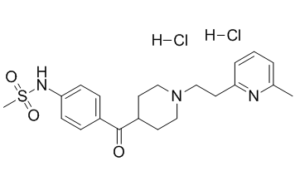
| SMILES | Cl[H].Cl[H].S(C([H])([H])[H])(N([H])C1C([H])=C([H])C(=C([H])C=1[H])C(C1([H])C([H])([H])C([H])([H])N(C([H])([H])C([H])([H])C2=C([H])C([H])=C([H])C(C([H])([H])[H])=N2)C([H])([H])C1([H])[H])=O)(=O)=O |
| InChi Key | ZQBNWMFBOSOOLX-UHFFFAOYSA-N |
| InChi Code | InChI=1S/C21H27N3O3S.2ClH/c1-16-4-3-5-19(22-16)12-15-24-13-10-18(11-14-24)21(25)17-6-8-20(9-7-17)23-28(2,26)27;;/h3-9,18,23H,10-15H2,1-2H3;2*1H |
| Chemical Name | N-[4-[1-[2-(6-methylpyridin-2-yl)ethyl]piperidine-4-carbonyl]phenyl]methanesulfonamide dihydrochloride |
| Synonyms | E-4031; E4031; E 4031; E-4031 dihydrochloride; E-4031; 1-(2-(6-Methyl-2-pyridyl)ethyl)-4-(4-methylsulfonylaminobenzoyl)piperidine dihydrochloride; N-[4-[1-[2-(6-methylpyridin-2-yl)ethyl]piperidine-4-carbonyl]phenyl]methanesulfonamide;dihydrochloride; E-4031 Free; 814AT2BQO7; CHEMBL536480; E4031 2HCl; E-4031 dihydrochloride |
| HS Tariff Code | 2934.99.9001 |
| Storage |
Powder-20°C 3 years 4°C 2 years In solvent -80°C 6 months -20°C 1 month Note: Please store this product in a sealed and protected environment, avoid exposure to moisture. |
| Shipping Condition | Room temperature (This product is stable at ambient temperature for a few days during ordinary shipping and time spent in Customs) |
Biological Activity
| Targets |
hERG potassium channel
E4031 diHCl is a highly selective inhibitor of the rapid component of the delayed rectifier potassium current (IKr), which is mediated by the human ether-à-go-go-related gene (hERG/Kv11.1) potassium channel; the IC50 for inhibiting IKr in rabbit sinoatrial nodal (SAN) myocytes is 100 nM [2], and the Ki value for canine ventricular myocyte IKr is 50 nM [1] E4031 diHCl has no significant inhibitory activity (IC50 > 10 μM) against the slow component of the delayed rectifier potassium current (IKs), sodium current (INa), or L-type calcium current (ICa) [1][2] |
| ln Vitro |
Maximum diastolic potential (MDP) of single SAN cells in New Zealand white rabbits is significantly de-longitudated by E-4031 (0.1 ~ 10 μM), resulting in the MDP de-longest action scaffold from -58.8±0.9 mV at 1 μM Extended to -24.5±1.8 mV and from -58.2±2.1 to -19.6±1.8 mV at 10 μM[2]. In a dose-dependent manner, E-4031 (0.1 ~ 10 μM) inhibits the depolarization process. In single SAN cells of New Zealand white rabbits, the partial external current and subsequent tail current (ITD) during the complex partial process resulted in an 88% [sup] reduction in ITD [2]. 1. Rabbit sinoatrial nodal (SAN) myocytes (Ref [2]): E4031 diHCl (10 nM–10 μM) dose-dependently inhibited IKr in isolated rabbit SAN myocytes (whole-cell patch-clamp); 100 nM of the drug achieved 80% inhibition of IKr tail current, and 1 μM completely blocked IKr. At 1 μM, it reduced SAN cell automaticity by 40% (pacemaker frequency from 120 ± 10 beats/min to 72 ± 8 beats/min), prolonged action potential duration at 90% repolarization (APD90) by 35%, and slowed diastolic depolarization rate (DDR) by 25%. No significant effect on INa or ICa was observed (IC50 > 10 μM) [2] 2. Canine ventricular myocytes (Ref [1]): In isolated canine ventricular myocytes (epicardial, midmyocardial M cells, endocardial), E4031 diHCl (500 nM) prolonged APD90 by 50% across the ventricular wall; APD90 values increased from 180 ± 15 ms (epicardium), 250 ± 20 ms (M cells), 200 ± 18 ms (endocardium) to 270 ± 22 ms, 375 ± 30 ms, 300 ± 25 ms, respectively. It also increased transmural dispersion of repolarization (TDR) by 45% [1] |
| ln Vivo |
Bepridil and E-4031 prolonged QT interval and ARI in all LV layers, though the magnitude of prolongation was greatest in Mid, increasing the transmural ARI dispersion, particularly during bradycardia. Compared with E-4031, bepridil caused mild, reverse use-dependent changes in ventricular repolarization, and less ARI dispersion than E-4031 during slow ventricular pacing. Both drugs increased ARI(max) and cycle length at 50% of ARI(max), though the changes were smaller after bepridil than after E-4031 administration. Bradycardia after the administration of each drug induced no VTA; however, sympathetic stimulation induced sustained polymorphic VTA in two of five dogs treated with E-4031 versus no dog treated with bepridil.
Conclusions: Unlike the pure I(kr) blocker, E-4031, bepridil exhibited weak properties of reverse use-dependency and protected against sympathetic stimulation-induced VTA. It may be an effective supplemental treatment for recipients of implantable cardioverter defibrillator.[2] The role of the delayed rectifier current (IK) in impulse generation was studied in single sinoatrial nodal myocytes of the rabbit. We used the class III antiarrhythmic drug E-4031, which blocks IK in rabbit ventricular myocytes. In single sinoatrial nodal cells, E-4031 (0.1 mumol/L) significantly prolonged cycle length and action potential duration, depolarized maximum diastolic potential, and reduced both the upstroke velocity of the action potential and the diastolic depolarization rate. Half of the cells were arrested completely. At higher concentrations (1 and 10 mumol/L), spontaneous activity ceased in all cells. Three ionic currents fundamental for pacemaking, ie, IK, the long-lasting inward calcium current (ICa,L), and the hyperpolarization-activated current (I(f)), were studied by using the whole-cell and amphotericin-perforated patch technique. E-4031 blocked part of the outward current during depolarizing steps as well as the tail current upon subsequent repolarization (ITD) in a dose-dependent manner. E-4031 (10 mumol/L) depressed ITD (88 +/- 4%) (n = 6), reduced peak ICa,L at 0 mV (29 +/- 15%) (n = 4), but did not affect I(f). Lower concentrations did not affect ICa,L. Additional use of 5 mumol/L nifedipine demonstrated that ITD is carried in part by a calcium-sensitive current. Interestingly, complete blockade of IK and ICa,L unmasked the presence of a background current component with a reversal potential of -32 +/- 5.4 mV (n = 8) and a conductance of 39.5 +/- 5.6 pS/pF, which therefore can contribute both to the initial part of repolarization and to full diastolic depolarization[2]. 1. Anesthetized dog model (Ref [1]): In pentobarbital-anesthetized adult dogs (15–20 kg), intravenous administration of E4031 diHCl (0.1 mg/kg bolus + 0.01 mg/kg/min continuous infusion) prolonged the surface ECG QT interval from 280 ± 20 ms to 420 ± 30 ms (50% increase) and the rate-corrected QT (QTc) interval by 40%. Programmed electrical stimulation induced sustained ventricular tachycardia (VT, >30 s) in 80% (8/10) of E4031-treated dogs, compared with only 10% (1/10) in vehicle-treated dogs. Compared with bepridil (1 mg/kg IV bolus), E4031 caused a more significant QT prolongation and higher VT inducibility (50% in bepridil group) [1] |
| Enzyme Assay |
1. IKr channel activity assay (rabbit SAN myocytes) (Ref [2]): Isolated rabbit SAN myocytes were placed in a perfusion chamber, and whole-cell voltage-clamp recordings were performed. The pipette solution contained KCl, MgATP, and EGTA, and the bath solution was Tyrode’s solution with NaCl, KCl, and CaCl2. IKr was activated by a voltage protocol (depolarization from -80 mV to +20 mV for 2 s, followed by repolarization to -40 mV to record tail current). Serial concentrations of E4031 diHCl (10 nM–10 μM) were added to the bath, and after 5 minutes of incubation, the voltage protocol was repeated to record IKr tail current amplitude. Inhibition rates were calculated, dose-response curves were generated, and the IC50 for IKr inhibition was determined [2] 2. IKr channel activity assay (canine ventricular myocytes) (Ref [1]): Canine ventricular myocytes (epicardial, M cells, endocardial) were isolated, and perforated patch-clamp recordings were performed to avoid intracellular solution loss. IKr was activated by a voltage protocol (prepolarization to -60 mV, depolarization to 0 mV for 3 s, repolarization to -50 mV to record tail current). E4031 diHCl (50 nM–1 μM) was added, tail current changes were recorded, and the Ki value for IKr inhibition was calculated [1] |
| Cell Assay |
Membrane potentials and currents were recorded by using both whole-cell21 and amphotericin–perforated patch techniques. The whole-cell method was used in only 5 of 10 cells in which the effects of 0.1 μmol/L E-4031 was studied. The amphotericin–perforated patch technique was used in all other experiments to reduce dilution of intracellular components, a possible cause of rundown of membrane currents. Electrodes were pulled from borosilicate glass (outer diameter, 1 mm; with a glass fiber inside the lumen) by using a vertical one-stage patch-electrode puller and were fire-polished. Electrode resistance varied between 3 and 5 MΩ[2]. 1. Rabbit SAN myocyte isolation and electrophysiology (Ref [2]): Adult New Zealand white rabbits (2–3 kg) were euthanized, and hearts were rapidly excised and placed in oxygenated cold Tyrode’s solution. Sinoatrial nodal tissue was dissected, cut into 1 mm³ pieces, and incubated in digestion solution containing collagenase and protease at 37°C for 20 minutes. The tissue was gently triturated to obtain a single-cell suspension, centrifuged, and resuspended in Tyrode’s solution with bovine serum albumin, then stored at 4°C for use. Isolated SAN myocytes were plated on glass-bottom dishes and allowed to adhere for 30 minutes. Spontaneous action potentials were recorded in current-clamp mode (no current injection) to measure basal pacemaker frequency, APD50/APD90, and DDR. E4031 diHCl (10 nM–10 μM) was added, and after 5 minutes of incubation, the above parameters were recorded again to analyze the drug’s effect on cellular electrophysiological properties [2] 2. Canine ventricular myocyte assay (Ref [1]): Canine ventricular myocytes were isolated via retrograde perfusion of the heart using a Langendorff system with collagenase-containing perfusate. Myocytes were divided into epicardial, midmyocardial (M cell), and endocardial groups. Action potentials (current-clamp) and IKr (voltage-clamp) were recorded via patch-clamp. After adding E4031 diHCl (500 nM), APD and IKr changes in transmural myocytes were detected, and transmural dispersion of repolarization (TDR) was calculated [1] |
| Animal Protocol |
Drug Administration[1] Bepridil, 4 mg/kg, was dissolved in sterile saline and administered intravenously in a single bolus.19 E-4013 was dissolved in sterile saline and administered intravenously in a bolus of 50 μg/kg, followed by a maintenance dose titrated between 5 and 20 μg/kg/min, to match the ARI prolongation induced by bepridil (280–360 ms) during pacing at 140 beats per minute (bpm).20 The data were collected during steady state, beginning 5 minutes after the administration of bepridil, or 5 minutes after the onset of a stable maintenance infusion of E-4031. Study Protocol and Data Collection[1] After the creation of complete AV block, the heart was paced at 100 bpm from the RV throughout the experiment. Completion of the following protocols of (1) and (2) was within 30 minutes. 1) Before the administration of bepridil or E-4031, the surface electrocardiogram, the transmural distribution of ARI, and ARI dispersion were recorded at a pacing cycle length of 428 ms (140 bpm), 500 ms (120 bpm), 600 ms (100 bpm), 750 ms (80 bpm), and 1,000 ms (60 bpm). The measurements were made at steady state, 30 seconds after the onset of each pacing rate. 2) To study the inducibility of VTA, (a) the back-up RV pacing rate was slowed to 40 bpm to allow lengthening of the cardiac cycle to 800–1,500 ms for 60 seconds, and (b) the left stellate ganglion was stimulated for 20 seconds during demand pacing at a cycle length of 750 ms. Completion of the following protocols of (3) was within 30 minutes. 3) These same protocols (1 and 2) were repeated after the administration of bepridil in five dogs and E-4031 in five other dogs. 1. Anesthetized dog experiment for ventricular arrhythmia (Ref [1]): Adult male beagle dogs (15–20 kg) were anesthetized with pentobarbital sodium (30 mg/kg IV), intubated and ventilated, and femoral artery/vein catheters were inserted for blood pressure monitoring and drug administration. Body surface ECG electrodes were placed to record standard leads, and an electrode catheter was inserted into the right ventricle via the right jugular vein for programmed electrical stimulation. E4031 diHCl was dissolved in normal saline (pH adjusted to 7.4 with lactic acid), administered as a 0.1 mg/kg IV bolus over 5 minutes, followed by a continuous infusion at 0.01 mg/kg/min for 2 hours. The control group received an equal volume of normal saline, and the bepridil group received a 1 mg/kg IV bolus followed by 0.1 mg/kg/min infusion. QT interval and QTc interval (Bazett’s formula) were recorded before drug administration and at 30, 60, 120 minutes post-administration. Programmed electrical stimulation (S1S1 basal pacing followed by 1–2 S2 premature stimuli) was performed to assess VT inducibility (sustained VT was defined as duration >30 s or VT requiring cardioversion). After the experiment, dogs were euthanized, and ventricular myocardial tissue was collected for isolated cell experiments [1] |
| Toxicity/Toxicokinetics |
1. Proarrhythmic toxicity (Ref [1]): Intravenous infusion of E4031 diHCl (0.1 mg/kg bolus + 0.01 mg/kg/min) in dogs significantly prolonged the QT interval and increased the inducibility of sustained ventricular tachycardia (80% induction rate), indicating a potential risk of torsades de pointes (TdP) [1] 2. Cytotoxicity (Ref [2]): Incubation of rabbit SAN myocytes with E4031 diHCl (10 nM–10 μM) for 2 hours resulted in >90% cell viability (trypan blue exclusion assay), with no significant cytotoxicity [2] |
| References |
[1]. Effects of bepridil versus E-4031 on transmural ventricular repolarization and inducibility of ventricular tachyarrhythmias in the dog. Pacing Clin Electrophysiol. 2010 Aug;33(8):950-9. [2]. Effects of delayed rectifier current blockade by E-4031 on impulse generation in single sinoatrial nodal myocytes of the rabbit. Circ Res. 1995 Apr;76(4):607-15. |
| Additional Infomation |
See also: E-4031 (annotation moved to). 1. E4031 diHCl is a benzimidazole-derived selective inhibitor of the rapid delayed rectifier potassium current (IKr/hERG/Kv11.1), primarily used as a research tool compound to investigate the role of IKr in myocardial repolarization and cardiac arrhythmias [1][2] 2. Mechanism of action: E4031 diHCl binds to the pore region of the hERG channel, blocking IKr efflux, prolonging myocardial cell action potential duration and ventricular repolarization, and thereby affecting myocardial electrical stability. In SAN cells, it slows diastolic depolarization rate and reduces automaticity by inhibiting IKr [1][2] 3. Clinical development: E4031 diHCl is only used as an experimental tool and has not been developed as a clinical antiarrhythmic drug due to its significant proarrhythmic risk (e.g., torsades de pointes). No FDA-approved indications or warning information have been reported [1][2] 4. Comparison with bepridil: In the dog model, E4031 diHCl shows highly selective inhibition of IKr, while bepridil inhibits IKr, IKs, and L-type calcium current simultaneously. Thus, E4031 causes more significant QT prolongation but higher VT inducibility, suggesting a greater proarrhythmic risk with selective IKr inhibition [1] |
Solubility Data
| Solubility (In Vitro) | H2O : ≥ 50 mg/mL (~105.39 mM) |
| Solubility (In Vivo) |
Solubility in Formulation 1: 100 mg/mL (210.77 mM) in PBS (add these co-solvents sequentially from left to right, and one by one), clear solution; with sonication. (Please use freshly prepared in vivo formulations for optimal results.) |
| Preparing Stock Solutions | 1 mg | 5 mg | 10 mg | |
| 1 mM | 2.1077 mL | 10.5387 mL | 21.0775 mL | |
| 5 mM | 0.4215 mL | 2.1077 mL | 4.2155 mL | |
| 10 mM | 0.2108 mL | 1.0539 mL | 2.1077 mL |