Y-27632 is a potent and selective inhibitor of ROCK1 (p160ROCK) with IC50 of 140 nM in a cell-free assay, it exhibits >200-fold selectivity over other kinases, including PKC, cAMP-dependent protein kinase, MLCK and PAK. Y27632 prevents dimethylnitrosamine-induced hepatic fibrosis in rats, increases apoptosis and disrupts the actin cortical mat in embryonic avian corneal epithelium, affects initial heart myofibrillogenesis in cultured chick blastoderm, promotes the proliferation and cell cycle progression of cultured astrocyte from spinal cord. Y-27632 suppresses the kinase activity of both ROCK-1 and ROCK-2 in vitro, and this compound inhibits the kinases by binding to the catalytic site of ROCK-1 and ROCK-2. Thus Y-27632 function on Rho-mediated stress fiber formation, the G1-S phase progression and cytokinesis.
Physicochemical Properties
| Molecular Formula | C14H21N3O | |
| Molecular Weight | 247.34 | |
| Exact Mass | 247.17 | |
| Elemental Analysis | C, 67.98; H, 8.56; N, 16.99; O, 6.47 | |
| CAS # | 146986-50-7 | |
| Related CAS # | Y-27632 dihydrochloride;129830-38-2; 331752-47-7 (HCl hydrate); 138381-45-0 (racemate HCl); 146986-50-7; 310898-86-3 (recamte free base) | |
| PubChem CID | 448042 | |
| Appearance | White to off-white solid powder | |
| Density | 1.1±0.1 g/cm3 | |
| Boiling Point | 462.6±15.0 °C at 760 mmHg | |
| Flash Point | 233.6±20.4 °C | |
| Vapour Pressure | 0.0±1.1 mmHg at 25°C | |
| Index of Refraction | 1.579 | |
| LogP | 1.54 | |
| Hydrogen Bond Donor Count | 2 | |
| Hydrogen Bond Acceptor Count | 3 | |
| Rotatable Bond Count | 3 | |
| Heavy Atom Count | 18 | |
| Complexity | 268 | |
| Defined Atom Stereocenter Count | 1 | |
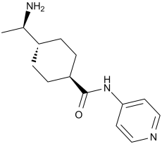
| SMILES | O=C([C@@H]1CC[C@]([C@H](N)C)([H])CC1)NC2=CC=NC=C2 |
|
| InChi Key | IYOZTVGMEWJPKR-IJLUTSLNSA-N | |
| InChi Code | InChI=1S/C14H21N3O/c1-10(15)11-2-4-12(5-3-11)14(18)17-13-6-8-16-9-7-13/h6-12H,2-5,15H2,1H3,(H,16,17,18)/t10-,11-,12-/m1/s1 | |
| Chemical Name | (1R,4r)-4-((R)-1-aminoethyl)-N-(pyridin-4-yl)cyclohexanecarboxamide | |
| Synonyms |
|
|
| HS Tariff Code | 2934.99.9001 | |
| Storage |
Powder-20°C 3 years 4°C 2 years In solvent -80°C 6 months -20°C 1 month Note: (1). This product requires protection from light (avoid light exposure) during transportation and storage.(2). Please store this product in a sealed and protected environment (e.g. under nitrogen), avoid exposure to moisture. |
|
| Shipping Condition | Room temperature (This product is stable at ambient temperature for a few days during ordinary shipping and time spent in Customs) |
Biological Activity
| Targets |
ROCK-I (Ki = 220 nM); ROCK-II (Ki = 300 nM); PKN (Ki = 3.1 μM); Citron kinase (Ki = 5.3 μM); PKCα (Ki = 73 μM); PKA (Ki = 25 μM)
Rho-associated coiled-coil kinase 1 (ROCK1) (Ki = 140 nM in ATP-competitive binding assay; IC50 = 220 nM in kinase activity assay) [1] Rho-associated coiled-coil kinase 2 (ROCK2) (Ki = 130 nM in ATP-competitive binding assay; IC50 = 300 nM in kinase activity assay) [1] Protein Kinase A (PKA)/Protein Kinase C (PKC)/Akt/ERK1/2 (no significant inhibition at concentrations up to 10 μM, IC50 > 10 μM) [1] Rho-associated kinase (ROCK) [2]-[8] |
| ln Vitro |
Y-27632 exhibits a 100-fold increase in concentration-inhibitory delay in the emergence of BrdU labeling, as well as an extension of the delay duration. In Swiss 3T3 cells treated with 10 and 100 μM Y-27632, respectively, delays of about 1 and 4 hours were noted [1]. Adipose tissue-derived stem cells (ADSCs) are encouraged by Y-27632 in the oral cavity of neurons. The 5.0 μM Y-27632 induction group had the greatest number of neural-like cells when compared to the 1.0 and 2.5 μM Y-27632 induction groups [2]. In epithelial cells, actin masks remodel and upregulate cytokines [8].\n \nY-27632 [(+)-(R)-trans-4-(1-aminoethyl)-N-(4-pyridyl)cyclohexanecarboxamide++ + dihydrochloride] is widely used as a specific inhibitor of the Rho-associated coiled-coil forming protein serine/threonine kinase (ROCK) family of protein kinases. This study examined the inhibition mechanism and profile of actions of Y-27632 and a related compound, Y-30141 [(+)-(R)-trans- 4-(1-aminoethyl)-N-(1H-pyrrolo[2, 3-b]pyridin-4-yl)cyclohexan-ecarboxamide dihydrochloride]. Y-27632 and Y-30141 inhibited the kinase activity of both ROCK-I and ROCK-II in vitro, and this inhibition was reversed by ATP in a competitive manner. This suggests that these compounds inhibit the kinases by binding to the catalytic site. Their affinities for ROCK kinases as determined by K(i) values were at least 20 to 30 times higher than those for two other Rho effector kinases, citron kinase and protein kinase PKN. [(3)H]Y-30141 was taken up by cells in a temperature- and time-dependent and saturable manner, and this uptake was competed with unlabeled Y-27632. No concentrated accumulation was found, suggesting that the uptake is a carrier-mediated facilitated diffusion. Y-27632 abolished stress fibers in Swiss 3T3 cells at 10 microM, but the G(1)-S phase transition of the cell cycle and cytokinesis were little affected at this concentration. Y-30141 was 10 times more potent than Y-27632 in inhibiting the kinase activity and stress fiber formation, and it caused significant delay in the G(1)-S transition and inhibition of cytokinesis at 10 microM. [1] \n \nY-27632 had the potency to induce neuronal-like differentiation in ADSCs in a dose-dependent manner. Moreover, the differentiation induced by Y-27632 was recovered upon drug withdraw. ADSCs treated with Y-27632 expressed neuronal markers such as NSE, MAP-2 and nestin while untreated ADSCs did not express these markers.\n\nConclusion: Selective ROCK inhibitor Y-27632 could potentiate the neuronal-like differentiation of ADSCs, suggesting that Y-27632 could be utilized to induce the differentiation of ADSCs to neurons and facilitate the clinical application of ADSCs in tissue engineering. [2] \n\nRobust control of human induced pluripotent stem cell (hIPSC) differentiation is essential to realize its patient-tailored therapeutic potential. Here, we demonstrate a novel application of Y-27632, a small molecule Rho-associated protein kinase (ROCK) inhibitor, to significantly influence the differentiation of hIPSCs in a lineage-specific manner. The application of Y-27632 to hIPSCs resulted in a decrease in actin bundling and disruption of colony formation in a concentration and time-dependent manner. Such changes in cell and colony morphology were associated with decreased expression of E-cadherin, a cell-cell junctional protein, proportional to the increased exposure to Y-27632. Interestingly, gene and protein expression of pluripotency markers such as NANOG and OCT4 were not downregulated by an exposure to Y-27632 up to 36h. Simultaneously, epithelial-to-mesenchymal (EMT) transition markers were upregulated with an exposure to Y-27632. These EMT-like changes in the cells with longer exposure to Y-27632 resulted in a significant increase in the subsequent differentiation efficiency towards mesendodermal lineage. In contrast, an inhibitory effect was observed when cells were subjected to ectodermal differentiation after prolonged exposure to Y-27632. Collectively, these results present a novel method for priming hIPSCs to modulate their differentiation potential with a simple application of Y-27632. [5] \n\n c-kit+ CSCs were cultured in CSC medium for 3-5 days followed by 48 hr treatment with 0 to 10 μM Y-27632 alone, 0 to 1.0 μM Dox alone, or Y-27632 followed by Dox (48 hrs). Cell viability, toxicity, proliferation, morphology, migration, Caspase-3 activity, expression levels of apoptotic-related key proteins and c-kit+ were examined. Results showed that 48 hr treatment with Y-27632 alone did not result in great changes in c-kit+ expression, proliferation, Caspase-3 activity, or apoptosis; however cell viability was significantly increased and cell migration was promoted. These effects likely involve the ROCK/Actin pathways. In contrast, 48 hr treatment with Dox alone dramatically increased Caspase-3 activity, resulting in cell death. Although Y-27632 alone did not affect the expression levels of apoptotic-related key factors (p-Akt, Akt, Bcl-2, Bcl-xl, Bax, cleaved Caspase-3, and Caspase-3) under basal conditions, it significantly inhibited the Dox-induced increase in cleaved Caspase-3 and reduced cell death under Dox treatment.\n\nConclusions: We conclude that preconditioning human CSCs with Y-27632 significantly reduces Dox-induced cell death and possibly involves the cleaved Caspase-3 and ROCK/Actin pathways. The beneficial effects of Y-27632 may be applied to stem cell-based therapy to increase cell survival rates after transplantation or to act as a cardiac protective agent for Dox-treated cancer patients. [6] \n\nActivation of the RhoA/ROCK signaling pathway has been shown to contribute to dissociation-induced apoptosis of embryonic and neural stem cells. We previously demonstrated that approximately 1 out of 40 Lin(-)Sca-1(+)CD49f(high) (LSC) prostate basal epithelial cells possess the capacities of stem cells for self-renewal and multi-lineage differentiation. We show here that treating LSC cells with the ROCK kinase inhibitor Y-27632 increases their cloning efficiency by 8 fold in an in vitro prostate colony assay. Y-27632 treatment allows prostate colony cells to replate efficiently, which does not occur otherwise. Y-27632 also increases the cloning efficiency of prostate stem cells in a prostate sphere assay and a dissociated prostate cell regeneration assay. The increased cloning efficiency is due to the suppression of the dissociation-induced, RhoA/ROCK activation-mediated apoptosis of prostate stem cells. Dissociation of prostate epithelial cells from extracellular matrix increases PTEN activity and attenuates AKT activity. Y-27632 treatment alone is sufficient to suppress cell dissociation-induced activation of PTEN activity. However, this does not contribute to the increased cloning efficiency, because Y-27632 treatment increases the sphere-forming unit of wild type and Pten null prostate cells to a similar extent. Finally, knocking down expression of both ROCK kinases slightly increases the replating efficiency of prostate colony cells, corroborating that they play a major role in the Y-27632 mediated increase in cloning efficiency. Our study implies that the numbers of prostate cells with stem/progenitor activity may be underestimated based on currently employed assays, supports that dissociation-induced apoptosis is a common feature of embryonic and somatic stem cells with an epithelial phenotype, and highlights the significance of environmental cues for the maintenance of stem cells. [7] \n\nWhole sheets of avian embryonic corneal epithelium were cultured in the presence of the ROCK inhibitor, Y27632 at 0, 0.03, 0.3, 3, or 10 microM before stimulating the cells with either collagen (COL) or LPA. Apoptosis was assessed by Caspase-3 activity assays and visualized with annexin V binding. The ROCK inhibitor blocked actin cortical mat reformation and disrupted the basal cell lateral membranes in a dose-dependent manner and increased the apoptosis marker annexin V. In addition, an in vitro caspase-3 activity assay was used to determine that caspase-3 activity was higher in epithelia treated with 10 microM Y-27632 than in those isolated without the basal lamina or epithelia stimulated with fibronectin, COL, or LPA. In conclusion, ECM molecules decreased apoptosis markers and inhibiting the ROCK pathway blocked ECM stimulated actin cortical mat reformation and increased apoptosis in embryonic corneal epithelial cells [8]. Y-27632 acts as a potent and selective ATP-competitive inhibitor of ROCK1 and ROCK2, with no significant inhibitory activity against other serine/threonine kinases (PKA, PKC, Akt, ERK1/2) or tyrosine kinases (EGFR, VEGFR2) at concentrations up to 10 μM; in cultured rat aortic smooth muscle cells (RASMCs), it dose-dependently inhibits RhoA-induced myosin light chain (MLC) phosphorylation (IC50 = 300 nM) and cell contraction (IC50 = 250 nM), and blocks LPA-induced stress fiber formation at 1 μM [1] Y-27632 (10 μM) promotes neuronal-like differentiation of adult human adipose tissue-derived stem cells (hADSCs) in vitro: it increases the expression of neuronal markers (β-III tubulin, NeuN, MAP2) detected by immunofluorescence and qRT-PCR (mRNA levels upregulated by 3.2-4.5-fold vs. control), and reduces the expression of stem cell marker CD44 (downregulated by 60% vs. control) after 7 days of treatment [2] Y-27632 (1-20 μM) dose-dependently attenuates doxorubicin (1 μM)-induced apoptosis of human cardiac stem cells (hCSCs): at 10 μM, it reduces the apoptotic rate from 45% (doxorubicin alone) to 12% (Annexin V/PI flow cytometry), upregulates anti-apoptotic protein Bcl-2 (2.1-fold vs. doxorubicin group) and downregulates pro-apoptotic protein Bax (0.4-fold vs. doxorubicin group) detected by Western blotting [6] Y-27632 (5-20 μM) suppresses dissociation-induced apoptosis of murine prostate stem/progenitor cells (mPSCs) and increases their cloning efficiency: at 10 μM, the apoptotic rate is reduced from 38% (control) to 11%, and the colony formation efficiency is increased from 5.2% to 18.7% in soft agar assays [7] Y-27632 (1-50 μM) dose-dependently induces apoptosis of embryonic avian corneal epithelial cells and disrupts the actin cortical mat: at 20 μM, the apoptotic rate reaches 35% (TUNEL assay), and actin filaments are fragmented and disorganized (phalloidin staining) [8] Y-27632 (10 μM) primes human induced pluripotent stem cells (hiPSCs) to differentiate towards mesendodermal lineage: it upregulates mesendodermal markers (Brachyury, Sox17) by 3.8-5.1-fold (qRT-PCR) and downregulates pluripotency markers (Oct4, Nanog) by 0.3-0.4-fold after 4 days of treatment, via modulation of epithelial-mesenchymal transition (EMT)-related genes (Snail, Twist) [5] |
| ln Vivo |
In comparison to the saline group, Y-27632 (5 and 10 mg/kg) considerably extended the onset time of myoclonic seizures. When Y-27632 (5 and 10 mg/kg) was administered, the onset of clonic convulsions was significantly delayed in comparison to the saline group [3]. Rats treated with dimethylnitrosamine (DMN) showed a significant reduction in liver weight and body weight (DMN-S group) when compared to the control group (SS group). Rats (DMN-Y group) were essentially shielded from this DMN-induced loss of body and liver weight by oral administration of Y27632 (30 mg/kg) [4]. \nY27632 treatment significantly decreased the occurrence of DMN-induced hepatic fibrosis and reduced the collagen and hydroxyproline content and alpha-SMA expression in the liver. [3]\n \n\nY27632 prevents body and liver weight loss induced by DMN [3] \nThe effect of oral administration of Y27632 on body and liver weights of rats with and without i.p. injection of DMN is shown in Fig. 1 and Table 1. Treatment with DMN caused a significant decrease in rat body and liver weight (DMN-S group) compared with control animals (S-S group). Oral Y27632 essentially prevented this DMN-induced rat body and liver weight loss (DMN-Y group). There was no significant difference between the S-S/S-Y and DMN-Y groups. There was no significant difference in serum ALT levels between the DMN-S and DMN-Y groups at the end of fourth week.\n \n\nY27632 reduces hepatic collagen and hydroxyproline content [3] \nThe significantly decreased deposition of hepatic collagen in the Y27632-treated group (the DMN-Y group) was confirmed by collagen assays (DMN-S, 808.5±122.4 μg/100 mg versus DMN-Y, 601.3±67.7 μg/100 mg, P<0.05) (Fig. 3A). Hepatic fibrosis was also quantified by the measurement of hepatic hydroxyproline. Here, it was found that the hydroxyproline content of the DMN-treated group (DMN-S) was significantly higher than that of the DMN and Y27632-treated group (DMN-Y) (DMN-S, 5.95±1.41 μmol/100 mg versus DMN-Y, 2.98±0.91 μmol/100 mg, P<0.05) (Fig. 3B). Furthermore, there was no significant difference in collagen and hydroxyproline contents between the DMN-untreated groups (S-S, S-Y) and the DMN and Y27632-treated group (DMN-Y). These results indicated that the treatment with Y27632 almost completely prevented the hepatic fibrosis induced by DMN.\n \n\nY27632 inhibits type I collagen mRNA expression [3] \nTo assess the level of inhibition of type I collagen mRNA transcription by Y27632 treatment, we examined the level of α1(I) collagen mRNA in DMN-treated liver by semi-quantitative RT-PCR. In the control group (the S-S group), even in the presence of only 0.02 fg competitor, a major competitor-specific band and a weak α1(I) collagen-specific band were detected in the competitive RT-PCR for α1(I) collagen (Fig. 4A, lane 4). In contrast, in the DMN-S group, DMN treatment (in the absence of Y27632) enhanced the transcription of the α1(I) collagen gene more than 100 times compared to the basal expression (S-S), and even in the presence of 2 fg competitor resulted in a band representing the α1(I) collagen product (Fig. 4B, lane 2). Treatment with Y27632 resulted in a more intense competitor band compared to DMN treatment (in the absence of Y27632); in the presence of 0.02 fg competitor, the α1(I) collagen PCR product was present at an intensity equal to that of the competitor (Fig. 4C, lane 4). These results demonstrate that treatment with Y27632 suppresses α1(I) collagen mRNA expression in the liver to the approximately 10% level of the DMN-induced expression.\n \n\nThe effects of fasudil and Y-27632 on the onset of myoclonic jerks induced by acute PTZ injection [4] \nThe effects of fasudil and Y-27632 on the onset of myoclonic jerks are shown in Figure 1. At the dose of 25 mg kg−1, fasudil significantly prolonged the onset time of myoclonic jerks induced by a single dose of PTZ (65 mg kg−1, i.p.) when compared with the saline group (P<0.05). However, it did not change the onset time at a dose of 5 mg kg−1. Furthermore, at the doses of 5 and 10 mg kg−1, Y-27632 significantly prolonged the onset time of myoclonic jerks when compared with those observed in the saline group (P<0.05).\n \n\nThe effects of fasudil and Y-27632 on the onset of clonic convulsions induced by acute PTZ injection [4] \nThe effects of fasudil and Y-27632 on the onset of clonic convulsions are shown in Figure 1. At the dose of 25 mg kg−1, fasudil significantly prolonged the onset time of clonic convulsions induced by a single dose of PTZ (65 mg kg−1, i.p.) when compared with the saline group (P<0.05). However, it did not change the onset time at the dose of 5 mg kg−1. Moreover, at the doses of 5 and 10 mg kg−1, Y-27632 significantly prolonged the onset time of clonic convulsions when compared with saline group (P<0.05).\n \n\nThe effects of fasudil and Y-27632 on tonic hindlimb extensions induced by acute PTZ injection [4] \nSeven out of ten mice in the PTZ-treated group had tonic hindlimb extensions and died after that. However, fasudil, in both doses, prevented the occurrence of tonic hindlimb extensions and death (data not shown). Similar to fasudil, Y-27632, in both doses, also prevented the occurrence of tonic hindlimb extensions and death (data not shown).\n \n\nThe effects of fasudil and Y-27632 on the duration of tonic hindlimb extensions and the recovery latency for righting reflex in MES series [4] \nBoth fasudil (25 mg kg−1) and Y-27632 (5 and 10 mg kg−1) significantly decreased the duration of tonic hindlimb extensions induced by a single application of MES when compared with the group treated with saline (Figure 2, P<0.05). Moreover, the Rho-kinase inhibitors fasudil and Y-27632 also significantly decreased the recovery latency for righting reflex when compared with the saline group (Figure 2, P<0.05).\n \n\nThe effects of acute single administration of fasudil or Y-27632 on the onset of myoclonic jerks and clonic convulsions in PTZ-kindled mice [4] \nNeither fasudil (25 mg kg−1) nor Y-27632 (5 and 10 mg kg−1) changed the onset times of myoclonic jerks and clonic convulsions when compared with the saline group (Figure 3).\n \n\nThe effects of repeated administration of fasudil or Y-27632 on the development of PTZ kindling [4] \nThe effects of repeated administration of fasudil or Y-27632 on the development of PTZ kindling are shown in Figure 4. PTZ injections by group interaction were found significant for saline, 25 mg kg−1 fasudil and 5 mg kg−1 Y-27632 groups (P<0.05). As shown in Figure 4, fasudil was not able to prevent the development of PTZ kindling, but it had a significant effect on the 2nd, 3rd and 4th days of the development by lowering the mean seizure stages when compared with the saline group (P<0.05). Unlike fasudil, Y-27632 was more potent and had a significant effect on the development of PTZ kindling from the beginning of the 2nd day, as compared with the saline group, by lowering the mean seizure stages (P<0.05).\n \n\nThe effects of fasudil and Y-27632 on the rota-rod performance [4] \nNeither fasudil (25 mg kg−1) nor Y-27632 (5 and 10 mg kg−1) changed motor coordination evaluated by performance on the rota-rod, at 20 r.p.m. (Table 1). Y-27632 (0.1-3 mg/kg, i.v.) dose-dependently relaxes the aorta and carotid artery in a rat model of phenylephrine (10 μg/kg, i.v.)-induced vasoconstriction, with an ED50 of 0.8 mg/kg for aortic relaxation; it also inhibits vasopressin-induced increase in blood pressure in rats (1 mg/kg, i.v.) by 40% vs. vehicle [1] Oral administration of Y-27632 (10-30 mg/kg/day) for 4 weeks significantly reduces liver fibrosis in a rat model of dimethylnitrosamine (DMN)-induced hepatic fibrosis (10 μg/kg, i.p., 3 times/week for 4 weeks): the 30 mg/kg dose decreases hepatic collagen content by 65% (Sirius Red staining), downregulates α-smooth muscle actin (α-SMA) and type I collagen expression by 0.3-0.4-fold (Western blotting/qRT-PCR), and reduces hepatic stellate cell (HSC) activation by 70% (immunohistochemistry) [3] In a mouse model of pentylenetetrazole (PTZ)-induced epilepsy (80 mg/kg, i.p.), intraperitoneal administration of Y-27632 (5-30 mg/kg) 30 minutes before PTZ injection dose-dependently increases the seizure latency (from 28 s to 75 s at 30 mg/kg) and reduces the seizure severity score (from 4.2 to 1.5 at 30 mg/kg); in a kainic acid-induced epilepsy model (30 mg/kg, i.p.), Y-27632 (20 mg/kg, i.p.) reduces the number of seizure episodes by 60% [4] |
| Enzyme Assay |
Recombinant ROCK-I, ROCK-II, PKN, or citron kinase is expressed in HeLa cells as Myc-tagged proteins by transfection using Lipofectamine, and is precipitated from the cell lysates by the use of 9E10 monoclonal anti-Myc antibody coupled to G protein-Sepharose. Recovered immunocomplexes are incubated with various concentrations of [32P]ATP and 10 mg of histone type 2 as substrates in the absence or presence of various concentrations of either Y-27632 or Y-30141 at 30°C for 30 min in a total volume of 30 μL of the kinase buffer containing 50 mM HEPES-NaOH, pH 7.4, 10 mM MgCl2, 5 mM MnCl2, 0.02% Briji 35, and 2 mM dithiothreitol. PKCa is incubated with 5 μM [32P]ATP and 200 μg/mL histone type 2 as substrates in the absence or presence of various concentrations of either Y-27632 or Y-30141 at 30°C for 10 min in a kinase buffer containing 50 mM Tris-HCl, pH 7.5, 0.5 mM CaCl2, 5 mM magnesium acetate, 25 μg/mL phosphatidyl serine, 50 ng/mL 12-O-tetradecanoylphorbol-13-acetate and 0.001% leupeptin in a total volume of 30 μL. Incubation is terminated by the addition of 10 μL of 43 Laemmli sample buffer. After boiling for 5 min, the mixture is subjected to SDS-polyacrylamide gel electrophoresis on a 16% gel. The gel is stained with Coomassie Brilliant Blue, and then dried. The bands corresponding to histone type 2 are excised, and the radioactivity is measured[1]. 1. ROCK1/ROCK2 kinase activity assay: Prepare recombinant human ROCK1 (residues 1-535) and ROCK2 (residues 1-543) kinase domain proteins, dilute to a final concentration of 5 nM in kinase reaction buffer (20 mM Tris-HCl pH 7.5, 10 mM MgCl2, 1 mM DTT, 0.01% BSA); incubate the enzyme with serial dilutions of Y-27632 (10⁻¹⁰-10⁻⁵ M) and ATP (100 μM) at 30°C for 10 minutes; add a biotinylated MLC peptide substrate (100 μM) and continue incubation for 60 minutes; terminate the reaction with 50 mM EDTA, add streptavidin-coated beads and anti-phospho-MLC antibody, and measure chemiluminescence using a microplate reader; calculate IC50 values via nonlinear regression analysis [1] 2. ATP-competitive binding assay for ROCK1: Immobilize recombinant ROCK1 kinase domain on a CM5 sensor chip via amine coupling; inject serial dilutions of Y-27632 (10⁻¹⁰-10⁻⁵ M) in running buffer containing 1 mM ATP at a flow rate of 30 μL/min; monitor resonance units (RU) for association (180 s) and dissociation (300 s) phases using surface plasmon resonance (SPR); fit the binding data to a one-site competitive model to calculate Ki values [1] |
| Cell Assay |
HeLa cells are plated at a density of 3×10⁴ cells per 3.5-cm dish. The cells are cultured in DMEM containing 10% FBS in the presence of 10 mM Thymidine for 16 h. After the cells are washed with DMEM containing 10% FBS, they are cultured for an additional 8 h, and then 40 ng/mL of Nocodazole is added. After 11.5 h of the Nocodazole treatment, various concentrations of Y-27632 (0-300 μM), Y-30141, or vehicle is added and the cells are incubated for another 30 min[1].\n \n\nADSCs were isolated from women undergoing plastic surgery and cultured. ADSCs were treated with different doses of Y-27632 and observed morphological changes under microscope. The expression of nestin, neuron specific enolase (NSE) and microtubule-associated protein-2 (MAP-2) in ADSCs treated with Y-27632 was detected by immunocytochemistry and Western blotting analysis. [2] \n\nCell isolation and culture [3] \nHSC from the liver of male Wistar rats that had not received DMN or Y-27632 were isolated and cultured as described previously. Experiments described in this study were performed on HSC between the second and fourth serial passages. In some experiments, HSC were used before the first passage.\n \n\nWestern blot analysis of α-SMA [3] \nHSC were cultured in 6 cm plastic tissue culture plates at a density of 1×10⁶ cells/ml with DMEM (20% FCS). After the medium was changed, HSC were maintained in either the presence or absence of Y-27632. The preparation of whole cell lysates and Western blot analysis were performed as previously reported.\n \n\nImmunocytochemistry [3] \nHSC were maintained in either the presence or absence of Y-27632 (30 μM) for 2 days. Immunocytochemistry was performed as previously reported. The slides were stained with FITC-conjugated goat anti-mouse IgG for α-SMA.\n\n \nTo enhance initial cell survival, hIPSCs were seeded in the presence of ROCK inhibitor, Y-27632 at 10 μM for 12 h, and the media was changed to regular maintenance media, mTeSR™1 for an additional 12 h to obtain a typical hIPSC cell/colony morphology. During the following pre-culture period, cells were exposed to Y-27632 with various concentrations and exposure durations, and subsequently subjected to gene and protein analyses.[5] \n \n\nCell Viability Assay Using Calcein-AM [6] \nAfter 2 days of culture in a 96-well plate, cells were treated with Y-27632 (0.0, 0.1, 1.0, and 10.0μM) or Doxorubicin (0.0, 0.2, 0.4, 0.6, 0.8, and 1.0μM) for 48hrs followed by Ham’s F12 wash and incubated with 2μM Calcein-AM for 30min in the dark at 37°C. Both fluorescent microscopy and a Spectra Max plate reader were used to obtain the original florescent images or the Relative Florescence Intensity (RFI) after staining. The Calcein positive cells (green) correspond to the live cells on the fluorescent images. RFI from the plate reader was used to calculate the ratios or fold changes of drug-treated groups over the control condition.\n \n\nCaspase-3 Activity Analysis [6] \nQuantiFluo Caspase-3 kit was used to measure Caspase-3 activity. Briefly, after 2 day treatments with Y-27632 (0.0, 0.1, 1.0, 10.0μM), Dox (0.0, 0.2, 0.4, 0.6, 0.8, 1μM), or Y-27632 (0.0, 0.1, 1.0, 10.0μM) plus Dox (0.6μM), both adherent and floating (dead) cells were collected. The cellular pellets were resuspended in 200μl lysis buffer (50mM HEPES, 100mM NaCl, 0.5% Triton X-100, pH7.2) on ice for 30min and the cell suspension was then rapidly frozen at -80°C until used. Protein content was assayed using Bradford reagents. To measure Caspase-3 activity, aliquots of cell extracts containing 20μg total protein were incubated in a reaction buffer containing 20mM HEPES (pH7.4), 0.1% CHAPS, 5mM DTT, 2mM EDTA, and 20μM of the Caspase substrate N-acetyl-Asp-Glu-Val-Asp-7 amino-4-methylcoumarin (Ac-DEVD-AMC) for 2hrs at room temperature (RT). The released AMC was measured as RFI using a 96-well Spectra Max fluorescence plate reader at Ex 360nm and Em 460nm.\n \n\nApoptotic and Necrotic Assay with Annexin V and Propidium (PI) [6] \nCSCs cultured in 12-well plates were treated with 10μM Y-27632 or Y-27632 plus 0.6μM Dox for an additional 2 days. Both adherent and floating (dead) cells were collected and combined for the apoptotic and necrotic assays using the Annexin V kit. Briefly, cells were washed twice with cold PBS and incubated with 100ul Annexin V-FITC buffer (10mM HEPES, 150mM NaCl, 5mM KCl, 1mM MgCl2, 2.5mM CaCl2 supplemented with 5 μg/ml PI, 5 μg/ml Hoechst 33342 and with Annexin V-FITC diluted 1:40, pH7.4) in the dark for 15min at RT. After two washes with 1X binding buffer (10mM HEPES, 150mM NaCl, 5mM KCl, 1mM MgCl2, 2.5mM CaCl2, pH7.4), the cells were cytospun on slides using a Cytospin 4 centrifuge. The fluorescence images were taken with an EVOS microscope or run with a BD FACSAIR flow cytometer. When Annexin V-FITC is used in conjunction with Propidium Iodide (PI), a vital dye, it is able to distinguish the apoptotic cells (Annexin V-FITC-positive, PI-negative) from necrotic/late-apoptotic (Annexin V-FITC-positive, PI-positive) cells.\n \n\nImmunocytochemical Staining Using C-kit Antibody [6] \nAfter a 4-day culture in the presence of 10μM Y-27632, cells were fixed with 4% PFA and followed by blocking in 1.5% BSA and 0.2% Tween in PBS at RT. The primary c-kit+ antibody at 1:100 was incubated at 4°C overnight followed by the secondary antibody (Alex Fluor 488 at 1:1000) and Hoechst co-incubated at RT for 2hrs. The fluorescence images were obtained using an EVOS microscope. C-kit+ cells were calculated from the total number of nuclei stained with Hoechst. Approximately 100–200 nuclei were counted per image and 6 images per experiment were averaged for statistical analysis.\n \n\nCell Proliferation Assay Using the EdU Kit [6] \nCSC proliferation was determined using the Click-iT EdU Alexa Fluor 488 Kit according to manufacturer’s protocol. Briefly, at the end of a 2-day treatment with Y-27632 in 12-well pates, 10μM final concentration of EdU (5-ethynyl-2'-deoxyuridine) was added to the Control (0μM Y27632) or Treated (10μM Y-27632) wells in triplicate. After an additional 16hrs culture in the 37°C incubator, the cells were washed with warm PBS and fixed/permeabilized with 4% PFA and 0.5% Triton X-100 for 20min at RT. The cells were continuously kept in the dark for 30min with 250μl of Click-iT reaction mixture (1X Click-iT reaction buffer, CuSO4, Alexa Fluor 488-azide, and reaction buffer additive). After being washed with 3% BSA and PBS, nuclei were counterstained with DAPI and the fluorescent images were taken using an EVOS fluorescence microscope. The % EdU positive cells were calculated among the total number of nuclei stained with DAPI. Approximately 200 nuclei were counted per image and five images per experiment were averaged for statistical analysis.\n\n 1. Rat aortic smooth muscle cell (RASMC) contraction assay: Isolate primary RASMCs and culture to passages 3-5; seed cells on collagen-coated coverslips and serum-starve for 24 hours; pretreat cells with Y-27632 (100 nM-10 μM) for 30 minutes, then stimulate with LPA (1 μM) to induce contraction; capture cell images under a phase-contrast microscope, measure cell area changes, and calculate the percentage inhibition of contraction [1] 2. MLC phosphorylation Western blot: Treat serum-starved RASMCs with Y-27632 (100 nM-10 μM) for 30 minutes, then stimulate with RhoA (1 μg/mL) for 10 minutes; extract total cellular protein, separate by SDS-PAGE, transfer to PVDF membranes, and probe with antibodies against phospho-MLC (Ser19), total MLC, and GAPDH; quantify band intensities to assess phosphorylation levels [1] 3. Human adipose-derived stem cell (hADSC) differentiation assay: Isolate hADSCs from human adipose tissue, culture in DMEM/F12 medium with 10% FBS; seed cells at 1×10⁴ cells/well in 24-well plates, treat with Y-27632 (1-20 μM) and neuronal induction medium for 7 days; perform immunofluorescence staining with anti-β-III tubulin, anti-NeuN, and anti-MAP2 antibodies (DAPI for nuclear staining), and quantify positive cells under a fluorescence microscope; extract total RNA and perform qRT-PCR to detect mRNA expression of neuronal and stem cell markers [2] 4. Human cardiac stem cell (hCSC) apoptosis assay: Isolate hCSCs from human atrial tissue, culture in stem cell medium; seed cells at 5×10⁴ cells/well in 6-well plates, pretreat with Y-27632 (1-20 μM) for 2 hours, then add doxorubicin (1 μM) and incubate for 48 hours; stain cells with Annexin V-FITC and PI, analyze apoptotic rate by flow cytometry; extract total protein and detect Bcl-2/Bax expression by Western blotting [6] 5. Murine prostate stem/progenitor cell (mPSC) cloning efficiency assay: Isolate mPSCs from mouse prostate tissue, culture in serum-free medium; seed single cells in 96-well plates at 1 cell/well, treat with Y-27632 (5-20 μM) for 14 days; count colony-forming units (CFUs) under a microscope to calculate cloning efficiency; stain cells with TUNEL kit to detect dissociation-induced apoptosis [7] 6. Avian corneal epithelial cell actin cytoskeleton assay: Isolate embryonic avian corneal epithelial cells (E7-E9 chick embryos), culture in MEM medium with 10% FBS; seed cells on glass coverslips, treat with Y-27632 (1-50 μM) for 24 hours; fix cells with 4% paraformaldehyde, stain with phalloidin-FITC to label F-actin, and observe actin filament organization under a confocal microscope; perform TUNEL assay to detect apoptotic cells [8] |
| Animal Protocol |
Dissolved in DMSO, and diluted in saline (Rat); 0.9% NaCl (Mice); 30 mg/kg/day (Rat); 0-10 mg/kg (mice); Oral for rats, IP for mice \nMale Wistar rats with spontaneous or induced hypertension; Swiss albino mice with Ehrlich ascites carcinoma \n\nY-27632 was given orally at 30 mg/kg daily for 4 weeks after the first injection of DMN. The degree of fibrosis was evaluated by image analysis and also by measurements of collagen and hydroxyproline content in the liver. The expression of alpha-smooth muscle actin (alpha-SMA) in the liver and in the primary cultured HSC was also evaluated. Semi-quantitative RT-PCR was performed to evaluate the expression of type I collagen mRNA in the liver.[3] \n\nEffects of Y-27632 (5-10 mg kg(-1)) and fasudil (5-25 mg kg(-1)) on duration of myoclonic jerks, clonic and tonic convulsions, tonic hindlimb extensions and percentage of tonic convulsion index, as well as recovery latency for righting reflex were investigated in mice stimulated with PTZ (65 mg kg(-1)) or MES (50 Hz, 50 mA and 0.4 s). These inhibitors were also tested on a model of kindling induced by PTZ (35 mg kg(-1), for 11 days). Membrane and cytosolic levels of RhoA protein were measured in brain homogenates from kindled mice.[4] \n\nTreatment of animals [3] \nDMN (1 g/ml) was diluted ten times with saline (final concentration 1%) and 10 mg/kg per day of DMN was injected intraperitoneally (i.p.) on the first 3 days of each week for 4 weeks as described. Y-27632 was dissolved in saline (final concentration 2%) and was given orally once per day at a dose of 30 mg/kg for 4 weeks starting on the day of the first injection of DMN. The dose of 30 mg/kg was selected according to a previous report that this dose corrected hypertension in several rat models without toxicity. Twenty rats were randomized into four experimental groups (n=5 in each group) as follows: (1) S-S (injection of saline i.p. and oral administration of saline); (2) S-Y (injection of saline i.p. and oral administration of Y27632); (3) DMN-S (DMN i.p. and oral administration of saline); (4) DMN-Y (DMN i.p. and oral administration of Y27632). The rats were weighed every week. They were sacrificed at the end of the fourth week and the liver was excised. In addition, a blood sample was taken immediately before the rats were sacrificed. \n \n\nAcute PTZ experiments [4] \nA group of animals were injected with a single dose of PTZ (65 mg kg−1) to investigate if the two Rho-kinase inhibitors, fasudil and Y-27632, changed the onset of PTZ seizures. Fasudil, Y-27632 or saline was given intraperitoneally 30 min before the PTZ injection. Each mouse was then observed for a 15-min period to measure the onset of the first myoclonic jerk, the onset of the first clonic convulsion and the occurrence of tonic hindlimb extension. Some of the animals died after tonic hindlimb extension, which is an expected outcome of acute PTZ injection. After the observation period, all animals were killed by halothane anaesthesia.\n \n\nPTZ kindling experiments [4] \nAnother group of mice were tested for PTZ kindling. Mice were injected with a sub-convulsive dose of PTZ (35 mg kg−1, i.p.) (on Mondays, Wednesdays and Fridays) of each week for a total of 11 injections. After each PTZ injection, mice were observed for 30 min and the occurrence of convulsive activity was recorded and classified using the scoring system of Fischer and Kittner (1998) as described below. After 30 min, the mice were then injected with either fasudil (25 mg kg−1, i.p.) or Y-27632 (5 mg kg−1, i.p.) and returned to their home cages until the next injection. Control mice for fasudil and Y-27632 received saline. An animal undergoing a stage 5 convulsion was considered to be fully kindled and was not further tested. The seizure stage rating scale was as follows: \nStage 0: no evidence of convulsive activity; \n\nStage 1: ear and facial twitching, head nodding; \n\nStage 2: myoclonic jerks; \n\nStage 3: forelimb clonuses with full rearing; \n\nStage4: generalized clonic convulsions with loss of righting reflex, rearing, jumping and falling down; and \n\nStage5: clonic-tonic convulsions with tonic hindlimb extensions. \n \nAnother group of kindled mice were injected with a single dose of either fasudil (25 mg kg−1) or Y-27632 (5–10 mg kg−1) to test if acute administration of these Rho-kinase inhibitors could change the seizure susceptibility in kindled mice. At the end of the kindling protocol, all animals were killed by cervical dislocation for the measurement of RhoA translocation in the brain by western blotting.\n \n\nMES experiments [4] \nTo investigate the possible effects of Rho-kinase inhibitors, fasudil and Y-27632, on the tonic hindlimb extensions induced by MES, a group of mice was exposed to an electroshock by ear-clip electrodes. Fasudil, Y-27632 or saline was injected 30 min before electroshock. Immediately after the electroshock, each mouse was put into a Plexiglas chamber to observe the duration of tonic hindlimb extension and the recovery latency for righting reflex for 10 min. After the observation period, all animals were killed by halothane anaesthesia.\n \n\nRota-rod performance test [4] \nA separate group of mice received either 25 mg kg−1 of fasudil or 10 mg kg−1 of Y-27632 and was examined in a rota-rod performance test to see if there was any failure of motor coordination induced by Rho-kinase inhibitors. \nY-27632 (5 or 10 mg kg−1) and PTZ (35 or 65 mg kg−1) were dissolved in 0.9% NaCl (saline) and injected intraperitoneally in a volume of 0.1 mL per 10 g body weight. Control animals received saline. 1. Rat vasoconstriction model: Use male Wistar rats (250-300 g); anesthetize with pentobarbital (50 mg/kg, i.p.) and cannulate the carotid artery for blood pressure measurement; administer Y-27632 (0.1, 0.3, 1, 3 mg/kg, i.v.) dissolved in 0.9% saline, or vehicle, 10 minutes before intravenous injection of phenylephrine (10 μg/kg); record aortic and carotid artery tension via a force transducer, and measure blood pressure changes for 30 minutes [1] 2. Rat hepatic fibrosis model: Use male Sprague-Dawley rats (200-250 g); induce hepatic fibrosis by intraperitoneal injection of dimethylnitrosamine (DMN, 10 μg/kg) 3 times per week for 4 weeks; administer Y-27632 (10, 20, 30 mg/kg/day, p.o.) dissolved in 0.5% methylcellulose, or vehicle, daily for the entire 4-week DMN treatment period; at the end of the experiment, collect liver tissue for collagen quantification (Sirius Red assay), immunohistochemistry (α-SMA staining), and qRT-PCR/Western blotting for fibrosis-related genes [3] 3. Mouse epilepsy model: Use male ICR mice (20-25 g); for the PTZ-induced seizure model, administer Y-27632 (5, 10, 20, 30 mg/kg, i.p.) dissolved in 10% DMSO + 90% saline, or vehicle, 30 minutes before intraperitoneal injection of PTZ (80 mg/kg); record seizure latency, severity (score 0-5), and duration for 30 minutes; for the kainic acid model, administer Y-27632 (20 mg/kg, i.p.) 30 minutes before intraperitoneal injection of kainic acid (30 mg/kg), and count seizure episodes for 2 hours [4] |
| ADME/Pharmacokinetics |
Y-27632 in rats: oral bioavailability = 58%, plasma Tmax = 1.2 hours (10 mg/kg p.o.), Cmax = 1.2 μg/mL, terminal half-life (t₁/₂) = 2.8 hours, volume of distribution (Vd) = 1.5 L/kg [1] Y-27632 is rapidly absorbed after oral administration, crosses the blood-brain barrier (brain/plasma concentration ratio = 0.25 at 1 hour post-dosing in mice) [4] Metabolism: Y-27632 is primarily metabolized in the liver via hydroxylation (major metabolite M1) and N-demethylation (minor metabolite M2), with 70% of the parent drug excreted in urine within 24 hours (10 mg/kg p.o. in rats) [1] |
| Toxicity/Toxicokinetics |
Acute toxicity: Intraperitoneal LD50 of Y-27632 in mice is 210 mg/kg, oral LD50 = 450 mg/kg [1] Subchronic toxicity: Oral administration of Y-27632 (30 mg/kg/day) to rats for 28 days causes no significant changes in body weight, food consumption, or serum ALT/AST/BUN/creatinine levels; histopathological analysis of liver, kidney, heart, and brain shows no abnormal lesions [1] Plasma protein binding: Y-27632 has a plasma protein binding rate of 42% in rat plasma and 38% in human plasma (ultrafiltration assay) [1] Cytotoxicity: Y-27632 shows no significant cytotoxicity to normal somatic cells (RASMCs, hADSCs, hCSCs) at concentrations ≤20 μM (cell viability >90% by MTT assay) [1,2,6]; at high concentrations (>50 μM), it induces cytotoxicity in embryonic avian corneal epithelial cells (viability <60%) [8] |
| References |
[1]. Pharmacological properties of Y-27632, a specific inhibitor of rho-associated kinases. Mol Pharmacol. 2000 May;57(5):976-83. [2]. Rho-associated coiled kinase inhibitor Y-27632 promotes neuronal-like differentiation of adult human adipose tissue-derived stem cells.Chin Med J (Engl). 2012 Sep;125(18):3332-5. [3]. A selective ROCK inhibitor, Y27632, prevents dimethylnitrosamine-induced hepatic fibrosis in rats. J Hepatol. 2001 Apr;34(4):529-36. [4]. Antiepileptic effects of two Rho-kinase inhibitors, Y-27632 and fasudil, in mice. Br J Pharmacol. 2008 Sep;155(1):44-51. [5]. ROCK inhibitor primes human induced pluripotent stem cells to selectively differentiate towardsmesendodermal lineage via epithelial-mesenchymal transition-like modulation. Stem Cell Res. 2016 Sep;17(2):222-227. [6]. Rho-Associated Kinase Inhibitor (Y-27632) Attenuates Doxorubicin-Induced Apoptosis of Human Cardiac Stem Cells. PLoS One. 2015;10(12):e0144513. [7]. ROCK inhibitor Y-27632 suppresses dissociation-induced apoptosis of murine prostate stem/progenitor cells and increases their cloning efficiency. PLoS One. 2011;6(3):e18271. [8]. ROCK inhibitor (Y27632) increases apoptosis and disrupts the actin cortical mat in embryonic avian corneal epithelium. Dev Dyn. 2004;229(3):579-590. |
| Additional Infomation |
Y-27632 is a monocarboxylic acid amide that is trans-[(1R)-1-aminoethyl]cyclohexanecarboxamide in which one of the nitrogens of the aminocarbony group is substituted by a pyridine nucleus. It has been shown to exhibit inhibitory activity against Rho-associated protein kinase (ROCK) enzyme. It has a role as an EC 2.7.11.1 (non-specific serine/threonine protein kinase) inhibitor. It is a monocarboxylic acid amide, a member of pyridines and a primary amino compound. Y-27632 is an inhibitor of Rho-associated protein kinase. It inhibits calcium sensitization to affect smooth muscle relaxation. Background: p160ROCK is a direct Rho target which mediates Rho-induced assembly of focal adhesions and stress fibers. We previously reported that Rho signaling pathways are involved in the activation of hepatic stellate cells (HSC) in vitro. The aim of the present study was to test the hypothesis that an inhibitor specific for p160ROCK (Y27632) could prevent experimental hepatic fibrosis induced by dimethylnitrosamine (DMN) in rats. Methods: Y27632 was given orally at 30 mg/kg daily for 4 weeks after the first injection of DMN. The degree of fibrosis was evaluated by image analysis and also by measurements of collagen and hydroxyproline content in the liver. The expression of alpha-smooth muscle actin (alpha-SMA) in the liver and in the primary cultured HSC was also evaluated. Semi-quantitative RT-PCR was performed to evaluate the expression of type I collagen mRNA in the liver. Results: Y27632 treatment significantly decreased the occurrence of DMN-induced hepatic fibrosis and reduced the collagen and hydroxyproline content and alpha-SMA expression in the liver. The expression of alpha-SMA in HSC was also suppressed in vitro. Conclusions: These findings indicate that inhibitors of the Rho-ROCK pathway might be useful therapeutically in hepatic fibrosis.[3] In this study, we report a previously unexplored field of interest where Y-27632 is identified as a potent small molecule to prime hIPSCs to selectively differentiate towards mesendodermal lineage. By effectively regulating the cell cytoskeleton and cell-cell junction proteins, Y-27632 induces hIPSCs to undergo EMT-like changes which predispose the cells to differentiate towards mesendodermal lineage. Simultaneously, such a disruption of actin and E-cadherin organization results in an inhibition of ectodermal differentiation. These results present a new methodology to enhance the directed differentiation of human PSCs towards mesendodermal target cell types. [5] In conclusion, the present study demonstrated that pretreatment of Y-27632 significantly reduces Caspase-3 activity and protects CSCs from Dox-induced apoptosis. The underlying mechanisms are still not completely understood, and probably involve direct and/or indirect interactions between ROCK and Caspase-3, ROCK and LIMK/ADF/Cofilin, or ROCK and p-MCL (Fig 8). The overall consequence or balance among these interactions may result in anti-apoptotic effects on human CSCs. In spite of the fact that many questions remain unanswered, Y-27632 deserves further evaluation in stem cell-based therapy in animal models and the final results may be transferable to human clinical trials.[6] Background: Recent clinical trials using c-kit+ human cardiac stem cells (CSCs) demonstrated promising results in increasing cardiac function and improving quality of life. However, CSC efficiency is low, likely due to limited cell survival and engraftment after transplantation. The Rho-associated protein kinase (ROCK) inhibitor, Y-27632, significantly increased cell survival rate, adhesion, and migration in numerous types of cells, including stem cells, suggesting a common feature of the ROCK-mediated apoptotic pathway that may also exist in human CSCs. In this study, we examine the hypothesis that pretreatment of human CSCs with Y-27632 protects cells from Doxorubicin (Dox) induced apoptosis.[6] Y-27632 is a synthetic pyridine derivative and the first selective small-molecule inhibitor of Rho-associated coiled-coil kinases (ROCKs), developed by Yoshitomi Pharmaceutical (now Astellas Pharma) in the 1990s [1] Mechanism of action: Y-27632 binds to the ATP-binding pocket of the ROCK kinase domain (type I competitive inhibition), blocking ROCK-mediated phosphorylation of downstream substrates (e.g., MLC, MYPT1, LIMK), which regulates actin cytoskeleton organization, cell contraction, migration, and survival [1] Therapeutic potential: Y-27632 has preclinical efficacy in fibrosis (hepatic, renal), cardiovascular diseases (vasospasm, hypertension), neurological disorders (epilepsy, neurodegeneration), and stem cell biology (differentiation, survival); it is widely used as a tool compound in basic research, but has not been approved for clinical use due to limited in vivo efficacy at therapeutic doses [1,3,4,5] Chemical properties: Y-27632 has a molecular formula of C₁₄H₂₁N₃O·2HCl, molecular weight of 336.26 g/mol, logP = 1.2, and is soluble in water (100 mM) and DMSO (50 mM) [1] |
Solubility Data
| Solubility (In Vitro) |
|
|||
| Solubility (In Vivo) |
Solubility in Formulation 1: ≥ 2.5 mg/mL (10.11 mM) (saturation unknown) in 10% DMSO + 40% PEG300 + 5% Tween80 + 45% Saline (add these co-solvents sequentially from left to right, and one by one), clear solution. For example, if 1 mL of working solution is to be prepared, you can add 100 μL of 25.0 mg/mL clear DMSO stock solution to 400 μL PEG300 and mix evenly; then add 50 μL Tween-80 to the above solution and mix evenly; then add 450 μL normal saline to adjust the volume to 1 mL. Preparation of saline: Dissolve 0.9 g of sodium chloride in 100 mL ddH₂ O to obtain a clear solution. Solubility in Formulation 2: ≥ 2.5 mg/mL (10.11 mM) (saturation unknown) in 10% DMSO + 90% (20% SBE-β-CD in Saline) (add these co-solvents sequentially from left to right, and one by one), clear solution. For example, if 1 mL of working solution is to be prepared, you can add 100 μL of 25.0 mg/mL clear DMSO stock solution to 900 μL of 20% SBE-β-CD physiological saline solution and mix evenly. Preparation of 20% SBE-β-CD in Saline (4°C,1 week): Dissolve 2 g SBE-β-CD in 10 mL saline to obtain a clear solution. Solubility in Formulation 3: ≥ 2.5 mg/mL (10.11 mM) (saturation unknown) in 10% DMSO + 90% Corn Oil (add these co-solvents sequentially from left to right, and one by one), clear solution. For example, if 1 mL of working solution is to be prepared, you can add 100 μL of 25.0 mg/mL clear DMSO stock solution to 900 μL of corn oil and mix evenly. Solubility in Formulation 4: 2 mg/mL (8.09 mM) in Saline (add these co-solvents sequentially from left to right, and one by one), clear solution; with ultrasonication (<60°C). Preparation of saline: Dissolve 0.9 g of sodium chloride in 100 mL ddH₂ O to obtain a clear solution. (Please use freshly prepared in vivo formulations for optimal results.) |
| Preparing Stock Solutions | 1 mg | 5 mg | 10 mg | |
| 1 mM | 4.0430 mL | 20.2151 mL | 40.4302 mL | |
| 5 mM | 0.8086 mL | 4.0430 mL | 8.0860 mL | |
| 10 mM | 0.4043 mL | 2.0215 mL | 4.0430 mL |