Y-27632 2HCl (Y27632; Y2 7632), the dihydrochloride salt of Y-27632, is a novel, potent and selective inhibitor of ROCK1 (p160ROCK) with potential antifibrotic and anticancer activity. It inhibits ROCK1 with an IC50 of 140 nM in a cell-free assay, and exhibits >200-fold selectivity over other closely related kinases, including PKC, cAMP-dependent protein kinase, MLCK and PAK. Y27632 prevents dimethylnitrosamine-induced hepatic fibrosis in rats, increases apoptosis and disrupts the actin cortical mat in embryonic avian corneal epithelium, affects initial heart myofibrillogenesis in cultured chick blastoderm, promotes the proliferation and cell cycle progression of cultured astrocyte from spinal cord. Y-27632 suppresses the kinase activity of both ROCK-1 and ROCK-2 in vitro, and this compound inhibits the kinases by binding to the catalytic site of ROCK-1 and ROCK-2. Thus Y-27632 function on Rho-mediated stress fiber formation, the G1-S phase progression and cytokinesis.
Physicochemical Properties
| Molecular Formula | C14H21N3O.2HCL | |
| Molecular Weight | 320.26 | |
| Exact Mass | 319.121 | |
| Elemental Analysis | C, 52.51; H, 7.24; Cl, 22.14; N, 13.12; O, 5.00 | |
| CAS # | 129830-38-2 | |
| Related CAS # | 146986-50-7; 129830-38-2 (2HCl); 331752-47-7 (HCl hydrate); 138381-45-0 (racemate HCl); 310898-86-3 (recamate free base) | |
| PubChem CID | 9901617 | |
| Appearance | White to off-white solid powder | |
| Melting Point | 258℃ | |
| LogP | 4.551 | |
| Hydrogen Bond Donor Count | 4 | |
| Hydrogen Bond Acceptor Count | 3 | |
| Rotatable Bond Count | 3 | |
| Heavy Atom Count | 20 | |
| Complexity | 268 | |
| Defined Atom Stereocenter Count | 1 | |
| SMILES | O=C([C@@H]1CC[C@]([C@H](N)C)([H])CC1)NC2=CC=NC=C2.[H]Cl.[H]Cl |
|
| InChi Key | IDDDVXIUIXWAGJ-LJDSMOQUSA-N | |
| InChi Code | InChI=1S/C14H21N3O.2ClH/c1-10(15)11-2-4-12(5-3-11)14(18)17-13-6-8-16-9-7-13;;/h6-12H,2-5,15H2,1H3,(H,16,17,18);2*1H/t10-,11-,12-;;/m1../s1 | |
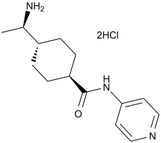
| Chemical Name | (1R,4r)-4-((R)-1-aminoethyl)-N-(pyridin-4-yl)cyclohexanecarboxamide dihydrochloride | |
| Synonyms |
|
|
| HS Tariff Code | 2934.99.9001 | |
| Storage |
Powder-20°C 3 years 4°C 2 years In solvent -80°C 6 months -20°C 1 month |
|
| Shipping Condition | Room temperature (This product is stable at ambient temperature for a few days during ordinary shipping and time spent in Customs) |
Biological Activity
| Targets |
ROCK-I (Ki = 220 nM); ROCK-II (Ki = 300 nM); PKN (Ki = 3.1 μM); Citron kinase (Ki = 5.3 μM); PKCα (Ki = 73 μM); PKA (Ki = 25 μM) Y-27632 2HCl specifically targets Rho-associated coiled kinase (ROCK) isoforms ROCK1 and ROCK2 (ROCK1 IC50 = 140 nM; ROCK2 IC50 = 130 nM) [4] Y-27632 2HCl shows no significant inhibition of other kinases (PKC, PKA, MLCK, ERK1/2: IC50 > 10 μM) [4] |
| ln Vitro |
Programming is enhanced and the quarter rate of direct conversion of mouse fibroblasts into neural cells is increased by Y-27632 dihydrochloride (GMP) (2 μM; 4–8 d) [1]. Human Y-27632 dihydrochloride (GMP) facilitates the differentiation of fibroblasts into pluripotent stem cells (hiPSCs) [2]. Primed hPSCs are converted into hEPS cells and human EPS cell proliferation is enhanced by Y-27632 dihydrochloride (GMP) (2–10 μM) [3]. Human EPS cells generated from blastocysts are induced by 10 μM of Y-27632 diHCl (GMP) [4].
Y-27632 [(+)-(R)-trans-4-(1-aminoethyl)-N-(4-pyridyl)cyclohexanecarboxamide++ + dihydrochloride] is widely used as a specific inhibitor of the Rho-associated coiled-coil forming protein serine/threonine kinase (ROCK) family of protein kinases. This study examined the inhibition mechanism and profile of actions of Y-27632 and a related compound, Y-30141 [(+)-(R)-trans- 4-(1-aminoethyl)-N-(1H-pyrrolo[2, 3-b]pyridin-4-yl)cyclohexan-ecarboxamide dihydrochloride]. Y-27632 and Y-30141 inhibited the kinase activity of both ROCK-I and ROCK-II in vitro, and this inhibition was reversed by ATP in a competitive manner. This suggests that these compounds inhibit the kinases by binding to the catalytic site. Their affinities for ROCK kinases as determined by K(i) values were at least 20 to 30 times higher than those for two other Rho effector kinases, citron kinase and protein kinase PKN. [(3)H]Y-30141 was taken up by cells in a temperature- and time-dependent and saturable manner, and this uptake was competed with unlabeled Y-27632. No concentrated accumulation was found, suggesting that the uptake is a carrier-mediated facilitated diffusion. Y-27632 abolished stress fibers in Swiss 3T3 cells at 10 microM, but the G(1)-S phase transition of the cell cycle and cytokinesis were little affected at this concentration. Y-30141 was 10 times more potent than Y-27632 in inhibiting the kinase activity and stress fiber formation, and it caused significant delay in the G(1)-S transition and inhibition of cytokinesis at 10 microM. [4] Y-27632 had the potency to induce neuronal-like differentiation in ADSCs in a dose-dependent manner. Moreover, the differentiation induced by Y-27632 was recovered upon drug withdraw. ADSCs treated with Y-27632 expressed neuronal markers such as NSE, MAP-2 and nestin while untreated ADSCs did not express these markers. Conclusion: Selective ROCK inhibitor Y-27632 could potentiate the neuronal-like differentiation of ADSCs, suggesting that Y-27632 could be utilized to induce the differentiation of ADSCs to neurons and facilitate the clinical application of ADSCs in tissue engineering. [3] In recombinant ROCK1/ROCK2 enzyme assays, Y-27632 2HCl dose-dependently inhibits kinase activity with IC50 values of 140 nM (ROCK1) and 130 nM (ROCK2), acting as a competitive inhibitor against ATP [4] - In rat hepatic stellate cells (HSCs), Y-27632 2HCl (1 μM) inhibits cell proliferation by 65% after 72 hours and reduces collagen type I synthesis by 58% compared to control. It also downregulates α-SMA (fibrosis marker) expression by 70% at protein level [1] - In adult human adipose tissue-derived stem cells (hADSCs), Y-27632 2HCl (10 μM) promotes neuronal-like differentiation, with increased expression of neuronal markers β-III tubulin (3.2-fold) and neurofilament 200 (2.8-fold) after 7 days. It also enhances cell morphology changes resembling neurons (80% of cells exhibit neurite outgrowth) [3] - In mouse cortical neurons, Y-27632 2HCl (5 μM) reduces glutamate-induced excitotoxicity, decreasing apoptotic cell death by 45% after 24 hours. It inhibits ROCK-mediated phosphorylation of MLC (myosin light chain) by 62% [2] - In smooth muscle cells, Y-27632 2HCl (0.5 μM) inhibits Ca²⁺-independent contraction by 75% and reduces ROCK-dependent phosphorylation of MLC (Ser19) by 68% [4] |
| ln Vivo |
Oral administration of Y-27632 2HCl at 30 mg/kg significantly decreases the blood pressure in a dose-dependent manner in spontaneous hypertensive rats, renal hypertensive rats, as well as deoxycorticosterone acetate (DOCA)-salt hypertensive rats. When Y-27632 2HCl is continuously administered at a rate of 0.55 μL per hour by implanted pumps for 11 days tumor cell invasion (MM1 cells expressing Val14-RhoA in rats) is significantly delayed. By inhibiting ROCK, Y-27632 2HCl treatment attenuates hypoxia-induced angiogenesis and vascular remodeling in the pulmonary circulation. Pretreatment with Y-27632 has a protective effect against tumor formation in albino mice with Ehrlich ascites carcinoma. Y27632 treatment significantly decreased the occurrence of DMN-induced hepatic fibrosis and reduced the collagen and hydroxyproline content and alpha-SMA expression in the liver. [1] Effects of Y-27632 (5-10 mg kg(-1)) and fasudil (5-25 mg kg(-1)) on duration of myoclonic jerks, clonic and tonic convulsions, tonic hindlimb extensions and percentage of tonic convulsion index, as well as recovery latency for righting reflex were investigated in mice stimulated with PTZ (65 mg kg(-1)) or MES (50 Hz, 50 mA and 0.4 s). These inhibitors were also tested on a model of kindling induced by PTZ (35 mg kg(-1), for 11 days). Membrane and cytosolic levels of RhoA protein were measured in brain homogenates from kindled mice.[2] Y27632 prevents body and liver weight loss induced by DMN [1] The effect of oral administration of Y27632 on body and liver weights of rats with and without i.p. injection of DMN is shown in Fig. 1 and Table 1. Treatment with DMN caused a significant decrease in rat body and liver weight (DMN-S group) compared with control animals (S-S group). Oral Y27632 essentially prevented this DMN-induced rat body and liver weight loss (DMN-Y group). There was no significant difference between the S-S/S-Y and DMN-Y groups. There was no significant difference in serum ALT levels between the DMN-S and DMN-Y groups at the end of fourth week. Y27632 reduces hepatic collagen and hydroxyproline content [1] The significantly decreased deposition of hepatic collagen in the Y27632-treated group (the DMN-Y group) was confirmed by collagen assays (DMN-S, 808.5±122.4 μg/100 mg versus DMN-Y, 601.3±67.7 μg/100 mg, P<0.05) (Fig. 3A). Hepatic fibrosis was also quantified by the measurement of hepatic hydroxyproline. Here, it was found that the hydroxyproline content of the DMN-treated group (DMN-S) was significantly higher than that of the DMN and Y27632-treated group (DMN-Y) (DMN-S, 5.95±1.41 μmol/100 mg versus DMN-Y, 2.98±0.91 μmol/100 mg, P<0.05) (Fig. 3B). Furthermore, there was no significant difference in collagen and hydroxyproline contents between the DMN-untreated groups (S-S, S-Y) and the DMN and Y27632-treated group (DMN-Y). These results indicated that the treatment with Y27632 almost completely prevented the hepatic fibrosis induced by DMN. Y27632 inhibits type I collagen mRNA expression [1] To assess the level of inhibition of type I collagen mRNA transcription by Y27632 treatment, we examined the level of α1(I) collagen mRNA in DMN-treated liver by semi-quantitative RT-PCR. In the control group (the S-S group), even in the presence of only 0.02 fg competitor, a major competitor-specific band and a weak α1(I) collagen-specific band were detected in the competitive RT-PCR for α1(I) collagen (Fig. 4A, lane 4). In contrast, in the DMN-S group, DMN treatment (in the absence of Y27632) enhanced the transcription of the α1(I) collagen gene more than 100 times compared to the basal expression (S-S), and even in the presence of 2 fg competitor resulted in a band representing the α1(I) collagen product (Fig. 4B, lane 2). Treatment with Y27632 resulted in a more intense competitor band compared to DMN treatment (in the absence of Y27632); in the presence of 0.02 fg competitor, the α1(I) collagen PCR product was present at an intensity equal to that of the competitor (Fig. 4C, lane 4). These results demonstrate that treatment with Y27632 suppresses α1(I) collagen mRNA expression in the liver to the approximately 10% level of the DMN-induced expression. The effects of fasudil and Y-27632 on the onset of myoclonic jerks induced by acute PTZ injection [2] The effects of fasudil and Y-27632 on the onset of myoclonic jerks are shown in Figure 1. At the dose of 25 mg kg−1, fasudil significantly prolonged the onset time of myoclonic jerks induced by a single dose of PTZ (65 mg kg−1, i.p.) when compared with the saline group (P<0.05). However, it did not change the onset time at a dose of 5 mg kg−1. Furthermore, at the doses of 5 and 10 mg kg−1, Y-27632 significantly prolonged the onset time of myoclonic jerks when compared with those observed in the saline group (P<0.05). The effects of fasudil and Y-27632 on the onset of clonic convulsions induced by acute PTZ injection [2] The effects of fasudil and Y-27632 on the onset of clonic convulsions are shown in Figure 1. At the dose of 25 mg kg−1, fasudil significantly prolonged the onset time of clonic convulsions induced by a single dose of PTZ (65 mg kg−1, i.p.) when compared with the saline group (P<0.05). However, it did not change the onset time at the dose of 5 mg kg−1. Moreover, at the doses of 5 and 10 mg kg−1, Y-27632 significantly prolonged the onset time of clonic convulsions when compared with saline group (P<0.05). The effects of fasudil and Y-27632 on tonic hindlimb extensions induced by acute PTZ injection [2] Seven out of ten mice in the PTZ-treated group had tonic hindlimb extensions and died after that. However, fasudil, in both doses, prevented the occurrence of tonic hindlimb extensions and death (data not shown). Similar to fasudil, Y-27632, in both doses, also prevented the occurrence of tonic hindlimb extensions and death (data not shown). The effects of fasudil and Y-27632 on the duration of tonic hindlimb extensions and the recovery latency for righting reflex in MES series [2] Both fasudil (25 mg kg−1) and Y-27632 (5 and 10 mg kg−1) significantly decreased the duration of tonic hindlimb extensions induced by a single application of MES when compared with the group treated with saline (Figure 2, P<0.05). Moreover, the Rho-kinase inhibitors fasudil and Y-27632 also significantly decreased the recovery latency for righting reflex when compared with the saline group (Figure 2, P<0.05). The effects of acute single administration of fasudil or Y-27632 on the onset of myoclonic jerks and clonic convulsions in PTZ-kindled mice [2] Neither fasudil (25 mg kg−1) nor Y-27632 (5 and 10 mg kg−1) changed the onset times of myoclonic jerks and clonic convulsions when compared with the saline group (Figure 3). The effects of repeated administration of fasudil or Y-27632 on the development of PTZ kindling [2] The effects of repeated administration of fasudil or Y-27632 on the development of PTZ kindling are shown in Figure 4. PTZ injections by group interaction were found significant for saline, 25 mg kg−1 fasudil and 5 mg kg−1 Y-27632 groups (P<0.05). As shown in Figure 4, fasudil was not able to prevent the development of PTZ kindling, but it had a significant effect on the 2nd, 3rd and 4th days of the development by lowering the mean seizure stages when compared with the saline group (P<0.05). Unlike fasudil, Y-27632 was more potent and had a significant effect on the development of PTZ kindling from the beginning of the 2nd day, as compared with the saline group, by lowering the mean seizure stages (P<0.05). The effects of fasudil and Y-27632 on the rota-rod performance [2] Neither fasudil (25 mg kg−1) nor Y-27632 (5 and 10 mg kg−1) changed motor coordination evaluated by performance on the rota-rod, at 20 r.p.m. (Table 1). In dimethylnitrosamine (DMN)-induced hepatic fibrosis rats, intraperitoneal administration of Y-27632 2HCl (10 mg/kg/day for 4 weeks) significantly attenuates liver fibrosis. Hepatic collagen content is reduced by 62% compared to vehicle controls, and α-SMA-positive HSCs are decreased by 55%. Liver function markers (ALT, AST) are normalized to near-normal levels [1] - In pentylenetetrazole (PTZ)-induced epileptic mice, intraperitoneal Y-27632 2HCl (10-30 mg/kg, single dose 30 minutes before PTZ) dose-dependently reduces seizure severity. The median seizure score decreases from 4.0 (vehicle) to 1.5 (30 mg/kg group), and seizure duration is shortened by 58% [2] - In kainic acid-induced epileptic mice, Y-27632 2HCl (15 mg/kg/day, ip, for 7 days) reduces hippocampal neuronal loss by 52% and inhibits microglial activation (Iba1-positive cells decreased by 48%) [2] |
| Enzyme Assay |
Recombinant ROCK-I, ROCK-II, PKN, or citron kinase is expressed in HeLa cells as Myc-tagged proteins by transfection using Lipofectamine, and is precipitated from the cell lysates by the use of 9E10 monoclonal anti-Myc antibody coupled to G protein-Sepharose. Recovered immunocomplexes are incubated with various concentrations of [32P]ATP and 10 mg of histone type 2 as substrates in the absence or presence of various concentrations of either Y-27632 or Y-30141 at 30°C for 30 min in a total volume of 30 μL of the kinase buffer containing 50 mM HEPES-NaOH, pH 7.4, 10 mM MgCl2, 5 mM MnCl2, 0.02% Briji 35, and 2 mM dithiothreitol. PKCa is incubated with 5 μM [32P]ATP and 200 μg/mL histone type 2 as substrates in the absence or presence of various concentrations of either Y-27632 or Y-30141 at 30°C for 10 min in a kinase buffer containing 50 mM Tris-HCl, pH 7.5, 0.5 mM CaCl2, 5 mM magnesium acetate, 25 μg/mL phosphatidyl serine, 50 ng/mL 12-O-tetradecanoylphorbol-13-acetate and 0.001% leupeptin in a total volume of 30 μL. Incubation is terminated by the addition of 10 μL of 43 Laemmli sample buffer. After boiling for 5 min, the mixture is subjected to SDS-polyacrylamide gel electrophoresis on a 16% gel. The gel is stained with Coomassie Brilliant Blue, and then dried. The bands corresponding to histone type 2 are excised, and the radioactivity is measured.[1] ROCK1/ROCK2 kinase activity assay: Purified recombinant human ROCK1 or ROCK2 was incubated with substrate peptide (MLC-derived) and Y-27632 2HCl (0.01-10 μM) in assay buffer (50 mM Tris-HCl, pH 7.5, 10 mM MgCl₂, 1 mM DTT, 0.1 mM ATP) at 30°C for 60 minutes. Phosphorylated substrate was detected by radiolabeled ATP counting, and IC50 values were calculated from dose-response curves [4] - Kinase selectivity assay: Y-27632 2HCl (10 μM) was screened against a panel of kinases (PKC, PKA, MLCK, ERK1/2, JNK) using respective substrate peptides and assay buffers. Kinase activity was quantified by radiolabeling or colorimetric methods, with no significant inhibition (>50% activity reduction) observed for off-target kinases [4] - ATP competition assay: ROCK2 was incubated with increasing concentrations of ATP (0.05-1 mM) and fixed Y-27632 2HCl (100 nM). Kinase activity was measured to confirm competitive binding to the ATP-binding pocket of ROCK [4] |
| Cell Assay |
HeLa cells are plated at a density of 3×10⁴ cells per 3.5-cm dish. The cells are cultured in DMEM containing 10% FBS in the presence of 10 mM Thymidine for 16 h. After the cells are washed with DMEM containing 10% FBS, they are cultured for an additional 8 h, and then 40 ng/mL of Nocodazole is added. After 11.5 h of the Nocodazole treatment, various concentrations of Y-27632 (0-300 μM), Y-30141, or vehicle is added and the cells are incubated for another 30 min. [1] ADSCs were isolated from women undergoing plastic surgery and cultured. ADSCs were treated with different doses of Y-27632 and observed morphological changes under microscope. The expression of nestin, neuron specific enolase (NSE) and microtubule-associated protein-2 (MAP-2) in ADSCs treated with Y-27632 was detected by immunocytochemistry and Western blotting analysis. [3] Cell isolation and culture [1] HSC from the liver of male Wistar rats that had not received DMN or Y-27632 were isolated and cultured as described previously. Experiments described in this study were performed on HSC between the second and fourth serial passages. In some experiments, HSC were used before the first passage. Western blot analysis of α-SMA [1] HSC were cultured in 6 cm plastic tissue culture plates at a density of 1×10⁶ cells/ml with DMEM (20% FCS). After the medium was changed, HSC were maintained in either the presence or absence of Y-27632. The preparation of whole cell lysates and Western blot analysis were performed as previously reported. Immunocytochemistry [1] HSC were maintained in either the presence or absence of Y-27632 (30 μM) for 2 days. Immunocytochemistry was performed as previously reported. The slides were stained with FITC-conjugated goat anti-mouse IgG for α-SMA. Hepatic stellate cell (HSC) fibrosis assay: Rat HSCs were seeded in 6-well plates at 2×10⁵ cells/well and activated with TGF-β1 (10 ng/mL) for 24 hours. Y-27632 2HCl (0.1-5 μM) was added, and cells were cultured for 72 hours. Cell proliferation was measured by MTT assay, collagen type I synthesis by ELISA, and α-SMA expression by Western blot [1] - hADSC neuronal differentiation assay: hADSCs were seeded in 6-well plates at 1×10⁴ cells/well and treated with Y-27632 2HCl (1-20 μM) in differentiation medium for 7 days. Neuronal markers (β-III tubulin, neurofilament 200) were detected by immunocytochemistry and Western blot. Neurite outgrowth was quantified by image analysis [3] - Neuronal excitotoxicity assay: Mouse cortical neurons were seeded in 96-well plates at 5×10³ cells/well and cultured for 7 days. Cells were pretreated with Y-27632 2HCl (1-10 μM) for 1 hour, then exposed to glutamate (100 μM) for 24 hours. Apoptosis was detected by Annexin V-FITC staining, and MLC phosphorylation by Western blot [2] - Smooth muscle cell contraction assay: Rat aortic smooth muscle cells were seeded in collagen gels and treated with Y-27632 2HCl (0.1-1 μM) for 1 hour. Ca²⁺-independent contraction was induced by thrombin (0.1 U/mL), and gel contraction was measured by area reduction after 24 hours [4] |
| Animal Protocol |
Dissolved in DMSO, and diluted in saline (Rat); 0.9% NaCl (Mice); 30 mg/kg/day (Rat); 0-10 mg/kg (mice); Oral for rats, IP for mice Male Wistar rats with spontaneous or induced hypertension; Swiss albino mice with Ehrlich ascites carcinoma Y27632 was given orally at 30 mg/kg daily for 4 weeks after the first injection of DMN. The degree of fibrosis was evaluated by image analysis and also by measurements of collagen and hydroxyproline content in the liver. The expression of alpha-smooth muscle actin (alpha-SMA) in the liver and in the primary cultured HSC was also evaluated. Semi-quantitative RT-PCR was performed to evaluate the expression of type I collagen mRNA in the liver.[1] Effects of Y-27632 (5-10 mg kg(-1)) and fasudil (5-25 mg kg(-1)) on duration of myoclonic jerks, clonic and tonic convulsions, tonic hindlimb extensions and percentage of tonic convulsion index, as well as recovery latency for righting reflex were investigated in mice stimulated with PTZ (65 mg kg(-1)) or MES (50 Hz, 50 mA and 0.4 s). These inhibitors were also tested on a model of kindling induced by PTZ (35 mg kg(-1), for 11 days). Membrane and cytosolic levels of RhoA protein were measured in brain homogenates from kindled mice.[2] Y-27632 was given orally at 30 mg/kg daily for 4 weeks after the first injection of DMN. The degree of fibrosis was evaluated by image analysis and also by measurements of collagen and hydroxyproline content in the liver. The expression of alpha-smooth muscle actin (alpha-SMA) in the liver and in the primary cultured HSC was also evaluated. Semi-quantitative RT-PCR was performed to evaluate the expression of type I collagen mRNA in the liver.[1] Effects of Y-27632 (5-10 mg kg(-1)) and fasudil (5-25 mg kg(-1)) on duration of myoclonic jerks, clonic and tonic convulsions, tonic hindlimb extensions and percentage of tonic convulsion index, as well as recovery latency for righting reflex were investigated in mice stimulated with PTZ (65 mg kg(-1)) or MES (50 Hz, 50 mA and 0.4 s). These inhibitors were also tested on a model of kindling induced by PTZ (35 mg kg(-1), for 11 days). Membrane and cytosolic levels of RhoA protein were measured in brain homogenates from kindled mice.[2] Treatment of animals [1] DMN (1 g/ml) was diluted ten times with saline (final concentration 1%) and 10 mg/kg per day of DMN was injected intraperitoneally (i.p.) on the first 3 days of each week for 4 weeks as described. Y-27632 was dissolved in saline (final concentration 2%) and was given orally once per day at a dose of 30 mg/kg for 4 weeks starting on the day of the first injection of DMN. The dose of 30 mg/kg was selected according to a previous report that this dose corrected hypertension in several rat models without toxicity. Twenty rats were randomized into four experimental groups (n=5 in each group) as follows: (1) S-S (injection of saline i.p. and oral administration of saline); (2) S-Y (injection of saline i.p. and oral administration of Y27632); (3) DMN-S (DMN i.p. and oral administration of saline); (4) DMN-Y (DMN i.p. and oral administration of Y27632). The rats were weighed every week. They were sacrificed at the end of the fourth week and the liver was excised. In addition, a blood sample was taken immediately before the rats were sacrificed. Acute PTZ experiments [2] A group of animals were injected with a single dose of PTZ (65 mg kg−1) to investigate if the two Rho-kinase inhibitors, fasudil and Y-27632, changed the onset of PTZ seizures. Fasudil, Y-27632 or saline was given intraperitoneally 30 min before the PTZ injection. Each mouse was then observed for a 15-min period to measure the onset of the first myoclonic jerk, the onset of the first clonic convulsion and the occurrence of tonic hindlimb extension. Some of the animals died after tonic hindlimb extension, which is an expected outcome of acute PTZ injection. After the observation period, all animals were killed by halothane anaesthesia. PTZ kindling experiments [2] Another group of mice were tested for PTZ kindling. Mice were injected with a sub-convulsive dose of PTZ (35 mg kg−1, i.p.) (on Mondays, Wednesdays and Fridays) of each week for a total of 11 injections. After each PTZ injection, mice were observed for 30 min and the occurrence of convulsive activity was recorded and classified using the scoring system of Fischer and Kittner (1998) as described below. After 30 min, the mice were then injected with either fasudil (25 mg kg−1, i.p.) or Y-27632 (5 mg kg−1, i.p.) and returned to their home cages until the next injection. Control mice for fasudil and Y-27632 received saline. An animal undergoing a stage 5 convulsion was considered to be fully kindled and was not further tested. The seizure stage rating scale was as follows: Stage 0: no evidence of convulsive activity; Stage 1: ear and facial twitching, head nodding; Stage 2: myoclonic jerks; Stage 3: forelimb clonuses with full rearing; Stage4: generalized clonic convulsions with loss of righting reflex, rearing, jumping and falling down; and Stage5: clonic-tonic convulsions with tonic hindlimb extensions. Another group of kindled mice were injected with a single dose of either fasudil (25 mg kg−1) or Y-27632 (5–10 mg kg−1) to test if acute administration of these Rho-kinase inhibitors could change the seizure susceptibility in kindled mice. At the end of the kindling protocol, all animals were killed by cervical dislocation for the measurement of RhoA translocation in the brain by western blotting. MES experiments [2] To investigate the possible effects of Rho-kinase inhibitors, fasudil and Y-27632, on the tonic hindlimb extensions induced by MES, a group of mice was exposed to an electroshock by ear-clip electrodes. Fasudil, Y-27632 or saline was injected 30 min before electroshock. Immediately after the electroshock, each mouse was put into a Plexiglas chamber to observe the duration of tonic hindlimb extension and the recovery latency for righting reflex for 10 min. After the observation period, all animals were killed by halothane anaesthesia. Rota-rod performance test [2] A separate group of mice received either 25 mg kg−1 of fasudil or 10 mg kg−1 of Y-27632 and was examined in a rota-rod performance test to see if there was any failure of motor coordination induced by Rho-kinase inhibitors. Y-27632 (5 or 10 mg kg−1) and PTZ (35 or 65 mg kg−1) were dissolved in 0.9% NaCl (saline) and injected intraperitoneally in a volume of 0.1 mL per 10 g body weight. Control animals received saline. DMN-induced hepatic fibrosis rat model: Adult male Wistar rats were injected intraperitoneally with DMN (10 mg/kg) three times weekly for 4 weeks to induce fibrosis. Concurrently, Y-27632 2HCl was administered intraperitoneally at 10 mg/kg/day for 4 weeks. Vehicle group received saline. Rats were euthanized, and liver tissues were collected for histopathological analysis (Masson's trichrome staining) and collagen content measurement [1] - PTZ-induced epileptic mouse model: Adult male ICR mice were pretreated with intraperitoneal Y-27632 2HCl (1, 10, 30 mg/kg) 30 minutes before intraperitoneal PTZ injection (80 mg/kg) to induce seizures. Seizure severity (Racine scale) and duration were recorded for 30 minutes [2] - Kainic acid-induced epileptic mouse model: Mice were injected intraperitoneally with kainic acid (15 mg/kg) to induce status epilepticus. Y-27632 2HCl (15 mg/kg/day) was administered intraperitoneally for 7 days starting 24 hours post-kainic acid injection. Hippocampal tissues were harvested for neuronal count and Iba1 immunostaining [2] |
| Toxicity/Toxicokinetics |
In vitro, Y-27632 2HCl shows low toxicity to normal cells (hADSCs IC50 > 50 μM; mouse cortical neurons IC50 > 40 μM) [2][3] - In in vivo studies, Y-27632 2HCl at tested doses (10-30 mg/kg, ip) causes no significant body weight loss (<5% vs. baseline) or overt lethality in rats and mice [1][2] - No significant changes in liver function (ALT, AST) or renal function (creatinine, BUN) were observed in Y-27632 2HCl-treated animals compared to vehicle controls [1][2] - Plasma protein binding rate of Y-27632 2HCl is 92-94% in rats (in vitro plasma binding assay) [4] - Mild hypotension was observed in rats at doses >50 mg/kg (ip) [4] |
| References |
[1]. A selective ROCK inhibitor, Y27632, prevents dimethylnitrosamine-induced hepatic fibrosis in rats. J Hepatol. 2001 Apr;34(4):529-36. [2]. Antiepileptic effects of two Rho-kinase inhibitors, Y-27632 and fasudil, in mice. Br J Pharmacol. 2008 Sep;155(1):44-51. [3]. Rho-associated coiled kinase inhibitor Y-27632 promotes neuronal-like differentiation of adult human adipose tissue-derived stem cells. Chin Med J (Engl). 2012 Sep;125(18):3332-5. [4]. Pharmacological properties of Y-27632, a specific inhibitor of rho-associated kinases. Mol Pharmacol. 2000 May;57(5):976-83. |
| Additional Infomation |
Background: Y-27632 is a specific inhibitor of Rho-associated coiled kinase (ROCK) and has been shown to promote the survival and induce the differentiation of a variety of cells types. However, the effects of Y-27632 on adult human adipose tissue-derived stem cells (ADSCs) are unclear. This study aimed to investigate the effects of Y-27632 on the neuronal-like differentiation of ADSCs.[3] Methods: ADSCs were isolated from women undergoing plastic surgery and cultured. ADSCs were treated with different doses of Y-27632 and observed morphological changes under microscope. The expression of nestin, neuron specific enolase (NSE) and microtubule-associated protein-2 (MAP-2) in ADSCs treated with Y-27632 was detected by immunocytochemistry and Western blotting analysis.[3] Results: Y-27632 had the potency to induce neuronal-like differentiation in ADSCs in a dose-dependent manner. Moreover, the differentiation induced by Y-27632 was recovered upon drug withdraw. ADSCs treated with Y-27632 expressed neuronal markers such as NSE, MAP-2 and nestin while untreated ADSCs did not express these markers.[3] Conclusion: Selective ROCK inhibitor Y-27632 could potentiate the neuronal-like differentiation of ADSCs, suggesting that Y-27632 could be utilized to induce the differentiation of ADSCs to neurons and facilitate the clinical application of ADSCs in tissue engineering.[3] Y-27632 [(+)-(R)-trans-4-(1-aminoethyl)-N-(4-pyridyl)cyclohexanecarboxamide++ + dihydrochloride] is widely used as a specific inhibitor of the Rho-associated coiled-coil forming protein serine/threonine kinase (ROCK) family of protein kinases. This study examined the inhibition mechanism and profile of actions of Y-27632 and a related compound, Y-30141 [(+)-(R)-trans- 4-(1-aminoethyl)-N-(1H-pyrrolo[2, 3-b]pyridin-4-yl)cyclohexan-ecarboxamide dihydrochloride]. Y-27632 and Y-30141 inhibited the kinase activity of both ROCK-I and ROCK-II in vitro, and this inhibition was reversed by ATP in a competitive manner. This suggests that these compounds inhibit the kinases by binding to the catalytic site. Their affinities for ROCK kinases as determined by K(i) values were at least 20 to 30 times higher than those for two other Rho effector kinases, citron kinase and protein kinase PKN. [(3)H]Y-30141 was taken up by cells in a temperature- and time-dependent and saturable manner, and this uptake was competed with unlabeled Y-27632. No concentrated accumulation was found, suggesting that the uptake is a carrier-mediated facilitated diffusion. Y-27632 abolished stress fibers in Swiss 3T3 cells at 10 microM, but the G(1)-S phase transition of the cell cycle and cytokinesis were little affected at this concentration. Y-30141 was 10 times more potent than Y-27632 in inhibiting the kinase activity and stress fiber formation, and it caused significant delay in the G(1)-S transition and inhibition of cytokinesis at 10 microM.[4] Y-27632 2HCl is a potent, selective small-molecule inhibitor of ROCK1 and ROCK2, key regulators of actin cytoskeleton organization and cell signaling [4] - Its mechanism of action involves binding to the ATP-binding pocket of ROCK, inhibiting phosphorylation of downstream substrates (MLC, LIMK) and blocking ROCK-mediated processes (cell contraction, proliferation, fibrosis, neuronal excitotoxicity) [1][2][4] - Y-27632 2HCl exhibits in vitro and in vivo efficacy in models of hepatic fibrosis and epilepsy, supporting its potential as a research tool for ROCK-related disorders [1][2] - It is widely used to study ROCK function in cell differentiation (neuronal, stem cell), fibrosis, smooth muscle contraction, and neuroprotection [3][4] - The compound does not show significant off-target kinase inhibition, ensuring high selectivity for ROCK isoforms [4] |
Solubility Data
| Solubility (In Vitro) |
|
|||
| Solubility (In Vivo) |
Note: Listed below are some common formulations that may be used to formulate products with low water solubility (e.g. < 1 mg/mL), you may test these formulations using a minute amount of products to avoid loss of samples. Injection Formulations (e.g. IP/IV/IM/SC) Injection Formulation 1: DMSO : Tween 80: Saline = 10 : 5 : 85 (i.e. 100 μL DMSO stock solution → 50 μL Tween 80 → 850 μL Saline) *Preparation of saline: Dissolve 0.9 g of sodium chloride in 100 mL ddH ₂ O to obtain a clear solution. Injection Formulation 2: DMSO : PEG300 :Tween 80 : Saline = 10 : 40 : 5 : 45 (i.e. 100 μL DMSO → 400 μLPEG300 → 50 μL Tween 80 → 450 μL Saline) Injection Formulation 3: DMSO : Corn oil = 10 : 90 (i.e. 100 μL DMSO → 900 μL Corn oil) Example: Take the Injection Formulation 3 (DMSO : Corn oil = 10 : 90) as an example, if 1 mL of 2.5 mg/mL working solution is to be prepared, you can take 100 μL 25 mg/mL DMSO stock solution and add to 900 μL corn oil, mix well to obtain a clear or suspension solution (2.5 mg/mL, ready for use in animals). Injection Formulation 4: DMSO : 20% SBE-β-CD in saline = 10 : 90 [i.e. 100 μL DMSO → 900 μL (20% SBE-β-CD in saline)] *Preparation of 20% SBE-β-CD in Saline (4°C,1 week): Dissolve 2 g SBE-β-CD in 10 mL saline to obtain a clear solution. Injection Formulation 5: 2-Hydroxypropyl-β-cyclodextrin : Saline = 50 : 50 (i.e. 500 μL 2-Hydroxypropyl-β-cyclodextrin → 500 μL Saline) Injection Formulation 6: DMSO : PEG300 : castor oil : Saline = 5 : 10 : 20 : 65 (i.e. 50 μL DMSO → 100 μLPEG300 → 200 μL castor oil → 650 μL Saline) Injection Formulation 7: Ethanol : Cremophor : Saline = 10: 10 : 80 (i.e. 100 μL Ethanol → 100 μL Cremophor → 800 μL Saline) Injection Formulation 8: Dissolve in Cremophor/Ethanol (50 : 50), then diluted by Saline Injection Formulation 9: EtOH : Corn oil = 10 : 90 (i.e. 100 μL EtOH → 900 μL Corn oil) Injection Formulation 10: EtOH : PEG300:Tween 80 : Saline = 10 : 40 : 5 : 45 (i.e. 100 μL EtOH → 400 μLPEG300 → 50 μL Tween 80 → 450 μL Saline) Oral Formulations Oral Formulation 1: Suspend in 0.5% CMC Na (carboxymethylcellulose sodium) Oral Formulation 2: Suspend in 0.5% Carboxymethyl cellulose Example: Take the Oral Formulation 1 (Suspend in 0.5% CMC Na) as an example, if 100 mL of 2.5 mg/mL working solution is to be prepared, you can first prepare 0.5% CMC Na solution by measuring 0.5 g CMC Na and dissolve it in 100 mL ddH2O to obtain a clear solution; then add 250 mg of the product to 100 mL 0.5% CMC Na solution, to make the suspension solution (2.5 mg/mL, ready for use in animals). Oral Formulation 3: Dissolved in PEG400 Oral Formulation 4: Suspend in 0.2% Carboxymethyl cellulose Oral Formulation 5: Dissolve in 0.25% Tween 80 and 0.5% Carboxymethyl cellulose Oral Formulation 6: Mixing with food powders Note: Please be aware that the above formulations are for reference only. InvivoChem strongly recommends customers to read literature methods/protocols carefully before determining which formulation you should use for in vivo studies, as different compounds have different solubility properties and have to be formulated differently. (Please use freshly prepared in vivo formulations for optimal results.) |
| Preparing Stock Solutions | 1 mg | 5 mg | 10 mg | |
| 1 mM | 3.1225 mL | 15.6123 mL | 31.2246 mL | |
| 5 mM | 0.6245 mL | 3.1225 mL | 6.2449 mL | |
| 10 mM | 0.3122 mL | 1.5612 mL | 3.1225 mL |