WYE-687 is a novel, potent, ATP-competitive and selective inhibitor of mTOR (mammalian target of rapamycin) with potential antitumor activity. With an IC50 of 7 nM, it inhibits mTOR, and WYE687 demonstrated strong antiproliferative activity in vitro and high antitumor efficacy in vivo.
Physicochemical Properties
| Molecular Formula | C28H32N8O3 | |
| Molecular Weight | 528.61 | |
| Exact Mass | 528.259 | |
| Elemental Analysis | C, 63.62; H, 6.10; N, 21.20; O, 9.08 | |
| CAS # | 1062161-90-3 | |
| Related CAS # | WYE-687 dihydrochloride;1702364-87-1 | |
| PubChem CID | 25229450 | |
| Appearance | White solid powder | |
| Density | 1.4±0.1 g/cm3 | |
| Boiling Point | 633.2±55.0 °C at 760 mmHg | |
| Flash Point | 336.7±31.5 °C | |
| Vapour Pressure | 0.0±1.9 mmHg at 25°C | |
| Index of Refraction | 1.708 | |
| LogP | 1.36 | |
| Hydrogen Bond Donor Count | 1 | |
| Hydrogen Bond Acceptor Count | 9 | |
| Rotatable Bond Count | 7 | |
| Heavy Atom Count | 39 | |
| Complexity | 785 | |
| Defined Atom Stereocenter Count | 0 | |
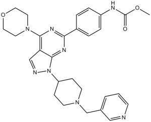
| SMILES | O=C(OC)NC1=CC=C(C2=NC(N3CCOCC3)=C4C(N(C5CCN(CC6=CC=CN=C6)CC5)N=C4)=N2)C=C1 |
|
| InChi Key | VDOCQQKGPJENHJ-UHFFFAOYSA-N | |
| InChi Code | InChI=1S/C28H32N8O3/c1-38-28(37)31-22-6-4-21(5-7-22)25-32-26(35-13-15-39-16-14-35)24-18-30-36(27(24)33-25)23-8-11-34(12-9-23)19-20-3-2-10-29-17-20/h2-7,10,17-18,23H,8-9,11-16,19H2,1H3,(H,31,37) | |
| Chemical Name | methyl N-[4-[4-morpholin-4-yl-1-[1-(pyridin-3-ylmethyl)piperidin-4-yl]pyrazolo[3,4-d]pyrimidin-6-yl]phenyl]carbamate | |
| Synonyms |
|
|
| HS Tariff Code | 2934.99.9001 | |
| Storage |
Powder-20°C 3 years 4°C 2 years In solvent -80°C 6 months -20°C 1 month |
|
| Shipping Condition | Room temperature (This product is stable at ambient temperature for a few days during ordinary shipping and time spent in Customs) |
Biological Activity
| Targets |
PI3K alpha (IC50 = 81 nM); PI3K gamma (IC50 = 3.11 μM); CDK2/CyclinE (IC50 = 430 nM); mTOR (IC50 = 7 nM); CK1 gamma1 (IC50 = 17.8 μM); p38 alpha (IC50 = 28.9 μM); mTORC1; mTORC2 Brefeldin A (BFA) targets ADP-ribosylation factor 1 (ARF1), inhibiting its guanine nucleotide exchange factor (GEF) activity, which is essential for Golgi apparatus structure maintenance; no IC50/Ki values for ARF1 are provided [1,6,7] - Brefeldin A (BFA) mediates ADP-ribosylation of C-terminal binding protein 1/Brefeldin A-ADP-ribosylated substrate (CtBP1/BARS), a key regulator of membrane fission; the EC50 for CtBP1/BARS ADP-ribosylation is not reported [4] - Brefeldin A (BFA) disrupts the microtubule and actin cytoskeletons by unknown direct targets, but indirectly alters cytoskeletal organization via Golgi dysfunction; no specific binding targets for cytoskeletal proteins are identified [1] - Brefeldin A (BFA) does not have a defined "drug target" in cancer stem cell (CSC) regulation, but inhibits CSC potential by suppressing the PI3K/Akt signaling pathway; no IC50 for PI3K/Akt is provided [2] |
| ln Vitro |
WYE-687 inhibits recombinant mTOR enzyme with an IC50 of 7 nM in the DELFIA measuring His6-S6K1 T389 phosphorylation[1]. The MTT cell survival assay results show that WYE-687 potently inhibits HL-60 cell survival in a dose-dependent manner when applied concentrations of WYE-687 (33–1000 nM) are used to treat HL-60 AML cells. WYE-687 exhibits a time-dependent response as well. Following application of WYE-687 (100-1000 nM) treatment, the proportion of dead (“trypan blue” positive) HL-60 cells significantly increases. By using the [H3] Thymidine integration assay, the WYE-687 has also been shown to inhibit the proliferation of HL-60 cells. WYE-687 is also antisurvival (or "cytotoxic") to U937, THP-1, and AML-193 AML cell lines, according to the results[2]. In human fibroblasts (WI-38 cells), treatment with Brefeldin A (BFA) (1 μg/mL, 1-4 hours) disrupts the Golgi apparatus (observed via immunofluorescence staining for Golgi marker GM130) and causes microtubule depolymerization: the number of intact microtubule filaments decreases by >60% after 2 hours, and actin stress fibers become fragmented (detected by phalloidin staining) [1] - In MDA-MB-231 human breast cancer cells: (1) Brefeldin A (BFA) (100 nM, 72 hours) reduces anchorage-independent survival, with soft agar clone formation rate decreasing from 32% (control) to 11%; (2) at 500 nM, it inhibits CSC potential, reducing sphere formation efficiency (SFE) from 8.5% to 2.1% (measured by sphere counting in serum-free medium); (3) 200 nM Brefeldin A (BFA) decreases cell migration by 58% (Transwell assay) and downregulates CSC markers CD44+/CD24- (flow cytometry: CD44+/CD24- cells from 35% to 12%) [2] - In K562 erythroleukemia cells, Brefeldin A (BFA) (500 nM, 24 hours) induces alternative mitophagy: Western blot shows a 2.3-fold increase in LC3-II (autophagy marker) and a 40% decrease in Tom20 (mitochondrial marker), with colocalization of LC3 and Tom20 (immunofluorescence) indicating mitophagosome formation [3] - In HeLa cell lysates, Brefeldin A (BFA) (10 μM, 30 minutes) induces ADP-ribosylation of recombinant CtBP1/BARS: radiometric assay shows 32P-ADP-ribose incorporation into CtBP1/BARS, and SDS-PAGE confirms a 1.8-fold increase in ADP-ribosylated CtBP1/BARS compared to control [4] - In induced pluripotent stem cells (iPSCs), Brefeldin A (BFA) (100 nM, 48 hours) enhances CRISPR/Cas9-mediated genome editing: transfection of sgRNA-Cas9 complex plus Brefeldin A (BFA) increases editing efficiency from 18% (control) to 39% (T7 endonuclease assay) without reducing cell viability (>90% vs. control) [5] - In Vero cells infected with HSV-1, Brefeldin A (BFA) (5 μg/mL, 1 hour post-infection) inhibits viral replication: viral titer (plaque assay) decreases from 10^6 PFU/mL (control) to 10^3 PFU/mL, and immunofluorescence shows reduced HSV-1 UL51 protein localization to the Golgi apparatus [6,7] - In HepG2 human hepatocellular carcinoma cells, Brefeldin A (BFA) -loaded nanomicelles (BFA-NMs) show dose-dependent cytotoxicity: IC50 of BFA-NMs is 8.2 μM (MTT assay, 72 hours), while free Brefeldin A (BFA) has an IC50 of 15.6 μM; BFA-NMs also induce 2.1-fold more apoptosis (Annexin V/PI staining) than free BFA [8] |
| ln Vivo |
U937 cells are inoculated into the flanks of SCID/beige mice. Mice are orally administered WYE-687 (5 or 25 mg/kg) daily for a total of 7 days when xenografted tumors have grown to a volume of about 100 mm3. The vehicle control, which consists of 5% ethanol, 2% Tween 80, and 5% polyethylene glycol-400, is also given to the mice at this point. Based on results from prior experiments and related research, the WYE-687 regimen used in this study. The in vivo activity of WYE-687 is dose-dependent and significantly reduces the growth of the U937 xenograft tumor in SCID mice when administered at doses of 5 or 25 mg/kg per day. At day 15, the tumors that were treated with WYE-687 at doses of 5 mg/kg and 25 mg/kg were 50% and 75% smaller, respectively, than the tumors that were under the control of the vehicle. The tumor weights of mice receiving WYE-687 treatment are also noticeably lower than those of the vehicle group. With minimal toxicities, oral administration of WYE-687 significantly slows the growth of the U937 leukemic xenograft tumor in SCID mice[2]. In nude mice bearing HepG2 xenografts (n=6 per group): (1) Intravenous injection of Brefeldin A (BFA) -loaded nanomicelles (BFA-NMs) at 20 mg/kg (every 3 days for 5 doses) results in 62.3% tumor growth inhibition (TGI) vs. 28.5% TGI for free Brefeldin A (BFA) (10 mg/kg); (2) BFA-NM group shows no significant body weight loss (<5% vs. control), while free BFA group has 8% weight loss; (3) Histopathology of liver/kidney tissues in BFA-NM group shows no obvious damage, whereas free BFA group has mild hepatocyte degeneration [8] |
| Enzyme Assay |
The standard inhibitor assays are carried out in 96-well plates for 2 hours at room temperature using 25 L containing 6 nM Flag-TOR(3.5) (estimated 5-10% purity), 1 μM His6-S6K, and 100 μM ATP. DELFIA uses the Euphospho-p70S6K T389 antibody to carry out and detect the assays. A commercially purchased batch of mTOR is used in some assays. For the inhibitor versus ATP matrix competition, mTOR kinase reactions are carried out in the presence of different concentrations of ATP (0, 25, 50, 100, 200, 400, and 800 M) as well as different concentrations of inhibitor. The assays have 12 nM Flag-TOR(3.5) and 1 M His-S6K, and they take 30 minutes to complete. DELFIA similarly detects the assay results and processes them to produce double-reciprocal plots[1]. CtBP1/BARS ADP-ribosylation assay [4]: 1. Recombinant human CtBP1/BARS protein (0.5 μg) is incubated in reaction buffer (50 mM Tris-HCl pH 7.5, 10 mM MgCl2, 1 mM DTT) with 10 μM Brefeldin A (BFA) and 5 μCi [32P]-NAD+ (as ADP-ribose donor) at 37°C for 30 minutes. 2. The reaction is stopped by adding 5× SDS loading buffer and boiling for 5 minutes. 3. Samples are separated by 12% SDS-PAGE, and the gel is dried. Radiolabeled ADP-ribosylated CtBP1/BARS is detected by autoradiography, and band intensity is quantified using ImageJ. The percentage of ADP-ribosylation is calculated relative to the control (without Brefeldin A (BFA)). |
| Cell Assay |
Acute myeloid leukemia (AML) cells and progenitor cells are plated onto 48-well tissue culture plates at a density of 1 ×10⁵ cells/well in 0.5 mL DMEM containing 10% FBS. Cells are then treated with WYE-687 at the corresponding concentrations (33-1000 nM) in the presence of 1 mCi/mL of tritiated thymidine. Cells are washed, DNA is precipitated with cold 10% trichloroacetic acid (TCA), solubilized with 1.0 M sodium hydroxide, and aliquots are counted by liquid-scintillation spectrometry to determine [H3] thymidine incorporation. According to the value of the untreated control group, the treatment group's value is normalized[2]. Microtubule/actin cytoskeleton staining assay [1]: 1. WI-38 fibroblasts are seeded on coverslips (1×10^4 cells/coverslip) and cultured overnight. Brefeldin A (BFA) is added at 0.1-5 μg/mL, and cells are incubated for 1-4 hours. 2. Cells are fixed with 4% paraformaldehyde (15 minutes, room temperature), permeabilized with 0.1% Triton X-100 (5 minutes), and blocked with 3% BSA (30 minutes). 3. For microtubules: incubate with anti-α-tubulin primary antibody (4°C, overnight), then Alexa Fluor 488-conjugated secondary antibody (1 hour, room temperature). For actin: incubate with Alexa Fluor 594-phalloidin (30 minutes, room temperature). Nuclei are stained with DAPI. 4. Images are captured by confocal microscopy, and the number of intact microtubule filaments/actin stress fibers per cell is counted (n=100 cells per group). - MDA-MB-231 cell migration assay [2]: 1. Transwell inserts (8 μm pores) are coated with Matrigel (1:10 dilution) for 1 hour at 37°C. MDA-MB-231 cells (5×10^4 cells/insert) in serum-free medium with 0-500 nM Brefeldin A (BFA) are added to the upper chamber; medium with 10% FBS is added to the lower chamber. 2. Cells are incubated for 24 hours, then non-migrated cells on the upper surface are removed with a cotton swab. Migrated cells on the lower surface are fixed with 4% paraformaldehyde, stained with 0.1% crystal violet, and counted under a microscope (5 fields per insert). Migration rate is calculated as (migrated cells in BFA group / migrated cells in control) × 100% [2] - HepG2 cell cytotoxicity assay [8]: 1. HepG2 cells are seeded in 96-well plates (2×10^3 cells/well) and cultured overnight. Free Brefeldin A (BFA) or BFA-NMs are added at concentrations of 1-50 μM (n=3 replicates per concentration). 2. After 72 hours, 20 μL MTT solution (5 mg/mL) is added to each well, and cells are incubated for 4 hours. The supernatant is removed, and 150 μL DMSO is added to dissolve formazan crystals. 3. Absorbance is measured at 570 nm, and cell viability is calculated as (A570 of BFA group / A570 of control) × 100%. IC50 is determined by GraphPad Prism using a four-parameter logistic model [8] - HSV-1 viral titer assay [7]: 1. Vero cells are infected with HSV-1 (MOI=0.1) for 1 hour, then treated with 0-10 μg/mL Brefeldin A (BFA). 2. After 24 hours, cell supernatants are collected, and serial dilutions (10^-1 to 10^-6) are prepared. Diluted supernatants are added to Vero cell monolayers (96-well plates) and incubated for 1 hour. 3. Cells are overlaid with 1% agarose in MEM, and plaques are counted after 72 hours. Viral titer is calculated as PFU/mL = (number of plaques × dilution factor) / volume of supernatant added [7] |
| Animal Protocol |
Mice: U937 cells(2×10⁶ cells/mice, suspended in 100 mL of culture medium) are injected into the right flanks of 6-week-old male CB17 severe combined immunodeficient (SCID)/beige mice, and cells are allowed to develop into palpable tumors .
WYE-687 (5 mg/kg body weight), WYE-687 (25 mg/kg body weight), or the vehicle control (5% ethanol, 2% Tween 80, and 5% polyethylene glycol-400) are given to the animals when tumors have grown to a volume of about 100 mm3 in size. Freshly prepared doses of WYE-687 and vehicle control are administered daily for 7 days straight via oral gavage. We measure tumor sizes. When the experiment is finished, the animals are put to death, and the tumors are taken out and weighed. HepG2 xenograft model in nude mice [8]: 1. Female BALB/c nude mice (6-8 weeks old) are used. HepG2 cells (5×10^6 cells in 0.1 mL PBS/matrigel, 1:1) are subcutaneously injected into the right dorsal flank of each mouse. 2. When tumors reach 100-150 mm³, mice are randomly divided into 4 groups (n=6 per group): (a) Control group (saline, intravenous injection); (b) Free Brefeldin A (BFA) group (10 mg/kg, dissolved in DMSO/saline 1:9, intravenous injection); (c) BFA-NMs low-dose group (10 mg/kg BFA equivalent, intravenous injection); (d) BFA-NMs high-dose group (20 mg/kg BFA equivalent, intravenous injection). 3. Treatments are administered every 3 days for a total of 5 doses. Tumor volume (length × width² × 0.5) and body weight are measured every 2 days. 4. After the last treatment, mice are euthanized. Tumors are excised, weighed, and fixed in 4% paraformaldehyde for histopathological analysis (H&E staining). Liver and kidney tissues are also collected for H&E staining and biochemical analysis (ALT, AST, BUN, Cr) [8] |
| Toxicity/Toxicokinetics |
In vitro toxicity: Brefeldin A (BFA) shows low cytotoxicity to normal human fibroblasts (WI-38 cells): viability remains >80% after 48 hours of treatment with 1 μg/mL Brefeldin A (BFA) [1]; in iPSCs, 100 nM Brefeldin A (BFA) does not reduce cell viability (>90% vs. control) [5] - In vivo toxicity (HepG2 xenograft model): (1) Brefeldin A (BFA) -loaded nanomicelles (20 mg/kg) cause no significant changes in body weight (<5% loss) or liver/kidney function (ALT: 35 ± 5 U/L vs. control 32 ± 4 U/L; AST: 82 ± 7 U/L vs. control 78 ± 6 U/L; BUN: 5.2 ± 0.4 mmol/L vs. control 4.9 ± 0.3 mmol/L; Cr: 45 ± 3 μmol/L vs. control 43 ± 2 μmol/L); (2) Free Brefeldin A (BFA) (10 mg/kg) causes 8% body weight loss and mild hepatocyte degeneration (H&E staining) [8] |
| References |
[1]. Cancer Res, 2009, 69(15), 6232-6240. |
| Additional Infomation |
Brefeldin A (BFA) is a naturally occurring macrocyclic lactone isolated from fungi (e.g., Eupenicillium brefeldianum), first identified for its ability to disrupt the Golgi apparatus [1,6] - The classic mechanism of Brefeldin A (BFA) is to inhibit ARF1 activation, leading to Golgi membrane fusion with the endoplasmic reticulum and disruption of protein secretion [1,6,7] - Brefeldin A (BFA) inhibits HSV-1 replication by blocking the Golgi-dependent trafficking of viral proteins (e.g., UL51), preventing viral particle assembly [6,7] - In cancer therapy, Brefeldin A (BFA) -loaded nanomicelles improve the solubility and tumor targeting of Brefeldin A (BFA), enhancing antitumor efficacy while reducing systemic toxicity [8] - Brefeldin A (BFA) enhances CRISPR editing in iPSCs by unknown mechanisms, possibly by modulating endosomal trafficking to increase sgRNA-Cas9 delivery to the nucleus [5] |
Solubility Data
| Solubility (In Vitro) |
|
|||
| Solubility (In Vivo) |
Solubility in Formulation 1: ≥ 2.5 mg/mL (4.73 mM) (saturation unknown) in 10% DMSO + 40% PEG300 + 5% Tween80 + 45% Saline (add these co-solvents sequentially from left to right, and one by one), clear solution. For example, if 1 mL of working solution is to be prepared, you can add 100 μL of 25.0 mg/mL clear DMSO stock solution to 400 μL PEG300 and mix evenly; then add 50 μL Tween-80 to the above solution and mix evenly; then add 450 μL normal saline to adjust the volume to 1 mL. Preparation of saline: Dissolve 0.9 g of sodium chloride in 100 mL ddH₂ O to obtain a clear solution. Solubility in Formulation 2: ≥ 2.5 mg/mL (4.73 mM) (saturation unknown) in 10% DMSO + 90% (20% SBE-β-CD in Saline) (add these co-solvents sequentially from left to right, and one by one), clear solution. For example, if 1 mL of working solution is to be prepared, you can add 100 μL of 25.0 mg/mL clear DMSO stock solution to 900 μL of 20% SBE-β-CD physiological saline solution and mix evenly. Preparation of 20% SBE-β-CD in Saline (4°C,1 week): Dissolve 2 g SBE-β-CD in 10 mL saline to obtain a clear solution. Solubility in Formulation 3: ≥ 2.5 mg/mL (4.73 mM) (saturation unknown) in 10% DMSO + 90% Corn Oil (add these co-solvents sequentially from left to right, and one by one), clear solution. For example, if 1 mL of working solution is to be prepared, you can add 100 μL of 25.0 mg/mL clear DMSO stock solution to 900 μL of corn oil and mix evenly. (Please use freshly prepared in vivo formulations for optimal results.) |
| Preparing Stock Solutions | 1 mg | 5 mg | 10 mg | |
| 1 mM | 1.8918 mL | 9.4588 mL | 18.9175 mL | |
| 5 mM | 0.3784 mL | 1.8918 mL | 3.7835 mL | |
| 10 mM | 0.1892 mL | 0.9459 mL | 1.8918 mL |