WST-8 is a novel, potent and water-soluble tetrazolium dye/salt used for assessing cell metabolic activity. At neutral pH and in the presence of the intermediate electron acceptor. It enhances sensitivity of the WST-8-based assay over the conventional MTS-based assay.
Physicochemical Properties
| Molecular Formula | C20H13N6NAO11S2 |
| Molecular Weight | 600.4706 |
| Exact Mass | 599.9981 |
| Elemental Analysis | C, 40.01; H, 2.18; N, 14.00; Na, 3.83; O, 29.31; S, 10.68 |
| CAS # | 193149-74-5 |
| Related CAS # | 193149-74-5 (sodium);755734-51-1 (free acid); |
| PubChem CID | 9894947 |
| Appearance | White to yellow or light yellow crystalline powder |
| LogP | 4.052 |
| Hydrogen Bond Donor Count | 0 |
| Hydrogen Bond Acceptor Count | 13 |
| Rotatable Bond Count | 4 |
| Heavy Atom Count | 40 |
| Complexity | 1080 |
| Defined Atom Stereocenter Count | 0 |
| SMILES | S(C1C([H])=C(C([H])=C([H])C=1C1=NN(C2C([H])=C([H])C(=C([H])C=2[H])[N+](=O)[O-])[N+](C2C([H])=C([H])C(=C([H])C=2OC([H])([H])[H])[N+](=O)[O-])=N1)S(=O)(=O)[O-])(=O)(=O)[O-].[Na+] |
| InChi Key | YCAKCISJXLQUEQ-UHFFFAOYSA-N |
| InChi Code | InChI=1S/C20H13N6O11S2.Na/c1-37-18-10-14(26(29)30)6-9-17(18)24-22-20(21-23(24)12-2-4-13(5-3-12)25(27)28)16-8-7-15(38(31,32)33)11-19(16)39(34,35)36/h2-11H,1H3/q-1+1 |
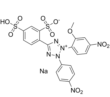
| Chemical Name | 4-(3-(2-methoxy-4-nitrophenyl)-2-(4-nitrophenyl)-2H-tetrazol-3-ium-5-yl)-3-sulfobenzenesulfonate, sodium salt |
| Synonyms | WST-8; WST 8; 270078-53-0; MFCD09264687; sodium 4-(3-(2-methoxy-4-nitrophenyl)-2-(4-nitrophenyl)-2H-tetrazol-3-ium-5-yl)benzene-1,3-disulfonate; sodium;4-[2-(2-methoxy-4-nitrophenyl)-3-(4-nitrophenyl)tetrazol-2-ium-5-yl]benzene-1,3-disulfonate; WST-8 Sodium; WST8. |
| HS Tariff Code | 2934.99.9001 |
| Storage |
Powder-20°C 3 years 4°C 2 years In solvent -80°C 6 months -20°C 1 month Note: Please store this product in a sealed and protected environment (e.g. under nitrogen), avoid exposure to moisture and light. |
| Shipping Condition | Room temperature (This product is stable at ambient temperature for a few days during ordinary shipping and time spent in Customs) |
Biological Activity
| Targets | water-soluble tetrazolium dye |
| ln Vitro | Emulsion polymerization was carried out at 48 and 72 hours to mediate the efficacy of G-CSF. A widely used MTS-based test was compared with a bioassay of the soluble tetrazolium dye WST-8 utilizing NFS-60 cells at a concentration of 7 × 10⁵ cells/mL vs 800 IU/mL of pelletized G-CSF at 24. Furthermore, the effectiveness of several commercially available polymer-mediated G-CSF formulations was evaluated using an optimized WST-8-based dye assay. Results indicated that while evaluating the efficacy of polymerized G-CSF using the NFS-60 cell line, the WST-8-based test offered more consistency than the conventional MTS-based assay [1]. |
| Enzyme Assay |
In vitro bioassay using NFS-60 cells[1] The in vitro bioassay for testing the potency of PEGylated G-CSF using MTS was based on the method outlined in European Pharmacopia (European Pharmacopoeia Citation6.3). The CCK-8 assay was performed as suggested by the manufacturer’s kit manual. The NFS-60 cells were grown in RPMI-1640 media containing 10% fetal bovine serum (FBS) and 50 µg/ml gentamycin. Before seeding, the cells were washed three times with RPMI-1640, and further separated from the solution by centrifugation at 1500 rpm for 7 min. The cells were counted using the Trypan blue dye exclusion method (Colotta et al., Citation1992; Strober, Citation2001). Various NFS-60 cell concentrations ranging from 4 × 10⁵ to 12 × 105 cells/ml were prepared in RPMI-1640 and a range of dilutions of PEGylated G-CSF were added to the cells in order to obtain the desired final concentrations ranging from 400 to 1200 IU/ml. Appropriate cell concentrations (7 × 10⁵ cells/ml) were dispensed in 96-well tissue culture plates with 50 µl/well volumes. To the cells, appropriate dilutions of PEGylated G-CSF (i.e., 800 IU/ml, as suggested by the European Pharmacopeia) were added to the wells. Six replicates were assigned for each cell concentration with respect to each dilution or concentration of PEGylated G-CSF. Six wells of negative controls with 50 µl of RPMI-1640 instead of PEGylated G-CSF were also included in the study. An internal standard was used in this study and the potency of the internal standard was considered to be 100%. The assay plates were incubated at 37 °C in a 5–7% CO2 incubator for 24, 48, 72, and 96 h time points. At each time interval, three plates were removed from the incubator and subjected to the MTS and WST-8 assays to study the cell proliferation. Water soluble tetrazolium dyes MTS/PMS (10 µl of 5 mg/ml) or WST-8 (10 µl as suggested in kit) were added to each well and incubated for an additional 4 h at 37 °C in a 5–7% CO2 incubator. Plates were read at absorbance (optical density) OD490 nm for MTS and OD450 nm for WST-8 in a microtitre plate reader (Molecular Devices, Silicon Valley, CA). Similarly, PEGylated G-CSF formulations from other manufacturers (Peg-Grafeel PGAS01512, Neulastim B1029B05, NeuPEG 5010043) were also tested with the same WST-8-based assay to determine the efficacy of the assay as well as the potency of the other commercially available PEGylated G-CSF preparations. |
| Cell Assay |
Study of PEGylated G-CSF concentration[1] For the dose–response relationship of PEGylated G-CSF on NFS-60 cells, 7 × 10⁵ cells/ml were stimulated with various concentrations of PEGylated G-CSF for 48 h and consequently proliferative responses were measured by MTS and WST-8-based assays. The concentrations of PEGylated G-CSF employed for stimulation of NFS-60 cells were 500, 600, 700, 800, 900, 1000, 1100, and 1200 IU/ml. The change in optical density (ΔOD) as a measure of proliferative responses was calculated by subtracting the OD of unstimulated cells by the OD of the stimulated cells. The dose-dependent enhancement in proliferative responses of the NFS-60 cells consequent to stimulation with PEGylated G-CSF was observed in 700–800 IU/ml in MTS assay and the narrow range of 600–700 IU/ml in WST-8 assay.[1] Study of cell concentration[1] To optimize the cell concentrations for utilization in cell-based bioassay for PEGylated G-CSF, various concentrations of NFS-60 cells ranging from 4 × 10⁵ to 12 × 105 cells/ml were seeded in 96-well cell culture plates and subsequently stimulated with 800 IU/ml of PEGylated G-CSF for 48 h. At the end of incubation period, the plates were assayed for proliferative responses by both the MTS and WST-8 assays.[1] Study of incubation time[1] To measure the optimum incubation time for assessing the proliferative responses of NFS-60 cells, 7 × 10⁵ NFS-60 cells/ml was stimulated with 800 IU/ml PEGylated G-CSF and assayed at various time intervals (24, 48, 72, and 96 h) by MTS and WST-8 assays. |
| References |
[1]. A sensitive WST-8-based bioassay for PEGylated granulocyte colony stimulating factor using the NFS-60 cell line. Pharm Biol. 2015 Jun;53(6):849-54. |
| Additional Infomation |
In the present study, the WST-8-based assay was found superior than the MTS-based assay for the quantification of the proliferative responses of the NFS-60 cells to PEGylated G-CSF. Further, our study demonstrates the potential application of WST-8-based bioassays for other biotherapeutic proteins of human and veterinary interest.[1] Context: Granulocyte colony stimulating factor (G-CSF) has been commonly used to treat neutropenia caused by chemotherapy, radiotherapy, and organ transplants. Improved in vitro efficacy of G-CSF has already been observed by conjugating it to polyethylene glycol (PEG). Objective: The in vivo bioassay using tetrazolium dye with the NFS-60 cell line has been recommended for G-CSF but no such monographs are available for PEGylated G-CSF in pharmacopeias. In the present study, the assay recommended for G-CSF was evaluated for its suitability to PEGylated G-CSF. Materials and methods: The generally used MTS [3-(4,5-dimethylthiazol-2-yl)-5-(3-carboxymethoxyphenyl)-2-(4-sulfophenyl)-2H-tetrazolium]-based assay was compared with a bioassay employing a water-soluble tetrazolium dye, WST-8 [2-(2-methoxy-4-nitrophenyl)-3-(4-nitrophenyl)-5-(2,4-disulfophenyl)-2H-tetrazolium], using NFS-60 cells at a concentration of 7 × 10(5) cells/ml against 800 IU/ml of PEGylated G-CSF at 24, 48, 72, and 72 h time points to determine the efficacy of PEGylated G-CSF. Further, the optimized WST-8 dye-based assay was used to test the potency of various commercially available PEGylated G-CSF preparations. Results: The results demonstrated enhanced sensitivity of the WST-8-based assay over the conventional MTS-based assay for determining the potency of PEGylated G-CSF using the NFS-60 cell line. Conclusion: Our study demonstrates the potential application of WST-8-based bioassays for other biotherapeutic proteins of human and veterinary interest.[1] |
Solubility Data
| Solubility (In Vitro) |
H2O : ~83.33 mg/mL (~138.77 mM) DMSO : ~10 mg/mL (~16.65 mM) |
| Solubility (In Vivo) |
Solubility in Formulation 1: ≥ 1 mg/mL (1.67 mM) (saturation unknown) in 10% DMSO + 40% PEG300 + 5% Tween80 + 45% Saline (add these co-solvents sequentially from left to right, and one by one), clear solution. For example, if 1 mL of working solution is to be prepared, you can add 100 μL of 10.0 mg/mL clear DMSO stock solution to 400 μL of PEG300 and mix evenly; then add 50 μL of Tween-80 to the above solution and mix evenly; then add 450 μL of normal saline to adjust the volume to 1 mL. Preparation of saline: Dissolve 0.9 g of sodium chloride in 100 mL ddH₂ O to obtain a clear solution. Solubility in Formulation 2: ≥ 1 mg/mL (1.67 mM) (saturation unknown) in 10% DMSO + 90% (20% SBE-β-CD in Saline) (add these co-solvents sequentially from left to right, and one by one), clear solution. For example, if 1 mL of working solution is to be prepared, you can add 100 μL of 10.0 mg/mL clear DMSO stock solution to 900 μL of 20% SBE-β-CD physiological saline solution and mix evenly. Preparation of 20% SBE-β-CD in Saline (4°C,1 week): Dissolve 2 g SBE-β-CD in 10 mL saline to obtain a clear solution. Solubility in Formulation 3: ≥ 1 mg/mL (1.67 mM) (saturation unknown) in 10% DMSO + 90% Corn Oil (add these co-solvents sequentially from left to right, and one by one), clear solution. For example, if 1 mL of working solution is to be prepared, you can add 100 μL of 10.0 mg/mL clear DMSO stock solution to 900 μL of corn oil and mix evenly. Solubility in Formulation 4: 100 mg/mL (166.54 mM) in PBS (add these co-solvents sequentially from left to right, and one by one), clear solution; with ultrasonication. (Please use freshly prepared in vivo formulations for optimal results.) |
| Preparing Stock Solutions | 1 mg | 5 mg | 10 mg | |
| 1 mM | 1.6654 mL | 8.3268 mL | 16.6536 mL | |
| 5 mM | 0.3331 mL | 1.6654 mL | 3.3307 mL | |
| 10 mM | 0.1665 mL | 0.8327 mL | 1.6654 mL |