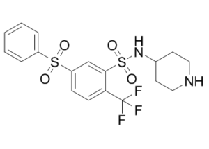
WAY 316606 is a novel and potent small molecule inhibitor of the secreted protein sFRP-1 (secreted frizzled-related protein) which is an endogenous antagonist of the secreted glycoprotein Wnt. Modulators of the Wnt pathway have been proposed as anabolic agents for the treatment of osteoporosis or other bone-related disorders. WAY-316606 bound to sFRP-1 with a K(D) of 0.08 microM and inhibited it with an EC(50) of 0.65 microM. Moreover, this compound increased total bone area in a murine calvarial organ culture assay at concentrations as low as 0.0001 microM. This work demonstrates the feasibility of developing small molecules that inhibit sFRP-1 and stimulate canonical Wnt signaling to increase bone formation. Canonical Wnt signaling has been demonstrated to increase bone formation, and Wnt pathway components are being pursued as potential drug targets for osteoporosis and other metabolic bone diseases. Deletion of the Wnt antagonist sFRP-1 in mice activates canonical signaling in bone and increases trabecular bone formation in aged animals.
Physicochemical Properties
| Molecular Formula | C18H19F3N2O4S2 | |
| Molecular Weight | 448.48 | |
| Exact Mass | 448.074 | |
| CAS # | 915759-45-4 | |
| Related CAS # | 915759-45-4;1781835-02-6 (HCl); | |
| PubChem CID | 16727102 | |
| Appearance | White to off-white solid powder | |
| LogP | 5.449 | |
| Hydrogen Bond Donor Count | 2 | |
| Hydrogen Bond Acceptor Count | 9 | |
| Rotatable Bond Count | 5 | |
| Heavy Atom Count | 29 | |
| Complexity | 747 | |
| Defined Atom Stereocenter Count | 0 | |
| InChi Key | ITBGJNVZJBVPLJ-UHFFFAOYSA-N | |
| InChi Code | InChI=1S/C18H19F3N2O4S2/c19-18(20,21)16-7-6-15(28(24,25)14-4-2-1-3-5-14)12-17(16)29(26,27)23-13-8-10-22-11-9-13/h1-7,12-13,22-23H,8-11H2 | |
| Chemical Name |
|
|
| Synonyms |
|
|
| HS Tariff Code | 2934.99.9001 | |
| Storage |
Powder-20°C 3 years 4°C 2 years In solvent -80°C 6 months -20°C 1 month |
|
| Shipping Condition | Room temperature (This product is stable at ambient temperature for a few days during ordinary shipping and time spent in Customs) |
Biological Activity
| Targets | WAY-316606's EC50 on U2-OS cells' Wnt-luciferase activity is 0.65 μM[1]. Frizzled-related protein (sFRP)-1 dye is bound by WAY-316606, which has a KD of 0.08 μM and an EC50 of 0.65 μM for sFRP-1 inhibition. Similar to KD of 1 μM, WAY-316606 binds to sFRP-2, but with a strength that is almost ten times weaker. WAY-316606 has an IC50 of 0.5 μM when it comes to a collagen binding assay that uses a fluorescent material and a sealed human sFRP-1 fingerprint in a competitive binding format [2]. |
| ln Vitro |
WAY-316606's EC50 on U2-OS cells' Wnt-luciferase activity is 0.65 μM[1]. Frizzled-related protein (sFRP)-1 dye is bound by WAY-316606, which has a KD of 0.08 μM and an EC50 of 0.65 μM for sFRP-1 inhibition. Similar to KD of 1 μM, WAY-316606 binds to sFRP-2, but with a strength that is almost ten times weaker. WAY-316606 has an IC50 of 0.5 μM when it comes to a collagen binding assay that uses a fluorescent material and a sealed human sFRP-1 fingerprint in a competitive binding format [2]. Adavivint (SM04690) is a potent and selective small-molecule inhibitor of canonical Wnt signaling, as demonstrated in a TCF/LEF reporter assay using SW480 colon cancer cells (EC50 = 19.5 nM). It was approximately 150-500 times more potent than other known Wnt pathway inhibitors (e.g., FH535, IWR-1, ICG001, iCRT14, KY02111, CX-4945). In human mesenchymal stem cells (hMSCs), Adavivint treatment led to a dose-dependent decrease in the expression of Wnt pathway genes (ASCL1, LEF1, TCF7L2, TCF7, C-MYC, AXIN2) and reduced β-catenin protein levels. Adavivint induced chondrogenic differentiation of hMSCs, evidenced by increased formation of Rhodamine B-stained chondrogenic nodules (EC50 = 10 nM), elevated expression of chondrocyte markers (SOX9, COMP, aggrecan, COL2A1, TIMP1, CD44, COL10A1), increased sulfated glycosaminoglycan (GAG) content, and positive staining for Alcian Blue and Safranin O after 21 days of culture. In cytokine-stimulated chondrocytes and hMSCs (using TNF-α + Oncostatin M or IL-1β), Adavivint (30 nM) inhibited the expression of catabolic enzymes (MMP-1, MMP-3, MMP-13, IHH) and reduced GAG breakdown and nitric oxide (NO) release. The compound showed minimal effects on non-canonical Wnt and BMP pathways in hMSCs.[1] |
| ln Vivo |
In neonatal calvarial osteometry, WAY-316606 resulted in an increase. With an EC50 of about 1 nM, WAY-316606 exhibits a dose-dependent increase in total bone area of up to 60%. Good solubility, moderate or low inhibition of cytochrome p450 isoenzymes (3A4, 2D6, 2C9), and good stability in locus and human liver microsomes (per species t1/2>60 minutes) are the characteristics of WAY-316606. After a single intravenous bolus dosage (2 mg/kg), female Sprague-Dawley WAY-316606 shows strong network clearance (77 mL/min/kg, higher hepatic blood flow), which led to a quick drug decrease in the central area despite drug routes [2]. In a rat model of knee osteoarthritis (OA) induced by anterior cruciate ligament transection plus partial medial meniscectomy (ACLT+pMMx), a single intra-articular (IA) injection of Adavivint (0.3 µg) administered one week post-surgery significantly improved joint morphology at 12 weeks post-treatment. This was evidenced by reduced Osteoarthritis Research Society International (OARSI) histopathology scores, increased Safranin O staining intensity, increased cartilage thickness, and elevated serum levels of the cartilage synthesis biomarker PIIANP at 3 weeks post-treatment. Adavivint treatment also increased the number of Doublecortin (Dcx)-positive articular chondrocytes and upregulated chondrogenic gene expression (Col2a1, aggrecan, COMP) in cartilage at 4 weeks post-treatment. Additionally, it reduced the expression of cartilage-degrading proteases (MMP-1, MMP-3, MMP-13, ADAMTS5) in cartilage and decreased circulating levels of cartilage oligomeric matrix protein (COMP) at 5 weeks post-treatment. The expression of Wnt pathway genes (e.g., GSK3β, Dvl1, Wnt3a, TCF7, Axin2, β-catenin) was decreased in cartilage from Adavivint-treated rats, while Wnt inhibitory genes (e.g., DKK1, WIF1) were upregulated.[1] |
| Enzyme Assay |
A cell-based TCF/LEF reporter assay was used for high-throughput screening to identify Wnt pathway inhibitors. SW480 cells, which have constitutively active canonical Wnt signaling due to an APC mutation, were transduced with a TCF/LEF-luciferase lentivirus. Compounds were added to cells, and after 48 hours, luciferase activity was measured using a luminescence reagent to quantify Wnt pathway inhibition. Counter-screening was performed using SW480 cells expressing an SV40-driven luciferase reporter to exclude non-specific inhibitors.[1] |
| Cell Assay |
For early chondrogenesis assessment, hMSCs were plated and treated with Adavivint for 5 days. Cells were then fixed and stained with Rhodamine B. The number of stained chondrogenic nodules, indicative of early cell condensation, was quantified using high-content imaging. For chondrocyte differentiation, hMSCs were cultured in incomplete chondrogenic differentiation medium and treated with Adavivint for 21 days. Differentiation was assessed by staining with Alcian Blue or Safranin O, immunocytochemistry for chondrocyte markers (e.g., Sox9, Type II collagen, aggrecan), and measurement of sulfated GAG content using a dimethylmethylene blue assay. To evaluate cartilage protection, differentiated chondrocytes or hMSCs were stimulated with pro-inflammatory cytokines (TNF-α + Oncostatin M or IL-1β) in the presence of Adavivint for 72 hours. Gene expression of catabolic enzymes (MMP-1, MMP-3, MMP-13, IHH) was analyzed by qRT-PCR. Secreted GAG and nitric oxide levels in the culture medium were measured using colorimetric assays.[1] |
| Animal Protocol |
Male Sprague-Dawley rats (10 weeks old) underwent surgical induction of OA via anterior cruciate ligament transection combined with partial medial meniscectomy (ACLT+pMMx). One week after surgery, rats were randomized into treatment groups. Adavivint (SM04690) was administered as a single intra-articular (IA) injection into the knee joint at a dose of 0.3 µg in a 50 µL volume. The vehicle control group received an equivalent volume of the formulation without the active compound. Animals were euthanized at various time points post-treatment (e.g., 4, 12, or 13 weeks). Knee joints were collected, fixed, decalcified, embedded in paraffin, and sectioned. Sections were stained with Safranin O/Fast Green for histological evaluation. Cartilage thickness, staining intensity, and OARSI scores were assessed. Serum was collected at multiple time points for biomarker analysis (PIIANP, COMP).[1] |
| ADME/Pharmacokinetics |
Following a single IA injection of Adavivint (0.3 µg) in rats, the compound showed prolonged residence time in the knee joint, remaining detectable for over 180 days. Systemic exposure was minimal, with plasma concentrations below the quantifiable limit (10 nM). No significant systemic absorption was observed.[1] |
| References |
[1]. Modulation of Wnt Signaling Through Inhibition of Secreted Frizzled-Related Protein I (sFRP-1) with N-Substituted Piperidinyl Diphenylsulfonyl Sulfonamides. Journal of Medicinal Chemistry (2009), 52(1), 105-116. [2]. A small molecule inhibitor of the Wnt antagonist secreted frizzled-related protein-1 stimulates bone formation. Bone. 2009 Jun;44(6):1063-8. |
| Additional Infomation |
WAY-316606 is a sulfonamide resulting from the formal condensation of the sulfonic acid group of 5-(phenylsulfonyl)-2-(trifluoromethyl)benzenesulfonic acid with the primary amino group of piperidin-4-amine. An inhibitor of secreted Frizzled-Related Protein-1 (sFRP-1). It has a role as a secreted frizzled-related protein 1 inhibitor. It is a sulfone, a sulfonamide, a member of (trifluoromethyl)benzenes, a member of piperidines and a secondary amino compound. Adavivint (SM04690) is a novel small-molecule inhibitor of the canonical Wnt/β-catenin signaling pathway, being developed as a potential disease-modifying osteoarthritis drug (DMOAD). Its proposed dual mechanism of action involves promoting chondrogenesis from mesenchymal stem cells and protecting existing cartilage from catabolic breakdown. The preclinical data supported the initiation of a phase 1 first-in-human clinical trial, where the compound appeared safe and well-tolerated (as mentioned in the text, with details "in press").[1] |
Solubility Data
| Solubility (In Vitro) |
|
|||
| Solubility (In Vivo) |
Solubility in Formulation 1: ≥ 2.5 mg/mL (5.57 mM) (saturation unknown) in 10% DMSO + 90% (20% SBE-β-CD in Saline) (add these co-solvents sequentially from left to right, and one by one), clear solution. For example, if 1 mL of working solution is to be prepared, you can add 100 μL of 25.0 mg/mL clear DMSO stock solution to 900 μL of 20% SBE-β-CD physiological saline solution and mix evenly. Preparation of 20% SBE-β-CD in Saline (4°C,1 week): Dissolve 2 g SBE-β-CD in 10 mL saline to obtain a clear solution. Solubility in Formulation 2: ≥ 2.5 mg/mL (5.57 mM) (saturation unknown) in 10% DMSO + 90% Corn Oil (add these co-solvents sequentially from left to right, and one by one), clear solution. For example, if 1 mL of working solution is to be prepared, you can add 100 μL of 25.0 mg/mL clear DMSO stock solution to 900 μL of corn oil and mix evenly. (Please use freshly prepared in vivo formulations for optimal results.) |
| Preparing Stock Solutions | 1 mg | 5 mg | 10 mg | |
| 1 mM | 2.2298 mL | 11.1488 mL | 22.2975 mL | |
| 5 mM | 0.4460 mL | 2.2298 mL | 4.4595 mL | |
| 10 mM | 0.2230 mL | 1.1149 mL | 2.2298 mL |