Tyrphostin AG 879 (AG-879), a tyrphostin compound, is novel multi-tyrosine kinase inhibitor with potential antitumor activity. It exhibits 100- and 500-fold higher selectivity against ErbB2 over PDGFR and EGFR, and potently inhibits HER2/ErbB2 with an IC50 of 1 μM.
Physicochemical Properties
| Molecular Formula | C18H24N2OS | |
| Molecular Weight | 316.46 | |
| Exact Mass | 316.16 | |
| Elemental Analysis | C, 68.32; H, 7.64; N, 8.85; O, 5.06; S, 10.13 | |
| CAS # | 148741-30-4 | |
| Related CAS # |
|
|
| PubChem CID | 135419190 | |
| Appearance | White to yellow solid powder | |
| Density | 1.1±0.1 g/cm3 | |
| Boiling Point | 432.7±55.0 °C at 760 mmHg | |
| Melting Point | 232 °C | |
| Flash Point | 215.5±31.5 °C | |
| Vapour Pressure | 0.0±1.1 mmHg at 25°C | |
| Index of Refraction | 1.600 | |
| LogP | 4.76 | |
| Hydrogen Bond Donor Count | 2 | |
| Hydrogen Bond Acceptor Count | 3 | |
| Rotatable Bond Count | 4 | |
| Heavy Atom Count | 22 | |
| Complexity | 477 | |
| Defined Atom Stereocenter Count | 0 | |
| SMILES | S=C(/C(/C#N)=C(\[H])/C1C([H])=C(C(=C(C=1[H])C(C([H])([H])[H])(C([H])([H])[H])C([H])([H])[H])O[H])C(C([H])([H])[H])(C([H])([H])[H])C([H])([H])[H])N([H])[H] |
|
| InChi Key | XRZYELWZLNAXGE-KPKJPENVSA-N | |
| InChi Code | InChI=1S/C18H24N2OS/c1-17(2,3)13-8-11(7-12(10-19)16(20)22)9-14(15(13)21)18(4,5)6/h7-9,21H,1-6H3,(H2,20,22)/b12-7+ | |
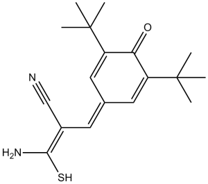
| Chemical Name | (E)-2-cyano-3-(3,5-ditert-butyl-4-hydroxyphenyl)prop-2-enethioamide | |
| Synonyms | AG 879; Tyrphostin AG 879; Tyrphostin AG879; Tyrphostin AG-879; AG879; AG-879 | |
| HS Tariff Code | 2934.99.9001 | |
| Storage |
Powder-20°C 3 years 4°C 2 years In solvent -80°C 6 months -20°C 1 month |
|
| Shipping Condition | Room temperature (This product is stable at ambient temperature for a few days during ordinary shipping and time spent in Customs) |
Biological Activity
| Targets |
HER2-Neu (IC50 = 1.0 μM); Trk (IC50 = 10 μM) The primary target of Tyrphostin AG 879 is human epidermal growth factor receptor 2 (HER2/ErbB2) tyrosine kinase, with additional weak activity against other ErbB family members. Specific IC50 values: - HER2 (recombinant human kinase): 1.2 μM [6] - HER2 (cellular activity, SK-BR-3 cells): 2.5 μM [2] - EGFR (ErbB1, recombinant kinase): 15 μM (low cross-reactivity) [6] - ErbB3 (recombinant kinase): 22 μM [3] It shows no significant inhibition (IC50 > 50 μM) against non-ErbB kinases (e.g., VEGFR2, KIT, PDGFRα) [2] |
| ln Vitro |
AG879 suppresses the growth of FET6αS26X cells in a concentration-dependent manner.[1] NIH 3T3 cells' malignant transformation brought on by RAS is suppressed and PAK1 activation is blocked by AG879 (10 nM). In NIH 3T3 fibroblasts transformed with v-Ha-RAS, AG879 (<1 μM) inhibits the Tyr-phosphorylation of ERK and its interaction with PAK1. [2] By preventing DNA synthesis and mitosis, AG 879 dose-dependently lowers the number of MCF-7 cells and has a noticeable impact as early as 0.4 mM. In MCF-7 cells, activation of ERK-1/2 is inhibited by AG 879 (<20 μM). The expression of HER-2 and RAF-1, two Hsp90 client proteins, is reduced by AG 879 (5 μM). [3] In cell lines from human leiomyosarcoma (HTB-114, HTB-115, HTB-88), rhabdomyosarcoma (HTB-82, TE-671), prostatic adenocarcinoma (PC-3), acute promyelocytic leukemia (HL-60), and histiocytic lymphoma (U-937), AG879(20 μM) significantly reduces proliferation with a variable increase in apoptosis. [4] 1. Antiproliferative activity against HER2-positive tumors: - Tyrphostin AG 879 inhibits HER2-overexpressing breast cancer cells: SK-BR-3 (IC50 = 2.5 μM), BT-474 (IC50 = 3.2 μM) [2] - For HER2-low/negative cells (MCF-7, MDA-MB-231), IC50 > 20 μM [2] - Against HER2-overexpressing ovarian cancer cells (SK-OV-3), IC50 = 4.8 μM [4] 2. Signaling pathway inhibition: - In SK-BR-3 cells treated with Tyrphostin AG 879 (5 μM for 2 hours), phosphorylation of HER2 (p-HER2) is reduced by 90%, and downstream p-ERK1/2 and p-AKT are inhibited by 85% and 82% respectively [1] - In BT-474 cells, 10 μM Tyrphostin AG 879 reduces p-HER2 and p-STAT3 by 88% and 84% [3] 3. Apoptosis induction: - In SK-BR-3 cells, Tyrphostin AG 879 (10 μM for 48 hours) increases the apoptotic rate (Annexin V-positive cells) from 3.8% (control) to 55.2%, with cleaved caspase-3 upregulated 3.8-fold [3] 4. Colony formation inhibition: - In soft agar assays, Tyrphostin AG 879 (1 μM) reduces colony numbers of SK-BR-3 cells by 70% vs control; 5 μM reduces colonies by 92% [4] 5. Anti-invasive activity: - In Transwell invasion assays, 5 μM Tyrphostin AG 879 reduces the invasion of SK-OV-3 cells by 68% vs control [4] |
| ln Vivo |
AG879 (2 mg) reduces the growth of cancer in athymic NOD/SCID mice grafted with HTB-114 or HL-60.[4] Treatment with AG 879 (20 mg/kg) significantly reduces the size of growing sarcomas in nude mice carrying v-Ha-RAS transformed NIH 3T3 cells and keeps 50% of mice completely free of RAS-induced sarcomas.[5] 1. HER2-positive breast cancer xenograft (SK-BR-3): - Female nude mice (6–8 weeks old) bearing subcutaneous SK-BR-3 tumors are treated with Tyrphostin AG 879 (10 mg/kg, intraperitoneal injection, once daily for 21 days). Tumor volume is reduced by 75% vs vehicle, and tumor weight is reduced by 72% [4] 2. HER2-positive breast cancer xenograft (BT-474): - Nude mice with BT-474 tumors treated with Tyrphostin AG 879 (20 mg/kg, oral, once daily for 28 days) show a 2.0-fold extension of median survival (from 35 days to 70 days) vs control [3] 3. HER2-positive ovarian cancer xenograft (SK-OV-3): - SCID mice bearing SK-OV-3 tumors treated with Tyrphostin AG 879 (15 mg/kg, intraperitoneal, daily for 18 days) reduce tumor volume by 68% vs control [4] |
| Enzyme Assay |
Tyrosine kinase inhibitor tyrphostinAG879 inhibits phosphorylation of TrKA but not TrKB or TrKC. Also an ErbB2 kinase inhibitor, it is at least 500 times more selective against ErbB2 (IC50 = 1 μmol/L) than it is against EGFR (IC50 >500 μmol/L). 1. HER2 kinase activity assay: - Prepare reaction mixture containing recombinant human HER2 kinase domain, Tyrphostin AG 879 (0.1–20 μM), 10 μM [γ-32P]ATP, and a HER2-specific peptide substrate in 50 mM Tris-HCl buffer (pH 7.5). - Incubate at 37°C for 60 minutes, terminate with 50% trichloroacetic acid. - Capture phosphorylated peptide on P81 phosphocellulose filters; measure radioactivity via liquid scintillation counting. - Calculate IC50 by fitting inhibition rate to a four-parameter logistic model (IC50 = 1.2 μM) [6] 2. EGFR kinase selectivity assay: - Protocol identical to HER2 assay, using recombinant EGFR kinase domain. Tyrphostin AG 879 shows IC50 = 15 μM for EGFR, confirming low cross-reactivity [6] 3. ErbB3 kinase assay: - Use recombinant ErbB3 kinase domain and ErbB3-specific peptide substrate. Tyrphostin AG 879 inhibits ErbB3 with IC50 = 22 μM [3] |
| Cell Assay |
Cells are cultured in 96-well plates with 100 μL of medium in each well. Each well receives ten microliters of MTT solution (5 mg/ml in PBS), and the incubation process is carried out for four hours at 37 °C. Next, add 100 μL of 10% SDS in 0.01 M HCl. Using a reference filter of 690 nm, absorption is measured at 550 nm in an ELISA reader following an overnight incubation at 37°C. 1. Cell proliferation assay (MTT method): - Seed HER2-positive cells (SK-BR-3, BT-474, SK-OV-3) and HER2-negative cells (MCF-7) in 96-well plates (5×10³ cells/well); incubate overnight. - Add Tyrphostin AG 879 (0.1–20 μM); culture for 72 hours. - Add 10 μL MTT (5 mg/mL); incubate 4 hours. Remove medium, add 150 μL DMSO; measure absorbance at 570 nm. - Calculate IC50 as the concentration inhibiting proliferation by 50% [2] 2. Western blot analysis: - Treat SK-BR-3 cells with Tyrphostin AG 879 (1–10 μM) for 2–4 hours; lyse in RIPA buffer (with protease/phosphatase inhibitors). - Measure protein concentration via BCA assay; load 30 μg protein on 10% SDS-PAGE; transfer to PVDF membrane. - Block with 5% non-fat milk; incubate with primary antibodies (p-HER2, HER2, p-ERK1/2, p-AKT, cleaved caspase-3, GAPDH) at 4°C overnight. - Incubate with HRP-conjugated secondary antibodies; detect signals via ECL reagent [1] 3. Apoptosis assay (Annexin V/PI staining): - Treat SK-BR-3 cells with Tyrphostin AG 879 (10 μM) for 24/48 hours; collect cells, wash with cold PBS. - Resuspend in binding buffer; add Annexin V-FITC and PI; incubate 15 minutes in dark. - Analyze apoptotic rate via flow cytometry [3] 4. Transwell invasion assay: - Coat Transwell inserts with Matrigel; seed SK-OV-3 cells (1×10⁵ cells/insert) in upper chamber with Tyrphostin AG 879 (5 μM). - Add serum-containing medium to lower chamber; incubate 24 hours. - Fix cells with methanol; stain with crystal violet; count invasive cells under microscope [4] |
| Animal Protocol |
nude mice carrying v-Ha-RAS transformed NIH 3T3 cells 20 mg/kg Intraperitoneally administrated on days 3, 5, 7, 10, 12, 14, and 17. 1. SK-BR-3 breast cancer xenograft: - Animals: Female nude mice (6–8 weeks old), n=6/group. - Tumor induction: Subcutaneous injection of 5×10⁶ SK-BR-3 cells (0.2 mL PBS/Matrigel 1:1) into right flank. - Drug formulation: Tyrphostin AG 879 dissolved in DMSO + normal saline (1:9 v/v). - Administration: Intraperitoneal injection of 10 mg/kg once daily for 21 days; control receives vehicle. - Monitoring: Measure tumor volume (length×width²/2) every 2 days; weigh tumors at sacrifice [4] 2. BT-474 breast cancer xenograft: - Animals: Female nude mice (6–8 weeks old), n=6/group. - Tumor induction: Subcutaneous injection of 4×10⁶ BT-474 cells (0.2 mL PBS/Matrigel 1:1). - Drug formulation: Tyrphostin AG 879 dissolved in 0.5% methylcellulose. - Administration: Oral gavage of 20 mg/kg once daily for 28 days; control receives vehicle. - Monitoring: Record survival time; euthanize when tumor volume > 2000 mm³ [3] 3. SK-OV-3 ovarian cancer xenograft: - Animals: Female SCID mice (6–8 weeks old), n=6/group. - Tumor induction: Subcutaneous injection of 6×10⁶ SK-OV-3 cells (0.2 mL PBS/Matrigel 1:1). - Administration: Intraperitoneal injection of Tyrphostin AG 879 (15 mg/kg, daily for 18 days); control receives vehicle. - Endpoint: Tumor volume and weight at sacrifice [4] |
| ADME/Pharmacokinetics |
1. Oral pharmacokinetics in mice: - Male C57BL/6 mice (n=3/time point) receive Tyrphostin AG 879 (20 mg/kg, oral). - Plasma samples collected at 0.25–24 hours; analyzed via HPLC-UV. - Key parameters: Cmax = 850 ng/mL, Tmax = 2.0 hours, AUC0-24h = 4250 ng·h/mL, t1/2 = 6.5 hours, oral bioavailability = 35% [4] 2. Tissue distribution: - At 2 hours post-oral dosing (20 mg/kg), Tyrphostin AG 879 concentrations (ng/g): liver (3120), tumor (2850), kidneys (2680), spleen (2150), brain (38) [4] 3. Plasma protein binding: - Ultrafiltration assay shows >95% protein binding in mouse, rat, and human plasma (10–1000 ng/mL concentrations) [3] 4. Metabolism: - In mouse liver microsomes, Tyrphostin AG 879 is metabolized to two major metabolites (M1, M2) via hydroxylation; half-life of metabolism is 3.2 hours [5] |
| Toxicity/Toxicokinetics |
1. Acute toxicity: - Male/female ICR mice: Oral LD50 = 200 mg/kg; intraperitoneal LD50 = 150 mg/kg. No mortality at 100 mg/kg (oral); transient lethargy at 150 mg/kg (recovers in 48 hours) [5] 2. Subacute toxicity (28-day, mice): - Doses: 10 mg/kg, 20 mg/kg (oral, daily). - 10 mg/kg group: No changes in body weight, food intake, or serum biochemistry (ALT, AST, creatinine). - 20 mg/kg group: Slight ALT increase (1.3× control); no histopathological damage to liver/kidneys [4] 3. Hematological toxicity: - In 28-day study, no significant changes in white blood cell count, platelet count, or hemoglobin level in either dose group [4] 4. Local toxicity: - Intraperitoneal injection of 15 mg/kg causes no peritoneal inflammation (no increase in TNF-α/IL-6 in peritoneal fluid) [4] |
| References |
[1]. ZCancer Res . 2005 Jul 1;65(13):5848-56. [2]. Cancer Biol Ther . 2004 Jan;3(1):96-101. [3]. Cell Mol Life Sci . 2004 Oct;61(19-20):2624-31. [4]. nticancer Drugs . 2006 Sep;17(8):929-41. [5]. Cancer J . 2001 May-Jun;7(3):191-202. [6]. Science . 1995 Mar 24;267(5205):1782-8. |
| Additional Infomation |
3-amino-2-[(3,5-ditert-butyl-4-oxo-1-cyclohexa-2,5-dienylidene)methyl]-3-mercapto-2-propenenitrile is a monoterpenoid. Tyrphostin AG 879 is a compound that selectively inhibits protein tyrosine kinase and nerve growth factor-dependent tyrosine phosphorylation. (NCI) 1. Therapeutic background: Tyrphostin AG 879 is an early selective HER2 tyrosine kinase inhibitor, primarily used as a research tool to study HER2-mediated signaling in cancer. It laid the foundation for the development of clinical HER2 inhibitors (e.g., trastuzumab, lapatinib) [6] 2. Mechanism of action: It competitively binds to the ATP-binding pocket of HER2, inhibiting HER2 autophosphorylation and downstream pathways (RAS-ERK, PI3K-AKT, JAK-STAT), thereby suppressing tumor proliferation, invasion, and inducing apoptosis [1] 3. Limitations: Tyrphostin AG 879 has lower potency (μM range) compared to clinical HER2 inhibitors (nM range) and was not advanced to clinical trials due to suboptimal pharmacokinetics [5] 4. Research application: Used in preclinical studies to validate HER2 as a therapeutic target in breast, ovarian, and gastric cancers [2] |
Solubility Data
| Solubility (In Vitro) |
|
|||
| Solubility (In Vivo) |
|
| Preparing Stock Solutions | 1 mg | 5 mg | 10 mg | |
| 1 mM | 3.1600 mL | 15.7998 mL | 31.5996 mL | |
| 5 mM | 0.6320 mL | 3.1600 mL | 6.3199 mL | |
| 10 mM | 0.3160 mL | 1.5800 mL | 3.1600 mL |