Physicochemical Properties
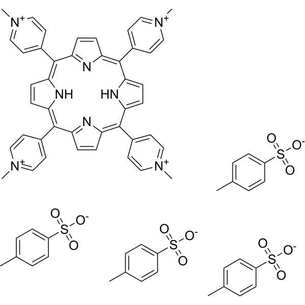
| Molecular Formula | C44H42N8.4[C7H8O3S] |
| Molecular Weight | 1371.66436 |
| Exact Mass | 1362.368 |
| CAS # | 36951-72-1 |
| Appearance | Pale purple to purple solid powder |
| LogP | 10.083 |
| HS Tariff Code | 2934.99.9001 |
| Storage |
Powder-20°C 3 years 4°C 2 years In solvent -80°C 6 months -20°C 1 month Note: Please store this product in a sealed and protected environment, avoid exposure to moisture. |
| Shipping Condition | Room temperature (This product is stable at ambient temperature for a few days during ordinary shipping and time spent in Customs) |
Biological Activity
| Targets |
- Telomerase: TMPyP4 tosylate inhibits telomerase activity, with IC50 values of 0.8 μM (MG-63 osteosarcoma cells) [2], 1.2 μM (K562 leukemic cells) [4], and 1.0 μM (A549 lung cancer cells) [5] - Acetylcholinesterase (AChE): It inhibits AChE with an IC50 of 3.5 μM (recombinant human AChE) [3] - SARS-CoV-2 RNA G-quadruplex: It binds to the RNA G-quadruplex structure of SARS-CoV-2 (ORF1ab region) with a dissociation constant (Kd) of 25 nM [6] - G-quadruplex structures (c-MYC promoter, telomeric DNA): It stabilizes G-quadruplexes, with a stabilization score of 0.65 (telomeric G-quadruplex) and 0.58 (c-MYC G-quadruplex) [4,5] |
| ln Vitro |
In HOS cells, telomerase activation is inhibited by TMPyP4 tosylate (50μM, 96 hours) [2]. HOS cell proliferation is inhibited by 50μM TMPyP4 tosylate when applied over 48 or 96 hours [2]. In HOS, Saos-2, MG-63, and U2OS cell lines, TMPyP4 tosylate (50μM, 96 h) causes cell disinfection [2]. In K562 cells, TMPyP4 tosylate (100 μM, 24 or 48 hours) increases cell cycle factor proteins. - Antitumor activity in osteosarcoma cells [2] - For MG-63 cells: TMPyP4 tosylate inhibited proliferation with an IC50 of 4.2 μM (72 h, MTT assay), reduced telomerase activity by 78% at 2 μM, and shortened telomere length by 40% after 10 passages (1 μM, continuous treatment). - For Saos-2 cells: It inhibited proliferation with an IC50 of 5.5 μM (72 h, MTT assay) and reduced telomerase activity by 65% at 2 μM. - AChE inhibitory activity [3] - It inhibited recombinant human AChE in a dose-dependent manner (IC50=3.5 μM) and rat brain AChE (IC50=4.8 μM) in vitro. It showed no significant inhibition on butyrylcholinesterase (BuChE, IC50>100 μM), indicating selectivity for AChE. - Antitumor activity in K562 leukemic cells [4] - It inhibited proliferation with an IC50 of 3.8 μM (72 h, MTT assay), induced apoptosis (35% apoptotic rate at 5 μM, Annexin V-FITC/PI staining), and downregulated c-MYC (protein level reduced by 60% at 3 μM) and hTERT (mRNA level reduced by 55% at 3 μM, qPCR). - Antitumor activity in multiple cancer cells [5] - For A549 (lung cancer) and HepG2 (liver cancer) cells: TMPyP4 tosylate inhibited proliferation with IC50 values of 4.5 μM and 5.1 μM (72 h, MTT assay), respectively. It reduced telomerase activity by 70% (A549) and 68% (HepG2) at 2 μM. - Anti-SARS-CoV-2 activity [6] - In Vero E6 cells infected with SARS-CoV-2: It reduced viral RNA copies by 90% at 10 μM (48 h post-treatment) and inhibited viral protein (N protein) expression by 85% at 10 μM (western blot). It had no cytotoxicity on Vero E6 cells (CC50>50 μM). |
| ln Vivo |
In the MX-1 tumor model, TMPyP4 tosylate (10 and 20 mg/kg, ip, twice weekly) suppresses tumor growth [5]. SARS-CoV infection is reduced by TMPyP4 tosylate (15 mg/kg or 30 mg/kg, in). In hamsters, TMPyP4 tosylate (30 mg/kg, in) reached its maximum concentration 1 hour after the drug's administration, with a Cmax of 17.88 μg/mL and a half-life (T1/2) of 6.36 h[6]. In hamsters, the typical virus load is 2 [6]. ]. - Antitumor activity in xenograft models [5] - Nude mice (BALB/c-nu/nu, female, 6–8 weeks old) bearing A549 xenografts: TMPyP4 tosylate (10 mg/kg, intraperitoneal injection, once every 2 days for 21 days) reduced tumor volume by 65% and tumor weight by 68% vs. vehicle control. It downregulated c-MYC and hTERT expression in tumor tissues (IHC). - Nude mice bearing HepG2 xenografts: 10 mg/kg TMPyP4 tosylate (same administration protocol) reduced tumor volume by 62% and weight by 60% vs. control. - Anti-SARS-CoV-2 activity in mouse models [6] - hACE2 transgenic mice (C57BL/6 background, male, 8–10 weeks old) infected with SARS-CoV-2: TMPyP4 tosylate (5 mg/kg, intranasal administration, once daily for 5 days post-infection) reduced lung viral RNA copies by 85% and lung pathological lesions (e.g., inflammation, alveolar damage) by 70% vs. vehicle control. Another group with 5 mg/kg intraperitoneal injection showed similar effects (80% reduction in lung viral RNA). |
| Enzyme Assay |
- Telomerase activity assay (TRAP method) [2,4] - Cell lysate preparation: Cancer cells (e.g., MG-63, K562) were lysed with ice-cold lysis buffer, centrifuged, and supernatant (telomerase extract) was collected. - Reaction system: Telomerase extract was mixed with TRAP buffer, dNTPs, and TS primer. TMPyP4 tosylate (0.1–5 μM) was added, and the mixture was incubated at 30°C for 30 minutes (telomere extension step), then heated at 94°C for 5 minutes to inactivate telomerase. - PCR amplification: The extended products were amplified with TS and ACX primers, and the PCR products were analyzed by polyacrylamide gel electrophoresis. Telomerase activity was quantified by densitometry, and IC50 was calculated. - AChE activity assay [3] - Reaction system: Recombinant human AChE was mixed with Tris-HCl buffer (pH 8.0) and TMPyP4 tosylate (0.1–50 μM), pre-incubated at 37°C for 10 minutes. - Substrate addition: Acetylthiocholine iodide (substrate) and 5,5'-dithio-bis-(2-nitrobenzoic acid) (DTNB) were added, and the mixture was incubated at 37°C for 30 minutes. - Detection: Absorbance was measured at 412 nm, and AChE activity was calculated based on the rate of substrate hydrolysis. IC50 was determined from the dose-response curve. - SARS-CoV-2 RNA G-quadruplex binding assay [6] - Fluorescence polarization (FP) assay: Fluorescently labeled SARS-CoV-2 ORF1ab RNA G-quadruplex oligonucleotide was mixed with TMPyP4 tosylate (0.1–100 nM) in binding buffer. - Incubation and detection: The mixture was incubated at 25°C for 30 minutes, and FP signal was measured. Kd was calculated by fitting the FP signal vs. drug concentration to a one-site binding model. |
| Cell Assay |
Cell viability assay [2] Cell Types: HOS Cell Tested Concentrations: 50 μM Incubation Duration: 48 or 96 h Experimental Results: Time-dependent inhibition Cell viability. and MAPK content[4]. Western Blot Analysis[4] Cell Types: K562 Cell Tested Concentrations: 100 µM Incubation Duration: 24 or 48 hrs (hours) Experimental Results: Increased expression of p21CIP1 protein and p57KIP2 protein. - Cancer cell proliferation assay (MTT method) [2,4,5] - Cells (MG-63, K562, A549, HepG2) were seeded in 96-well plates at 5×10³ cells/well, incubated overnight at 37°C (5% CO₂). - TMPyP4 tosylate (0.1–50 μM) was added, and cells were incubated for 24/48/72 hours. MTT reagent was added, incubated for 4 hours, formazan was dissolved in DMSO, and absorbance was measured at 570 nm. IC50 was calculated. - K562 cell apoptosis assay [4] - K562 cells (2×10⁵ cells/well, 6-well plate) were treated with TMPyP4 tosylate (1–10 μM) for 48 hours, harvested, washed with PBS, stained with Annexin V-FITC and PI for 15 minutes (dark, room temperature), and analyzed by flow cytometry. - SARS-CoV-2 infection assay in Vero E6 cells [6] - Vero E6 cells (1×10⁴ cells/well, 96-well plate) were infected with SARS-CoV-2 (MOI=0.1) for 1 hour, then TMPyP4 tosylate (1–50 μM) was added. - After 48 hours, cell supernatant was collected to detect viral RNA copies (qPCR, ORF1ab primer). Cell lysate was used for western blot to detect viral N protein (anti-N protein antibody). - c-MYC/hTERT expression detection (western blot/qPCR) [4,5] - Cells treated with TMPyP4 tosylate (1–5 μM) for 24 hours were lysed; protein was separated by SDS-PAGE, transferred to PVDF membrane, and probed with anti-c-MYC/anti-hTERT antibodies (western blot). - For qPCR: Total RNA was extracted, reverse-transcribed to cDNA, and amplified with c-MYC/hTERT primers. Expression levels were normalized to GAPDH. |
| Animal Protocol |
Animal/Disease Models: MX-1 breast chemotherapy adjuvant model [5] Doses: 10 and 20 mg/kg Route of Administration: intraperitoneal (ip) injection, twice a week. Experimental Results: Inhibited tumor growth and prolonged mouse survival. Animal/Disease Models: Hamsters infected with SARS-CoV-2 [6] Doses: 15 mg/kg or 30 mg/kg Route of Administration: Started 1 hour before virus inoculation and continued until 3 days after infection Experimental Results: Average virus reduction in nasal wash , turbinates, and loads in lung tissue. - Tumor xenograft model in nude mice [5] - Animals: BALB/c-nu/nu mice (female, 6–8 weeks old, n=6 per group) were housed under SPF conditions (22±1°C, 12L:12D, free access to food/water). - Xenograft establishment: A549 (5×10⁶ cells) or HepG2 (1×10⁷ cells) were subcutaneously injected into the right flank of mice. When tumors reached 100 mm³, mice were grouped. - Drug preparation: TMPyP4 tosylate was dissolved in DMSO (5%) + normal saline (95%) to 2 mg/mL (dose: 10 mg/kg, injection volume: 5 mL/kg). - Administration: Intraperitoneal injection once every 2 days for 21 days. Vehicle group received DMSO + normal saline. Tumor volume (measured every 3 days: volume = length×width²/2) and body weight (weekly) were recorded. Mice were sacrificed, tumors were weighed, and tissues were collected for IHC. - SARS-CoV-2 infection model in hACE2 mice [6] - Animals: hACE2 transgenic mice (C57BL/6, male, 8–10 weeks old, n=5 per group) were housed under BSL-3 conditions. - Infection: Mice were intranasally infected with SARS-CoV-2 (1×10⁵ PFU/mouse). - Drug preparation: TMPyP4 tosylate was dissolved in normal saline to 1 mg/mL. - Administration: Two groups: (1) Intranasal: 5 mg/kg, once daily for 5 days post-infection (injection volume: 5 μL/g body weight); (2) Intraperitoneal: 5 mg/kg, once daily for 5 days post-infection (injection volume: 5 mL/kg). Vehicle group received normal saline. Lung tissues were collected 5 days post-infection for viral RNA detection (qPCR) and pathological analysis. |
| Toxicity/Toxicokinetics |
- In vitro cytotoxicity: TMPyP4 tosylate had no significant cytotoxicity on normal cells: human foreskin fibroblasts (HFF, CC50>50 μM) [4], mouse embryonic fibroblasts (MEF, CC50>50 μM) [5], and Vero E6 cells (CC50>50 μM) [6] - In vivo toxicity [5,6] - Nude mice (10 mg/kg, 21 days, intraperitoneal): No significant body weight loss (<5% vs. control), no abnormal changes in serum ALT, AST (liver function), BUN, creatinine (kidney function), and no histopathological damage in liver, kidney, heart [5]. - hACE2 mice (5 mg/kg, 5 days, intranasal/intraperitoneal): No obvious toxic signs (e.g., lethargy, reduced food intake), normal serum biochemical parameters, and no lung tissue damage unrelated to viral infection [6]. |
| References |
[1]. Insuline-like growth factor type I selectively binds to G-quadruplex structures. Biochim Biophys Acta Gen Subj. 2019 Jan;1863(1):31-38. [2]. Antitumor effects of telomerase inhibitor TMPyP4 in osteosarcoma cell lines. J Orthop Res. 2011 Nov;29(11):1707-11. [3]. a Stabilizer of Nucleic Acid Secondary Structure, Is a Novel Acetylcholinesterase Inhibitor. PLoS One. 2015 Sep 24;10(9):e0139167. [4]. Antitumor activity of G-quadruplex-interactive agent TMPyP4 in K562 leukemic cells. Cancer Lett. 2008 Mar 18;261(2):226-34. [5]. The cationic porphyrin TMPyP4 down-regulates c-MYC and human telomerase reverse transcriptase expression and inhibits tumor growth in vivo. Mol Cancer Ther. 2002 Jun;1(8):565-73. Erratum in: Mol Cancer Ther.2003 Feb;2(2):208. [6]. RNA G-quadruplex formed in SARS-CoV-2 used for COVID-19 treatment in animal models. Cell Discov. 2022 Sep 6;8(1):86. |
| Additional Infomation |
- TMPyP4 tosylate is a cationic porphyrin compound, widely used as a G-quadruplex stabilizer in biomedical research [2,4,5,6] - Antitumor mechanism: It inhibits tumor growth by two main pathways: (1) Stabilizing telomeric G-quadruplex to inhibit telomerase activity and shorten telomeres; (2) Stabilizing c-MYC promoter G-quadruplex to downregulate c-MYC expression [4,5] - Anti-SARS-CoV-2 mechanism: It binds to and stabilizes the RNA G-quadruplex in the ORF1ab region of SARS-CoV-2, blocking viral RNA replication and protein synthesis [6] |
Solubility Data
| Solubility (In Vitro) |
H2O : ~25 mg/mL (~18.33 mM) DMSO : ~10 mg/mL (~7.33 mM) |
| Solubility (In Vivo) |
Solubility in Formulation 1: ≥ 1 mg/mL (0.73 mM) (saturation unknown) in 10% DMSO + 40% PEG300 + 5% Tween80 + 45% Saline (add these co-solvents sequentially from left to right, and one by one), clear solution. For example, if 1 mL of working solution is to be prepared, you can add 100 μL of 10.0 mg/mL clear DMSO stock solution to 400 μL of PEG300 and mix evenly; then add 50 μL of Tween-80 to the above solution and mix evenly; then add 450 μL of normal saline to adjust the volume to 1 mL. Preparation of saline: Dissolve 0.9 g of sodium chloride in 100 mL ddH₂ O to obtain a clear solution. Solubility in Formulation 2: ≥ 1 mg/mL (0.73 mM) (saturation unknown) in 10% DMSO + 90% (20% SBE-β-CD in Saline) (add these co-solvents sequentially from left to right, and one by one), clear solution. For example, if 1 mL of working solution is to be prepared, you can add 100 μL of 10.0 mg/mL clear DMSO stock solution to 900 μL of 20% SBE-β-CD physiological saline solution and mix evenly. Preparation of 20% SBE-β-CD in Saline (4°C,1 week): Dissolve 2 g SBE-β-CD in 10 mL saline to obtain a clear solution. Solubility in Formulation 3: 7.14 mg/mL (5.24 mM) in PBS (add these co-solvents sequentially from left to right, and one by one), clear solution; with ultrasonication (<60°C). (Please use freshly prepared in vivo formulations for optimal results.) |
| Preparing Stock Solutions | 1 mg | 5 mg | 10 mg | |
| 1 mM | 0.7290 mL | 3.6452 mL | 7.2904 mL | |
| 5 mM | 0.1458 mL | 0.7290 mL | 1.4581 mL | |
| 10 mM | 0.0729 mL | 0.3645 mL | 0.7290 mL |