Spastazoline is a first-in-class, potent and selective spastin (a microtubule-severing AAA protein) inhibitor, with an IC50 of 99 nM for Human spastin. Spastazoline shows no effect on ATPase activity of a recombinant human VPS4. Spastin is a microtubule-severing AAA (ATPases associated with diverse cellular activities) protein needed for cell division and intracellular vesicle transport.
Physicochemical Properties
| Molecular Formula | C20H30N8 |
| Molecular Weight | 382.505802631378 |
| Exact Mass | 382.259 |
| Elemental Analysis | C, 62.80; H, 7.91; N, 29.29 |
| CAS # | 2351882-11-4 |
| PubChem CID | 135397891 |
| Appearance | Typically exists as Off-white to gray solid at room temperature |
| LogP | 4.1 |
| Hydrogen Bond Donor Count | 4 |
| Hydrogen Bond Acceptor Count | 6 |
| Rotatable Bond Count | 5 |
| Heavy Atom Count | 28 |
| Complexity | 521 |
| Defined Atom Stereocenter Count | 1 |
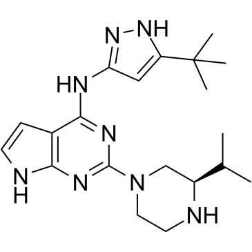
| SMILES | CC(C)[C@@H]1CN(CCN1)C2=NC3=C(C=CN3)C(=N2)NC4=NNC(=C4)C(C)(C)C |
| InChi Key | HNOLTAKAPDKZRY-AWEZNQCLSA-N |
| InChi Code | InChI=1S/C20H30N8/c1-12(2)14-11-28(9-8-21-14)19-24-17-13(6-7-22-17)18(25-19)23-16-10-15(26-27-16)20(3,4)5/h6-7,10,12,14,21H,8-9,11H2,1-5H3,(H3,22,23,24,25,26,27)/t14-/m0/s1 |
| Chemical Name | (R)-N-(5-(tert-Butyl)-1H-pyrazol-3-yl)-2-(3-isopropylpiperazin-1-yl)-7H-pyrrolo[2,3-d]pyrimidin-4-amine |
| Synonyms | Spastazoline; 2351882-11-4; (R)-N-(5-(tert-butyl)-1H-pyrazol-3-yl)-2-(3-isopropylpiperazin-1-yl)-7H-pyrrolo[2,3-d]pyrimidin-4-amine; CHEMBL5190401; Spastazoline?; SCHEMBL22035095; EX-A3095; BDBM50587865; |
| HS Tariff Code | 2934.99.9001 |
| Storage |
Powder-20°C 3 years 4°C 2 years In solvent -80°C 6 months -20°C 1 month |
| Shipping Condition | Room temperature (This product is stable at ambient temperature for a few days during ordinary shipping and time spent in Customs) |
Biological Activity
| Targets |
Human spastin (IC50 = 99 nM)[1] Spastazoline targets AAA protein spastin (ATPase activity IC50 = 1.7 μM; binding Ki = 0.9 μM) [1] |
| ln Vitro |
HeLa cell division is influenced by spastazoline (10 μM; 4.5 h) by increasing the amount of cells with intercellular bridges [1]. These data, along with computational docking, guided improvements in compound potency and selectivity and led to spastazoline, a pyrazolyl-pyrrolopyrimidine-based cell-permeable probe for spastin. These studies also identified spastazoline resistance-conferring point mutations in spastin. Spastazoline, along with matched inhibitor-sensitive and inhibitor-resistant cell lines we generated, were used in parallel experiments to dissect spastin-specific phenotypes in dividing cells. Together, our findings suggest how chemical probes for AAA proteins, along with inhibitor resistance-conferring mutations, can be designed and used to dissect dynamic cellular processes[1]. 1. Spastin ATPase activity inhibition: - Spastazoline inhibits the ATPase activity of recombinant human spastin in a concentration-dependent manner, with an IC50 of 1.7 μM (malachite green ATPase assay) [1] - It shows no significant inhibition of other AAA proteins (e.g., VPS4A, p97) at concentrations up to 20 μM, confirming spastin selectivity [1] 2. Spastin-mediated microtubule severing inhibition: - Spastazoline (5 μM) reduces spastin-induced microtubule severing by 75% in vitro, as visualized by total internal reflection fluorescence (TIRF) microscopy [1] - At 10 μM, it almost completely blocks microtubule severing (inhibition rate >90%) without affecting microtubule polymerization or stability [1] 3. Cellular spastin function inhibition: - Spastazoline (2.5, 5, 10 μM) inhibits spastin-dependent microtubule remodeling in HeLa cells, leading to increased microtubule bundle formation (immunofluorescence analysis) [1] - It reduces the mobility of spastin on microtubules (single-molecule tracking) with a 2.3-fold decrease in diffusion coefficient at 5 μM [1] - No significant effect on cell viability or proliferation at concentrations up to 20 μM (MTT assay) [1] |
| ln Vivo | Mice with SCI in all groups exhibited paralysis in the right hindlimb after hemisection which confirmed the successful establishment of the lateral hemisection SCI model. For mice in the SCI control group, the BMS scores gradually improved to ~4 by 16 DPI. In contrast, the scores of mice in the FC-A group indicated an improvement in locomotor function which was impaired in the spastazoline group. Moreover, the improvement in locomotor function following the administration of FC-A diminished when spastazoline was administered (Figure 7E). We further analyzed the footprint of mice with SCI and calculated the length or width of strides (Figure 7—figure supplement 3). We found that stride length improved in mice of the FC-A group but it was reduced in the spastazoline group although not significantly. The improvement induced by FC-A diminished when co-treated with spastazoline (Figure 7G). Notably, there was no significant difference in stride width between the groups (Figure 7H). Similar effects were observed in the foot fault scan experiment. The faults of the right hindlimb significantly decreased in the FC-A group and increased in the spastazoline group (Figure 7F). The improvement of hindlimb fault in mice administered with FC-A diminished following the application of spastazoline. We also explored whether the stability of MTs was altered by the administration of FC-A and spastazoline in the spinal cord injury model. We found that the ratio of acetylated tubulin to total tubulin was significantly increased in the SCI group, decreased in the FC-A group, and upregulated in the spastazoline group, which further confirmed the successful delivery of drugs to the lesion site (Figure 7—figure supplement 4). These findings suggested that FC-A was effectively delivered to the spinal cord tissue and improved the locomotor function of mice with SCI, and spastin activation is the prerequisite the repair after spinal cord injury. Reference: Elife. 2024 Jan 17;12:RP90184. |
| Enzyme Assay |
Spastin is a microtubule-severing AAA (ATPases associated with diverse cellular activities) protein needed for cell division and intracellular vesicle transport. Currently, we lack chemical inhibitors to probe spastin function in such dynamic cellular processes. To design a chemical inhibitor of spastin, we tested selected heterocyclic scaffolds against wild-type protein and constructs with engineered mutations in the nucleotide-binding site that do not substantially disrupt ATPase activity. These data, along with computational docking, guided improvements in compound potency and selectivity and led to spastazoline, a pyrazolyl-pyrrolopyrimidine-based cell-permeable probe for spastin. These studies also identified spastazoline-resistance-conferring point mutations in spastin. Spastazoline, along with the matched inhibitor-sensitive and inhibitor-resistant cell lines we generated, were used in parallel experiments to dissect spastin-specific phenotypes in dividing cells. Together, our findings suggest how chemical probes for AAA proteins, along with inhibitor resistance-conferring mutations, can be designed and used to dissect dynamic cellular processes[1]. 1. Malachite green spastin ATPase activity assay: - Recombinant human spastin (catalytic domain) is diluted in assay buffer to a final concentration of 50 nM [1] - Spastazoline is serially diluted (0.1 μM to 20 μM) and mixed with spastin, followed by incubation at 37°C for 30 minutes [1] - ATP is added to a final concentration of 1 mM to initiate the reaction, which is incubated for another 60 minutes at 37°C [1] - Malachite green reagent is added to stop the reaction, and absorbance at 620 nm is measured to detect released inorganic phosphate (Pi) [1] - IC50 is calculated by fitting the dose-response curve of Pi production inhibition [1] 2. Fluorescence polarization (FP) binding assay: - Fluorescein-labeled Spastazoline derivative is prepared and diluted in binding buffer to 10 nM [1] - Recombinant spastin (catalytic domain) is serially diluted (0.5 nM to 500 nM) and mixed with the labeled ligand [1] - The mixture is incubated at room temperature for 1 hour, and fluorescence polarization is measured at excitation 485 nm and emission 535 nm [1] - Binding affinity (Ki) is calculated using a one-site binding model to fit the polarization changes [1] 3. TIRF microscopy-based microtubule severing assay: - Fluorescently labeled microtubules are polymerized in vitro and immobilized on glass coverslips coated with anti-tubulin antibody [1] - Purified spastin (100 nM) is mixed with Spastazoline (0.5-10 μM) or vehicle, then added to the immobilized microtubules [1] - Microtubule severing events are visualized in real-time using TIRF microscopy for 30 minutes [1] - The number of severing events per microtubule length is quantified to calculate inhibition rate [1] |
| Cell Assay |
Rat cortical neurons were seeded in 96-well plates (30,000 cells per well), and then a plastic P10 pipet tip was used to scratch across the center of the well. Half the media was aspirated out and replaced with fresh ones every three days until the 7th DIV. The wells were immediately filled with chemicals after scratching. The cultures were fixed and stained with anti-βIII tubulin (1:1000, ab18207) the following day. Images of axons stained with βIII tubulin were collected, imported in ImageJ, and the proportion of neurites stained by βIII tubulin in the central 70% of the scratch was calculated.Reference: Elife. 2024 Jan 17;12:RP90184. 1. HeLa cell microtubule remodeling assay: - HeLa cells are seeded on glass coverslips in DMEM medium supplemented with fetal bovine serum and antibiotics, cultured overnight [1] - Cells are treated with Spastazoline (2.5, 5, 10 μM) or vehicle for 4 hours [1] - Cells are fixed with 4% paraformaldehyde, permeabilized with 0.1% Triton X-100, and stained with anti-α-tubulin antibody (FITC-conjugated) and DAPI (nuclear stain) [1] - Microtubule morphology is observed using confocal microscopy, and the number of microtubule bundles per cell is quantified [1] 2. Single-molecule spastin tracking assay: - HeLa cells are transfected with GFP-tagged spastin expression plasmid and cultured for 24 hours [1] - Transfected cells are treated with Spastazoline (5 μM) or vehicle for 2 hours [1] - Spastin mobility on microtubules is tracked using total internal reflection fluorescence microscopy (TIRF-M) with 100 ms frame intervals [1] - Diffusion coefficients are calculated from the trajectory data using particle tracking software [1] 3. Cell viability assay (MTT): - HeLa cells are seeded in 96-well plates at 5×10^3 cells per well and cultured overnight [1] - Cells are treated with Spastazoline (0.1 μM to 20 μM) for 24 hours [1] - MTT reagent is added, incubated for 4 hours, and formazan crystals are dissolved in DMSO [1] - Absorbance at 570 nm is measured to calculate cell viability relative to the control group [1] |
| Animal Protocol | After the establishment of the spinal cord injury model, the mice within the injury group underwent random allocation into cages following a double-blind methodology. Another experimenter, blinded to the groupings, then administered the indicated drug treatment randomly to the mice within the injury group. An equal volume of normal saline was intraperitoneally injected into SCI control mice. Mice were intraperitoneally injected with FC-A at a dose of 15 mg/kg starting immediately after SCI and every other day until the end of the experiment. Mice were intraperitoneally injected with spastazoline at a dose of 20 mg/kg, immediately after SCI then every other day until the end of experiments. |
| References |
[1]. Designing a chemical inhibitor for the AAA protein spastin using active site mutations. Nat Chem Biol. 2019 Feb 18. |
| Additional Infomation | Axon regeneration is abortive in the central nervous system following injury. Orchestrating microtubule dynamics has emerged as a promising approach to improve axonal regeneration. The microtubule severing enzyme spastin is essential for axonal development and regeneration through remodeling of microtubule arrangement. To date, however, little is known regarding the mechanisms underlying spastin action in neural regeneration after spinal cord injury. Here, we use glutathione transferase pulldown and immunoprecipitation assays to demonstrate that 14-3-3 interacts with spastin, both in vivo and in vitro, via spastin Ser233 phosphorylation. Moreover, we show that 14-3-3 protects spastin from degradation by inhibiting the ubiquitination pathway and upregulates the spastin-dependent severing ability. Furthermore, the 14-3-3 agonist Fusicoccin (FC-A) promotes neurite outgrowth and regeneration in vitro which needs spastin activation. Western blot and immunofluorescence results revealed that 14-3-3 protein is upregulated in the neuronal compartment after spinal cord injury in vivo. In addition, administration of FC-A not only promotes locomotor recovery, but also nerve regeneration following spinal cord injury in both contusion and lateral hemisection models; however, the application of spastin inhibitor spastazoline successfully reverses these phenomena. Taken together, these results indicate that 14-3-3 is a molecular switch that regulates spastin protein levels, and the small molecule 14-3-3 agonist FC-A effectively mediates the recovery of spinal cord injury in mice which requires spastin participation. Reference: Elife. 2024 Jan 17;12:RP90184. |
Solubility Data
| Solubility (In Vitro) | DMSO : ~100 mg/mL (~261.43 mM) |
| Solubility (In Vivo) |
Solubility in Formulation 1: ≥ 5.25 mg/mL (13.73 mM) (saturation unknown) in 10% DMSO + 40% PEG300 + 5% Tween80 + 45% Saline (add these co-solvents sequentially from left to right, and one by one), clear solution. For example, if 1 mL of working solution is to be prepared, you can add 100 μL of 52.5 mg/mL clear DMSO stock solution to 400 μL PEG300 and mix evenly; then add 50 μL Tween-80 to the above solution and mix evenly; then add 450 μL normal saline to adjust the volume to 1 mL. Preparation of saline: Dissolve 0.9 g of sodium chloride in 100 mL ddH₂ O to obtain a clear solution. Solubility in Formulation 2: ≥ 5.25 mg/mL (13.73 mM) (saturation unknown) in 10% DMSO + 90% (20% SBE-β-CD in Saline) (add these co-solvents sequentially from left to right, and one by one), clear solution. For example, if 1 mL of working solution is to be prepared, you can add 100 μL of 52.5 mg/mL clear DMSO stock solution to 900 μL of 20% SBE-β-CD physiological saline solution and mix evenly. Preparation of 20% SBE-β-CD in Saline (4°C,1 week): Dissolve 2 g SBE-β-CD in 10 mL saline to obtain a clear solution. Solubility in Formulation 3: ≥ 5.25 mg/mL (13.73 mM) (saturation unknown) in 10% DMSO + 90% Corn Oil (add these co-solvents sequentially from left to right, and one by one), clear solution. For example, if 1 mL of working solution is to be prepared, you can add 100 μL of 52.5 mg/mL clear DMSO stock solution to 900 μL of corn oil and mix evenly. (Please use freshly prepared in vivo formulations for optimal results.) |
| Preparing Stock Solutions | 1 mg | 5 mg | 10 mg | |
| 1 mM | 2.6143 mL | 13.0716 mL | 26.1431 mL | |
| 5 mM | 0.5229 mL | 2.6143 mL | 5.2286 mL | |
| 10 mM | 0.2614 mL | 1.3072 mL | 2.6143 mL |