Physicochemical Properties
| Molecular Formula | C16H24NO5 |
| Molecular Weight | 310.3655 |
| Exact Mass | 310.16 |
| Elemental Analysis | C, 61.92; H, 7.79; N, 4.51; O, 25.77 |
| CAS # | 18696-26-9 |
| Related CAS # | Sinapine thiocyanate;7431-77-8;Sinapine hydroxide;122-30-5 |
| PubChem CID | 5280385 |
| Appearance | Off-white to yellow solid |
| Melting Point | 171.0-173.5ºC (dec.) |
| Source | Seeds of the cruciferous species |
| LogP | 1.672 |
| Hydrogen Bond Donor Count | 1 |
| Hydrogen Bond Acceptor Count | 5 |
| Rotatable Bond Count | 8 |
| Heavy Atom Count | 22 |
| Complexity | 363 |
| Defined Atom Stereocenter Count | 0 |
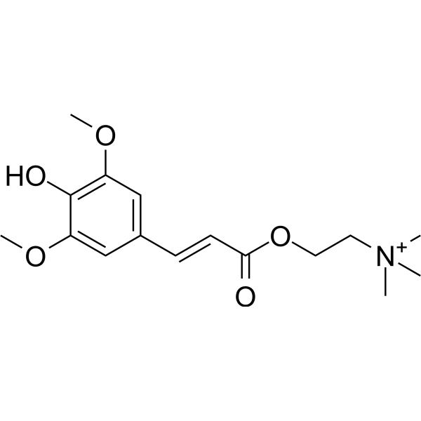
| SMILES | O(C(/C(/[H])=C(\[H])/C1C([H])=C(C(=C(C=1[H])OC([H])([H])[H])O[H])OC([H])([H])[H])=O)C([H])([H])C([H])([H])[N+](C([H])([H])[H])(C([H])([H])[H])C([H])([H])[H] |
| InChi Key | HUJXHFRXWWGYQH-UHFFFAOYSA-O |
| InChi Code | InChI=1S/C16H23NO5/c1-17(2,3)8-9-22-15(18)7-6-12-10-13(20-4)16(19)14(11-12)21-5/h6-7,10-11H,8-9H2,1-5H3/p+1 |
| Chemical Name | Ethanaminium, 2-((3-(4-hydroxy-3,5-dimethoxyphenyl)-1-oxo-2-propenyl)oxy)-N,N,N-trimethyl- |
| HS Tariff Code | 2934.99.9001 |
| Storage |
Powder-20°C 3 years 4°C 2 years In solvent -80°C 6 months -20°C 1 month Note: Please store this product in a sealed and protected environment (e.g. under nitrogen), avoid exposure to moisture and light. |
| Shipping Condition | Room temperature (This product is stable at ambient temperature for a few days during ordinary shipping and time spent in Customs) |
Biological Activity
| Targets |
Acetylcholinesterase (AChE); P-glycoprotein (P-gp)
Sinapine: Acetylcholinesterase (AChE) (IC₅₀ = 1.25 mM for AChE inhibition) [4] Sinapine: P-glycoprotein (P-gp) (downregulates P-gp expression in Caco-2 cells) [3] |
| ln Vitro |
In cardiomyocytes, sinapine (6 or 60 μM; 1 hour) counteracts the oxidative stress caused by H2O2 and antimycin A on the mitochondria [1]. With relatively low toxicity, sinapine (10-200 μM; 24 h) suppresses Caco-2 cell proliferation in a dose-dependent manner [3]. By causing a reduction in P-glycoprotein (P-gp), sinapine (10-200 μM; 24 hours) stimulates the accumulation of doxorubicin in Caco-2 cells [3]. The phosphorylation of ERK1/2 and FRS2α is considerably reduced by sinapine (10-200 μM; 24 hours) [3]. 1. In H9c2 cardiomyocytes exposed to H₂O₂ (200 μM) for 24 h to induce mitochondrial oxidative stress, pretreatment with Sinapine (10 μM, 2 h) significantly reduced intracellular reactive oxygen species (ROS) levels (ROS fluorescence intensity decreased by ~45% compared with H₂O₂-only group), restored mitochondrial membrane potential (ΔΨm, JC-1 red/green fluorescence ratio increased from 0.62 to 1.18), inhibited cytochrome c release from mitochondria to cytoplasm (protein level decreased by ~50%), and reduced cardiomyocyte apoptosis (apoptotic rate decreased from 32% to 11%); in contrast, sinapic acid (10 μM) showed no such protective effects [1] 2. In human colon adenocarcinoma Caco-2 cells treated with Sinapine at concentrations of 0.1–1 mM for 24/48/72 h, the compound exhibited concentration- and time-dependent antiproliferative activity; at 1 mM for 72 h, cell viability decreased to 62% of the control group; additionally, Sinapine (0.5 mM, 48 h) downregulated P-gp protein expression by ~35% and mRNA expression by ~40% in Caco-2 cells, as detected by western blot and RT-PCR respectively [3] 3. In in vitro antioxidant activity assays, Sinapine (extracted from rapeseed pomace) showed dose-dependent free radical scavenging capacity: it scavenged DPPH radicals with an IC₅₀ of 0.83 mM, and ABTS⁺ radicals with an IC₅₀ of 0.76 mM; it also exhibited ferric reducing antioxidant power (FRAP) of 0.32 mM Fe²⁺ equivalent at a concentration of 1 mM [4] 4. In AChE inhibition assays, Sinapine inhibited AChE activity with an IC₅₀ of 1.25 mM; the inhibition was non-competitive (no significant competition with acetylcholine substrate, as shown by Lineweaver-Burk plots) [4] |
| ln Vivo |
By altering the makeup of the intestinal microbiota in mice, sinapine lowers the incidence of non-alcoholic fatty liver disease [2]. 1. In C57BL/6J mice fed a high-fat diet (HFD) for 12 weeks to induce non-alcoholic fatty liver disease (NAFLD), oral administration of Sinapine (50 mg/kg body weight/day, 8 weeks) significantly improved liver steatosis: hepatic triglyceride (TG) content decreased by ~42%, total cholesterol (TC) content decreased by ~35%, and serum alanine transaminase (ALT) and aspartate transaminase (AST) levels decreased by ~38% and ~40% respectively compared with HFD-only group; the compound also modulated gut microbiota composition, increasing the abundance of beneficial bacteria (Bifidobacterium increased by ~65%, Akkermansia increased by ~58%) and reducing the abundance of pathogenic bacteria (Desulfovibrio decreased by ~45%); additionally, it upregulated hepatic AMPK phosphorylation (by ~50%) and downregulated SREBP-1c expression (by ~32%) to improve lipid metabolism [2] 2. In growing rats fed a diet supplemented with Sinapine (0.15% and 0.3% of diet weight, 4 weeks), the compound at 0.3% dose reduced daily food intake by ~12% and increased feed conversion ratio (FCR) by ~8% compared with control group; it had no significant effect on protein digestibility, but increased fat digestibility by ~6% at 0.15% dose; no significant changes in body weight gain were observed in either dose group [5] |
| Enzyme Assay |
1. For AChE inhibition assay of Sinapine: Prepare the reaction system containing AChE solution, buffer (pH 8.0) and serial concentrations of Sinapine (0.25–2.5 mM); pre-incubate the mixture at 37°C for 15 min; add acetylthiocholine iodide (substrate) to initiate the reaction and incubate for another 30 min at 37°C; add DTNB (color-developing reagent) to terminate the reaction and measure absorbance at 412 nm with a microplate reader; calculate the inhibition rate of each concentration and fit the data to obtain the IC₅₀ value; conduct Lineweaver-Burk plot analysis by varying substrate concentrations at fixed Sinapine concentrations to determine the inhibition type [4] 2. For antioxidant activity (DPPH radical scavenging) assay of Sinapine: Prepare DPPH solution (0.1 mM) in ethanol; mix equal volumes of DPPH solution with serial concentrations of Sinapine (0.2–2 mM); incubate the mixture in the dark at room temperature for 30 min; measure absorbance at 517 nm with a spectrophotometer; calculate the scavenging rate using the formula [(A_control - A_sample)/A_control] × 100% and determine the IC₅₀ value for DPPH scavenging [4] 3. For ABTS⁺ radical scavenging assay of Sinapine: Generate ABTS⁺ radical cation by reacting ABTS solution with potassium persulfate and incubating at room temperature for 12–16 h in the dark; dilute the ABTS⁺ solution with buffer to obtain an absorbance of 0.70 ± 0.02 at 734 nm; mix ABTS⁺ solution with serial concentrations of Sinapine (0.2–2 mM); incubate for 6 min at room temperature; measure absorbance at 734 nm and calculate the scavenging rate to determine the IC₅₀ value [4] |
| Cell Assay |
The Caco-2 cells are seeded into a 96-well plate with 8000 cells/well for 24 h. After incubation with different doses of Sinapine (0-200 μM), doxorubicin, or both for 24 h, the medium is discarded. Cell survival after exposure to Sinapine alone or a combination of Sinapine and the anti-tumor agent doxorubicin is examined by MTT colorimetric assay[3]. 1. For H9c2 cardiomyocyte mitochondrial oxidative stress protection assay: Seed H9c2 cells in culture plates and culture until 70–80% confluence; pretreat cells with Sinapine (1, 5, 10 μM) or sinapic acid (10 μM) for 2 h; induce oxidative stress by adding H₂O₂ (200 μM) and incubate for 24 h; detect intracellular ROS using a fluorescent ROS probe and flow cytometry; assess mitochondrial membrane potential with JC-1 fluorescent dye and fluorescence microscopy; measure cytochrome c release by western blot (lyse cells to separate mitochondrial and cytoplasmic fractions, probe with anti-cytochrome c antibody); evaluate apoptosis by Annexin V-FITC/PI double staining and flow cytometry [1] 2. For Caco-2 cell antiproliferation and P-gp expression assay: Seed Caco-2 cells in 96-well plates (for viability) or 6-well plates (for gene/protein detection) and culture until logarithmic growth phase; treat cells with Sinapine (0.1, 0.5, 1 mM) for 24, 48, 72 h; detect cell viability using MTT assay at each time point; for P-gp detection, collect cells after 48 h of treatment with 0.5 mM Sinapine, extract total RNA for RT-PCR to measure P-gp mRNA levels (normalize to GAPDH), and extract total protein for western blot to quantify P-gp protein expression (normalize to β-actin) [3] |
| Animal Protocol |
Rats: Sixty male Sprague-Dawley rats (95 g) are randomly allotted to 6 groups of 10 rats each and reared in individual cages. Six groups of 10 growing rats each are fed ad libitum for 15 days one of six diets: diet A, rapeseed (3.80 g of sinapine/kg DM); diet B, ethanol/water-extracted rapeseed (0.48 g of sinapine); diet C, control diet; diet G, control diet+3.74 g of extracted sinapine; diet H, control diet + 3.72 g of sinapine+other phenolic compounds; or diet I, control diet+the hydrolysis products of sinapine and other phenolic compounds. The rats are weighed at 8 a.m. on days 1, 4, 8, 11 and 15 of the trial. After sacrifice the gut contents are eliminated to permit determination of empty body weight gain (EBWG). The distribution, refusal and intake of each rat are recorded every day[4]. 1. For NAFLD mouse model assay: Use 6-week-old male C57BL/6J mice, randomly divided into control group (normal chow), HFD group (high-fat diet), and Sinapine-treated group (HFD + 50 mg/kg/day Sinapine); construct the NAFLD model by feeding HFD for 12 weeks, and administer Sinapine via oral gavage daily for the last 8 weeks of the HFD feeding period (the compound was dissolved in normal saline to prepare the administration solution); at the end of the experiment, collect mouse serum to detect ALT, AST, TG, TC levels; harvest liver tissue to measure hepatic lipid content, perform H&E staining for histopathological analysis of steatosis, and extract liver protein for western blot to detect AMPK and SREBP-1c expression; collect fecal samples to analyze gut microbiota composition via 16S rRNA gene sequencing [2] 2. For growing rat food intake and nutrient utilization assay: Use weaned male rats, randomly divided into control group (basal diet) and two Sinapine-supplemented groups (basal diet + 0.15% Sinapine, basal diet + 0.3% Sinapine); feed the rats with corresponding diets for 4 weeks, record daily food intake and weekly body weight gain; collect fecal and urine samples during the last 3 days of the experiment to determine nutrient digestibility (protein, fat, carbohydrate); calculate feed conversion ratio (body weight gain/food intake) for each group [5] |
| Toxicity/Toxicokinetics |
1. In H9c2 cardiomyocytes, Sinapine at concentrations up to 10 μM showed no significant cytotoxicity (cell viability >95% after 24 h of treatment without H₂O₂ stimulation) [1] 2. In Caco-2 cells, Sinapine at concentrations ≤0.5 mM for 48 h had no obvious cytotoxicity (cell viability >85%); only at 1 mM for 72 h did it exhibit mild antiproliferative effects (no acute cytotoxicity or apoptosis induction) [3] 3. In mice treated with 50 mg/kg/day Sinapine for 8 weeks, no significant changes in serum BUN, creatinine (renal function markers) or liver function markers (except for reduced ALT/AST in NAFLD model) were observed, and no histopathological damage to kidney or heart was detected [2] 4. In rats fed with 0.3% Sinapine for 4 weeks, no obvious toxic symptoms (such as diarrhea, weight loss, organ damage) were observed, indicating good in vivo tolerance [5] |
| References |
[1]. Boulghobra D, et, al. Sinapine, but not sinapic acid, counteracts mitochondrial oxidative stress in cardiomyocytes. Redox Biol. 2020 Jul;34:101554. [2]. Li Y, et, al. Sinapine reduces non-alcoholic fatty liver disease in mice by modulating the composition of the gut microbiota. Food Funct. 2019 Jun 19;10(6):3637-3649. [3]. Guo Y, et al. Sinapine as an active compound for inhibiting the proliferation of Caco-2 cells via downregulation of P-glycoprotein. Food Chem Toxicol. 2014 May;67:187-92. [4]. Yates K, et, al. Determination of sinapine in rapeseed pomace extract: Its antioxidant and acetylcholinesterase inhibition properties. Food Chem. 2019 Mar 15;276:768-775. [4]. Valorization of rapeseed meal. 5. Effects of sinapine and other phenolic compounds on food intake and nutrient utilization in growing rats. Reprod Nutr Dev. 1987;27(4):781-90. |
| Additional Infomation |
Sinapine is an acylcholine in which the acyl group specified is sinapoyl. It has a role as a photosynthetic electron-transport chain inhibitor, an antioxidant and a plant metabolite. It is functionally related to a trans-sinapic acid. 1. Sinapine is a natural alkaloid ester derived from sinapic acid, mainly found in rapeseed, mustard seeds, and their byproducts (e.g., rapeseed pomace, rapeseed meal); it is one of the main phenolic compounds in these plant materials [4][5] 2. The mechanism of Sinapine in protecting cardiomyocytes from mitochondrial oxidative stress involves reducing ROS accumulation, stabilizing mitochondrial membrane potential, inhibiting cytochrome c release, and blocking the mitochondrial apoptotic pathway; notably, its protective effect is specific, as its metabolite sinapic acid does not have the same activity [1] 3. Sinapine ameliorates NAFLD in HFD-fed mice primarily through modulating gut microbiota composition (enriching beneficial bacteria and reducing harmful bacteria), which in turn improves hepatic lipid metabolism via the gut-liver axis, and also directly regulates hepatic AMPK/SREBP-1c signaling to inhibit lipogenesis [2] 4. The antiproliferative effect of Sinapine on Caco-2 cells is associated with the downregulation of P-gp, a transmembrane efflux pump that mediates multidrug resistance in tumor cells, suggesting potential application in reversing colon cancer drug resistance [3] 5. Sinapine has dual biological activities of antioxidant and AChE inhibition, making it a potential natural lead compound for the prevention of neurodegenerative diseases (e.g., Alzheimer's disease) [4] |
Solubility Data
| Solubility (In Vitro) | DMSO : ~100 mg/mL (~322.21 mM) |
| Solubility (In Vivo) |
Solubility in Formulation 1: ≥ 2.08 mg/mL (6.70 mM) (saturation unknown) in 10% DMSO + 40% PEG300 + 5% Tween80 + 45% Saline (add these co-solvents sequentially from left to right, and one by one), clear solution. For example, if 1 mL of working solution is to be prepared, you can add 100 μL of 20.8 mg/mL clear DMSO stock solution to 400 μL PEG300 and mix evenly; then add 50 μL Tween-80 to the above solution and mix evenly; then add 450 μL normal saline to adjust the volume to 1 mL. Preparation of saline: Dissolve 0.9 g of sodium chloride in 100 mL ddH₂ O to obtain a clear solution. Solubility in Formulation 2: ≥ 2.08 mg/mL (6.70 mM) (saturation unknown) in 10% DMSO + 90% (20% SBE-β-CD in Saline) (add these co-solvents sequentially from left to right, and one by one), clear solution. For example, if 1 mL of working solution is to be prepared, you can add 100 μL of 20.8 mg/mL clear DMSO stock solution to 900 μL of 20% SBE-β-CD physiological saline solution and mix evenly. Preparation of 20% SBE-β-CD in Saline (4°C,1 week): Dissolve 2 g SBE-β-CD in 10 mL saline to obtain a clear solution. (Please use freshly prepared in vivo formulations for optimal results.) |
| Preparing Stock Solutions | 1 mg | 5 mg | 10 mg | |
| 1 mM | 3.2220 mL | 16.1098 mL | 32.2196 mL | |
| 5 mM | 0.6444 mL | 3.2220 mL | 6.4439 mL | |
| 10 mM | 0.3222 mL | 1.6110 mL | 3.2220 mL |