STO-609 acetate is a novel, potent, specific and cell-permeable inhibitor of the Ca2+/Calmodulin-dependent protein kinase kinase(CaM-KK) that inhibits the activities of recombinant CaM-KKα and CaM-KKβ isoforms with Ki values of 80 and 15 ng/ml, respectively, it also inhibits their autophosphorylation activities. STO-609 inhibits the activities of recombinant CaM-KK alpha and CaM-KK beta isoforms, with K(i) values of 80 and 15 ng/ml, respectively, and also inhibits their autophosphorylation activities. Comparison of the inhibitory potency of the compound against various protein kinases revealed that STO-609 is highly selective for CaM-KK without any significant effect on the downstream CaM kinases (CaM-KI and -IV), and the IC(50) value of the compound against CaM-KII is approximately 10 microg/ml. STO-609 inhibits constitutively active CaM-KK alpha (glutathione S-transferase (GST)-CaM-KK-(84-434)) as well as the wild-type enzyme.
Physicochemical Properties
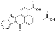
| Molecular Formula | C21H14N2O5 | |
| Molecular Weight | 374.35 | |
| Exact Mass | 374.09 | |
| Elemental Analysis | C, 67.38; H, 3.77; N, 7.48; O, 21.37 | |
| CAS # | 1173022-21-3 | |
| Related CAS # | STO-609;52029-86-4 | |
| PubChem CID | 16760660 | |
| Appearance | Typically exists as solid at room temperature | |
| LogP | 3.381 | |
| Hydrogen Bond Donor Count | 2 | |
| Hydrogen Bond Acceptor Count | 6 | |
| Rotatable Bond Count | 1 | |
| Heavy Atom Count | 28 | |
| Complexity | 596 | |
| Defined Atom Stereocenter Count | 0 | |
| SMILES | C1=CC=C2C(=C1)N=C3C4=CC=C(C5=C4C(=CC=C5)C(=O)N23)C(=O)O.CC(=O)O |
|
| InChi Key | WNRSTFUVBWNELX-UHFFFAOYSA-N | |
| InChi Code | InChI=1S/C19H10N2O3.C2H4O2/c22-18-13-5-3-4-10-11(19(23)24)8-9-12(16(10)13)17-20-14-6-1-2-7-15(14)21(17)18;1-2(3)4/h1-9H,(H,23,24);1H3,(H,3,4) | |
| Chemical Name | acetic acid;11-oxo-3,10-diazapentacyclo[10.7.1.02,10.04,9.016,20]icosa-1(20),2,4,6,8,12,14,16,18-nonaene-17-carboxylic acid | |
| Synonyms |
|
|
| HS Tariff Code | 2934.99.9001 | |
| Storage |
Powder-20°C 3 years 4°C 2 years In solvent -80°C 6 months -20°C 1 month |
|
| Shipping Condition | Room temperature (This product is stable at ambient temperature for a few days during ordinary shipping and time spent in Customs) |
Biological Activity
| Targets | Ca2+/Calmodulin-dependent protein kinase kinase(CaM-KK); CaM-KKα (Ki = 80 ng/mL); CaM-KKβ (Ki = 15 ng/mL) | ||
| ln Vitro | STO-609 suppresses both the autophosphorylation and activity of recombinant CaM-KKα and CaM-KKβ isoforms, with Ki values of 80 and 15 ng/mL, respectively. STO-609 has an IC50 value of 10 μg/mL against CaM-KII and is highly selective for CaM-KK with no discernible effect on the downstream CaM kinases (CaM-KI and -IV). Both the wild-type enzyme and constitutively active CaM-KKα are inhibited by STO-609. STO-609 suppresses the Ca2+-induced activation of CaM-KIV in transfected HeLa cells in a dose-dependent manner. At a concentration of 1μg/mL (80% inhibitory rate), STO-609 significantly lowers the endogenous activity of CaM-KK in SH-SY5Y neuroblastoma cells[1]. | ||
| ln Vivo |
|
||
| Enzyme Assay |
In Vitro Assay for CaM-KK Activity [1] Purified recombinant CaM-KKs (CaM-KKα, 0.28 μg/ml; CaM-KKβ, 0.52 μg/ml; constitutively active CaM-KK, 0.3 μg/ml) were incubated with 10 μg of GST-CaM-KI-(1–293)-K49E at 30 °C for 5 min in a solution (25 μl) containing 50 mm HEPES (pH 7.5), 10 mm Mg(Ac)2, 1 mm DTT, various concentrations (50–400 μm) of [γ-32P]ATP (650–6500 cpm/pmol) with various concentrations of STO-609 (0–10 μg/ml in Me2SO at a final concentration of 4%) in the presence of either 1 mm EGTA (for constitutively active CaM-KK) or 1 mm CaCl2, 2 μm CaM. The reaction was initiated by the addition of [γ-32P]ATP and terminated by spotting aliquots (15 μl) onto phosphocellulose paper followed by several washes with 75 mm phosphoric acid. Phosphate incorporation into GST-CaM-KI-(1–293)-K49E was determined by liquid scintillation counting of the filters. A 5-min reaction was chosen to determine CaM-KK activity based on the time course experiment described recently. Specific activities of CaM-KKα, CaM-KKβ, and constitutively active CaM-KK in the absence of STO-609 were calculated to be 723 ± 7 μmol/min/mg, 338 ± 18 μmol/min/mg, and 927 ± 40 μmol/min/mg, respectively. Autophosphorylation of CaM-KKα and -β [1] Purified recombinant CaM-KKα and -β (0.8 μg) were assayed at 30 °C for 5 min in a solution (25 μl) containing 50 mm HEPES (pH 7.5), 10 mm Mg(Ac)2, 1 mm DTT, 50 μm [γ-32P]ATP (6500 cpm/pmol) with various concentrations of STO-609 (0–10 μg/ml in Me2SO at a final concentration of 4%) in the presence of either 1 mm EGTA (for CaM-KKα and CaM-KKβ) or 1 mmCaCl2, 2 μm CaM (for CaM-KKα). The reaction was initiated by the addition of [γ-32P]ATP and terminated by the addition of SDS-PAGE sample buffer. The samples were subjected to SDS-10% PAGE followed by autoradiography. 32P incorporation into CaM-KK was estimated by densitometric scanning of the x-ray film. In Vitro Assay for CaM-KI, -II, and -IV and MLCK Activities [1] CaM-KI (2.5 μg/ml), CaM-KII (0.75 μg/ml), CaM-KIV (9 μg/ml), and MLCK (0.6 μg/ml) were incubated with 40 μm syntide-2 or 50 μm MLC peptide (for MLCK) at 30 °C for 5 min in a solution (25 μl) containing 50 mm HEPES (pH 7.5), 10 mm Mg(Ac)2, 1 mm DTT, 50 μm [γ-32P]ATP (4500 cpm/pmol) with various concentrations of STO-609 (0–10 μg/ml in Me2SO at a final concentration of 4%) in the presence of 1 mm CaCl2, 2 μm CaM. Protein kinase activity was measured by the phosphocellulose filter method as described above. Specific activities of CaM-KI, CaM-KII, CaM-KIV, and MLCK in the absence of STO-609 were calculated to be 24 ± 1 μmol/min/mg, 122 ± 3 μmol/min/mg, 48 ± 1 μmol/min/mg, and 178 ± 6 μmol/min/mg, respectively. In Vitro Assay for PKA, PKC, and p42 MAP Kinase Activities [1] PKA (8 μg/ml), PKC (25 ng/ml), and p42 MAP kinase (2 μg/ml) were incubated with 100 μm kemptamide (for PKA), 100 μm neurogranin peptide (for PKC, Promega), or 0.4 mg/ml myelin basic protein (for p42 MAP kinase) at 30 °C for 5 min in a solution (25 μl) containing 50 mm HEPES (pH 7.5), 10 mm Mg(Ac)2, 1 mm DTT, 50 μm [γ-32P]ATP (4500 cpm/pmol) with various concentrations of STO-609 (0–10 μg/ml in Me2SO at a final concentration of 4%) in the absence (for PKA and p42 MAP kinase) or presence of 1 mm CaCl2, 0.4 mg/ml phosphatidylserine, and 0.1 mg/ml bovine serum albumin. Protein kinase activity was measured by the phosphocellulose filter method as described above. Specific activities of PKA, PKC and p42 MAP kinase in the absence of STO-609 were calculated to be 22 ± 1 μmol/min/mg, 7.522 ± 0.062 mmol/min/mg, and 181 ± 3 μmol/min/mg, respectively. Protein kinase activity was measured under linear conditions based on the results obtained from titration experiments for each enzyme. Kinase Assays [1] The AMPK peptide, including the sequence surrounding the phosphorylation site of AMPK (167GEFLRTSCGSP177), was synthesized at the Support Unit for Bio-material Analysis in the RIKEN Brain Science Institute (BSI) Research Resources Center (RRC). Appropriate quantities of the purified CaMKKβ KD and full-length CaMKKβ were each incubated in the presence or absence of 500 μm AMPK peptide at 30 °C, in a reaction solution (20 μl) containing 50 mm HEPES (pH 7.5), 300 mm NaCl, 1 mm DTT, 10 mm MgCl2, 400 μm ATP, and 10% glycerol, with or without 0.5 μm STO-609. For the full-length CaMKKβ, 5 μm calmodulin and 1 mm CaCl2 were added to the reaction solution. ATP consumption was determined by using a Kinase-GloTM Max luminescent kinase assay (Promega) kit, which quantifies the amount of ATP in the reaction solution. Glow-type luminescence was recorded after 10 min, using a FusionTM universal microplate analyzer |
||
| Cell Assay |
Transient Expression and Immunoprecipitation of HA-CaM-KIV [1] HeLa cells were maintained in Dulbecco’s modified Eagle’s medium containing 10% fetal bovine serum. Cells were subcultured in 6-cm dishes 12 h before transfection. The cells were then transferred to serum-free medium and treated with a mixture of either 3 μg of pME18s plasmid DNA or 3 μg of HA (hemagglutinin-tagged)-CaM-KIV and 20 μg of LipofectAMINE Reagent in 2.5 ml of medium. After 20 h of incubation, the cells were further cultured in serum-free medium for 6 h in either the absence or presence of various concentrations of STO-609 (0.01–10 μg/ml in Me2SO at a final concentration of 0.5%) and then treated with or without 1 μm ionomycin for 5 min. Stimulation was terminated by the addition of 1 ml of lysis buffer (150 mm NaCl, 20 mm Tris-HCl (pH 7.5), 2 mm EDTA, 2 mm EGTA, 1% Nonidet P-40, 10% glycerol, 0.2 mm phenylmethylsulfonyl fluoride, 10 mg/liter leupeptin, 10 mg/liter trypsin inhibitor, and 1 μm microcystin LR), and the cells were lysed for 30 min on ice. The cell extract was collected and centrifuged at 15,000 × g for 15 min, the supernatant was precleared with 40 μl of Protein G-Sepharose (50% slurry) for 2 h at 4 °C, and the supernatant was mixed with 4 μg of anti-HA antibody for 3 h. 40 μl of Protein G-Sepharose was then applied to the extract and incubated overnight. The immunoprecipitated resin was washed three times with 1 ml of the lysis buffer as described above and then washed with 1 ml of kinase buffer (50 mm HEPES (pH 7.5), 10 mmMg(Ac)2, 1 mm DTT, 1 mm EGTA, and 1 μm microcystin LR). Protein G-Sepharose with immunoprecipitated HA-CaM-KIV was subjected to the protein kinase assay (50 μl reaction volume) in the presence of 1 mm EGTA using syntide-2 as a substrate as described above. To estimate the amount of immunoprecipitated HA-CaM-KIV, SDS-PAGE sample buffer (50 μl) was added to immunoprecipitated samples and then heated at 95 °C for 10 min. After centrifugation, 10 μl of the sample was subjected to SDS-10% PAGE followed by Western blotting using anti-CaM-KIV antibody (1:2000). Expression of Ca2+/CaM-independent CaM-KIV in SH-SY5Y Neuroblastoma Cells by Infection with Recombinant Adenovirus [1] Recombinant adenoviruses carrying cDNAs encoding Ca2+/CaM-independent CaM-KIV (305HMDT-DEDD), a kinase-deficient mutant (305HMDT-DEDD, K71E), or a constitutively active CaM-KK-(1–434) (3) were constructed as follows. Briefly, CaM-KIV mutants and constitutively active CaM-KK cDNAs in pME18s plasmid were digested, blunt-ended, and then ligated into pShuttle (CLONTECH). Recombinant viruses were obtained from HEK293 cells using the Adeno-X Expression System (CLONTECH) according to the manufacturer’s protocol. For virus infection, confluent SY5Y cells in 6-well culture plates were infected with viruses at a multiplicity of infection of 10 plaque-forming units/cell at 37 °C for 1 h. After infection, virus was aspirated, and the cells were further cultured in RPMI medium containing 10% fetal bovine serum for 12 h. The cells were then serum-starved for 6 h in either the absence or presence of various concentrations of STO-609 (0.01–10 μg/ml in Me2SO at a final concentration of 0.5%). The cells were stimulated with 1 μm ionomycin for 10 min (or not subjected to ionomycin treatment) in the absence or presence of various concentrations of STO-609 and then lysed, and the extract was subjected to SDS-7.5% PAGE followed by Western blotting using anti-CaM-KIV antibody. The intensity of the immunoreactive band was measured by densitometric scanning of the x-ray film. |
||
| Animal Protocol |
|
||
| References |
[1]. STO-609, a specific inhibitor of the Ca(2+)/calmodulin-dependent protein kinase kinase. J Biol Chem. 2002 May 3;277(18):15813-8. [2]. Crystal structure of the Ca2+/calmodulin-dependent protein kinase kinase in complex with the inhibitor STO-609. J Biol Chem. 2011 Jun 24;286(25):22570-9. |
||
| Additional Infomation |
LSM-3164 is a naphthoic acid. STO-609, a selective inhibitor of Ca(2+)/calmodulin-dependent protein kinase kinase (CaM-KK) was synthesized, and its inhibitory properties were investigated both in vitro and in vivo. STO-609 inhibits the activities of recombinant CaM-KK alpha and CaM-KK beta isoforms, with K(i) values of 80 and 15 ng/ml, respectively, and also inhibits their autophosphorylation activities. Comparison of the inhibitory potency of the compound against various protein kinases revealed that STO-609 is highly selective for CaM-KK without any significant effect on the downstream CaM kinases (CaM-KI and -IV), and the IC(50) value of the compound against CaM-KII is approximately 10 microg/ml. STO-609 inhibits constitutively active CaM-KK alpha (glutathione S-transferase (GST)-CaM-KK-(84-434)) as well as the wild-type enzyme. Kinetic analysis indicates that the compound is a competitive inhibitor of ATP. In transfected HeLa cells, STO-609 suppresses the Ca(2+)-induced activation of CaM-KIV in a dose-dependent manner. In agreement with this observation, the inhibitor significantly reduces the endogenous activity of CaM-KK in SH-SY5Y neuroblastoma cells at a concentration of 1 microg/ml (approximately 80% inhibitory rate). Taken together, these results indicate that STO-609 is a selective and cell-permeable inhibitor of CaM-KK and that it may be a useful tool for evaluating the physiological significance of the CaM-KK-mediated pathway in vivo as well as in vitro.[1] In summary, we have recently developed a potent and relatively selective inhibitor of CaM-KK, STO-609, which can permeate cells and which is a competitive inhibitor of ATP. Recent studies demonstrate that CaM-KK is a regulatory protein kinase for CaM-KI and CaM-KIVin vitro and in transfected cells; however, this property has not been demonstrated directly in vivo. We have shown in this report that STO-609 suppresses CaM-KK activity resulting in the inhibition of downstream CaM-KIV activity in intact cells, although it cannot inhibit downstream CaM kinase activities in vitro. Thus STO-609 could be a useful tool for evaluating the regulatory roles of CaM-KK for various physiological functions of the CaM kinase cascade such as the regulation of gene expression mediated by the CaM-KIV pathway. Furthermore, STO-609 could be used to distinguish between the functions of the two CaM-KK isoforms because the sensitivity of CaM-KKβ to the compound is ∼5-fold higher than that of the α isoform. The mechanism of differential sensitivity of CaM-KK isoforms to STO-609 is not clear, but it is not likely due to their differences in affinity for ATP because the apparent Km values of CaM-KKα and CaM-KKβ for ATP are indistinguishable (∼33 μm, Fig. 3B). Furthermore, physiological function(s) controlled by the CaM-KK/CaM-KI cascade have not been well studied, in contrast to what is known for CaM-KIV, and this question may be addressed with the use of STO-609. [1] Ca(2+)/calmodulin (CaM)-dependent protein kinase (CaMK) kinase (CaMKK) is a member of the CaMK cascade that mediates the response to intracellular Ca(2+) elevation. CaMKK phosphorylates and activates CaMKI and CaMKIV, which directly activate transcription factors. In this study, we determined the 2.4 Å crystal structure of the catalytic kinase domain of the human CaMKKβ isoform complexed with its selective inhibitor, STO-609. The structure revealed that CaMKKβ lacks the αD helix and that the equivalent region displays a hydrophobic molecular surface, which may reflect its unique substrate recognition and autoinhibition. Although CaMKKβ lacks the activation loop phosphorylation site, the activation loop is folded in an active-state conformation, which is stabilized by a number of interactions between amino acid residues conserved among the CaMKK isoforms. An in vitro analysis of the kinase activity confirmed the intrinsic activity of the CaMKKβ kinase domain. Structure and sequence analyses of the STO-609-binding site revealed amino acid replacements that may affect the inhibitor binding. Indeed, mutagenesis demonstrated that the CaMKKβ residue Pro(274), which replaces the conserved acidic residue of other protein kinases, is an important determinant for the selective inhibition by STO-609. Therefore, the present structure provides a molecular basis for clarifying the known biochemical properties of CaMKKβ and for designing novel inhibitors targeting CaMKKβ and the related protein kinases. [2] In conclusion, this study has demonstrated the unique structural properties of the CaMKKβ KD, which provide a molecular basis for understanding the known biochemical properties of CaMKKβ and the distinct STO-609 sensitivity between the CaMKKα and CaMKKβ isoforms. Our structure of the CaMKKβ·STO-609 complex also provides a structural basis for designing novel inhibitors to specifically block CaMKKβ and related protein kinases.[2] |
Solubility Data
| Solubility (In Vitro) |
|
|||
| Solubility (In Vivo) |
Note: Listed below are some common formulations that may be used to formulate products with low water solubility (e.g. < 1 mg/mL), you may test these formulations using a minute amount of products to avoid loss of samples. Injection Formulations (e.g. IP/IV/IM/SC) Injection Formulation 1: DMSO : Tween 80: Saline = 10 : 5 : 85 (i.e. 100 μL DMSO stock solution → 50 μL Tween 80 → 850 μL Saline) *Preparation of saline: Dissolve 0.9 g of sodium chloride in 100 mL ddH ₂ O to obtain a clear solution. Injection Formulation 2: DMSO : PEG300 :Tween 80 : Saline = 10 : 40 : 5 : 45 (i.e. 100 μL DMSO → 400 μLPEG300 → 50 μL Tween 80 → 450 μL Saline) Injection Formulation 3: DMSO : Corn oil = 10 : 90 (i.e. 100 μL DMSO → 900 μL Corn oil) Example: Take the Injection Formulation 3 (DMSO : Corn oil = 10 : 90) as an example, if 1 mL of 2.5 mg/mL working solution is to be prepared, you can take 100 μL 25 mg/mL DMSO stock solution and add to 900 μL corn oil, mix well to obtain a clear or suspension solution (2.5 mg/mL, ready for use in animals). Injection Formulation 4: DMSO : 20% SBE-β-CD in saline = 10 : 90 [i.e. 100 μL DMSO → 900 μL (20% SBE-β-CD in saline)] *Preparation of 20% SBE-β-CD in Saline (4°C,1 week): Dissolve 2 g SBE-β-CD in 10 mL saline to obtain a clear solution. Injection Formulation 5: 2-Hydroxypropyl-β-cyclodextrin : Saline = 50 : 50 (i.e. 500 μL 2-Hydroxypropyl-β-cyclodextrin → 500 μL Saline) Injection Formulation 6: DMSO : PEG300 : castor oil : Saline = 5 : 10 : 20 : 65 (i.e. 50 μL DMSO → 100 μLPEG300 → 200 μL castor oil → 650 μL Saline) Injection Formulation 7: Ethanol : Cremophor : Saline = 10: 10 : 80 (i.e. 100 μL Ethanol → 100 μL Cremophor → 800 μL Saline) Injection Formulation 8: Dissolve in Cremophor/Ethanol (50 : 50), then diluted by Saline Injection Formulation 9: EtOH : Corn oil = 10 : 90 (i.e. 100 μL EtOH → 900 μL Corn oil) Injection Formulation 10: EtOH : PEG300:Tween 80 : Saline = 10 : 40 : 5 : 45 (i.e. 100 μL EtOH → 400 μLPEG300 → 50 μL Tween 80 → 450 μL Saline) Oral Formulations Oral Formulation 1: Suspend in 0.5% CMC Na (carboxymethylcellulose sodium) Oral Formulation 2: Suspend in 0.5% Carboxymethyl cellulose Example: Take the Oral Formulation 1 (Suspend in 0.5% CMC Na) as an example, if 100 mL of 2.5 mg/mL working solution is to be prepared, you can first prepare 0.5% CMC Na solution by measuring 0.5 g CMC Na and dissolve it in 100 mL ddH2O to obtain a clear solution; then add 250 mg of the product to 100 mL 0.5% CMC Na solution, to make the suspension solution (2.5 mg/mL, ready for use in animals). Oral Formulation 3: Dissolved in PEG400 Oral Formulation 4: Suspend in 0.2% Carboxymethyl cellulose Oral Formulation 5: Dissolve in 0.25% Tween 80 and 0.5% Carboxymethyl cellulose Oral Formulation 6: Mixing with food powders Note: Please be aware that the above formulations are for reference only. InvivoChem strongly recommends customers to read literature methods/protocols carefully before determining which formulation you should use for in vivo studies, as different compounds have different solubility properties and have to be formulated differently. (Please use freshly prepared in vivo formulations for optimal results.) |
| Preparing Stock Solutions | 1 mg | 5 mg | 10 mg | |
| 1 mM | 2.6713 mL | 13.3565 mL | 26.7130 mL | |
| 5 mM | 0.5343 mL | 2.6713 mL | 5.3426 mL | |
| 10 mM | 0.2671 mL | 1.3356 mL | 2.6713 mL |