Physicochemical Properties
| Molecular Formula | C11H8N2S |
| Molecular Weight | 200.259 |
| Exact Mass | 200.041 |
| CAS # | 40172-65-4 |
| PubChem CID | 94880 |
| Appearance | Gray to brown solid powder |
| Density | 1.403g/cm3 |
| Boiling Point | 417.1ºC at 760 mmHg |
| Melting Point | 184-188ºC |
| Flash Point | 206.1ºC |
| Index of Refraction | 1.83 |
| LogP | 3.612 |
| Hydrogen Bond Donor Count | 1 |
| Hydrogen Bond Acceptor Count | 3 |
| Rotatable Bond Count | 0 |
| Heavy Atom Count | 14 |
| Complexity | 221 |
| Defined Atom Stereocenter Count | 0 |
| InChi Key | FECQXVPRUCCUIL-UHFFFAOYSA-N |
| InChi Code | InChI=1S/C11H8N2S/c12-11-13-10-8-4-2-1-3-7(8)5-6-9(10)14-11/h1-6H,(H2,12,13) |
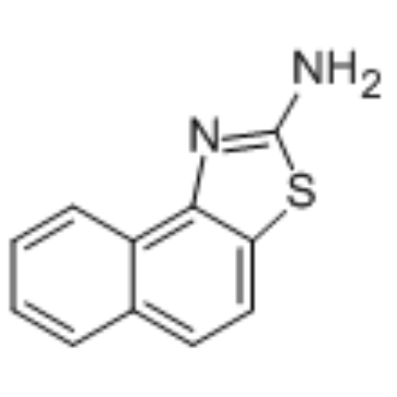
| Chemical Name | benzo[e][1,3]benzothiazol-2-amine |
| Synonyms | SKA31 SKA 31 SKA-31 |
| HS Tariff Code | 2934.99.9001 |
| Storage |
Powder-20°C 3 years 4°C 2 years In solvent -80°C 6 months -20°C 1 month |
| Shipping Condition | Room temperature (This product is stable at ambient temperature for a few days during ordinary shipping and time spent in Customs) |
Biological Activity
| ln Vitro | More specifically than other ion channels, SKA-31 inhibits KCa2/3 channels and activates them more effectively than PK 26124 [1]. In HCT-116 cells and HCT-8 cells, the IC50 values for SKA-31 are 5.3 μM and 46.9 μM, respectively [2]. SKA-31 (5.3 μM; 0-96 hours) decreases the swelling of HCT-116 cells [2]. HCT-116 cells are activated by SKA-31 (5 μM), and SKA-31 at 45 μM raises the proportion of G0/G1 phase cells in HCT-116 and HCT-8 cell lines at concentrations of 5 μM and 45 μM, respectively[2]. SKA-31 decreases CDDP-induced Akt phosphorylation and increases Caspase 3 activation [2]. Additionally effective at preventing HCT-116 cell proliferation are SKA-31 and CDDP [2]. |
| ln Vivo | SKA-31 exhibits favorable pharmacokinetic characteristics and no acute effects [1]. SKA-31 activates KCa3.1 and KCa2.3 in vascular endothelial cells and increases myocardial choline Induced endothelium-derived epidermal hyperplatelet (EDHF)-mediated vasodilation [1]. It also enhances native KCa3.1 and KCa2.3 in carotid endothelial cells. 1.6 μM and 225 nM, in that order[1]. SKA-31 (1-30 mg/kg; ip) decreases glycemic MAP in normal wild-type mice during a 24-day period, but not KCa3.1. It also improves EDHF-type vasodilation and lowers glycemia in mice. |
| Cell Assay |
Cell viability assay [2] Cell Types: HCT-116 cells, HCT-8 cells Tested Concentrations: Incubation Duration: 24 hrs (hours) Experimental Results: Cell viability was diminished in HCT-116 and HCT-8, with IC50s of 5.3 μM and 46.9 μM respectively. Cell proliferation assay[2] Cell Types: HCT-116 Cell Tested Concentrations: 5.3 μM Incubation Duration: 0-96 hrs (hours) Experimental Results: HCT-116 cell proliferation diminished when IC50S value was added at time zero. Apoptosis analysis [2] Cell Types: HCT-116 cells, HCT-8 cells Tested Concentrations: 5 μM (HCT-116 cells), 45 μM (HCT-8 cells) Incubation Duration: 24 hrs (hours) Experimental Results: Triggered HCT-116 cells apoptosis, and the effect was smaller in HCT-8 cells. Cell cycle analysis[2] Cell Types: HCT-116 cells, HCT-8 Cell Tested Concentrations: 5 μM (HCT-116), 45 μM (HCT-8) Incubation Duration: 24 hrs (hours) Experimental Results: G0/% increase in cells HCT-116 and G1 phase of HCT-8 cell line. Western Blot Analysis [2] Cell Types: HCT-116 cells Tested Concentrations: Incubation Duration: 24 hrs (hours) Experimental Results: When HCT-116 cells were co-treated with CDDP, Caspase 3 was fur |
| Animal Protocol |
Animal/Disease Models: 16-25 weeks of mice[1] Doses: 1 mg/kg, 10 mg/kg, 30 mg/kg Route of Administration: intraperitoneal (ip) injection Experimental Results: -/-) Mouse (-/-) MAP[1] . Normotensive wild-type mice had lower MAP over 24 hrs (hrs (hours)), but not KCa3.1(-/-) mice(-/-). |
| References |
[1]. Naphtho[1,2-d]thiazol-2-ylamine (SKA-31), a new activator of KCa2 and KCa3.1 potassium channels, potentiates the endothelium-derived hyperpolarizing factor response and lowers blood pressure. Mol Pharmacol. 2009 Feb;75(2):281-95. [2]. The combined activation of KCa3.1 and inhibition of Kv11.1/hERG1 currents contribute to overcome CDDP resistance in colorectal cancer cells. Br J Cancer. 2018 Jan; 118(2): 200–212. |
Solubility Data
| Solubility (In Vitro) | DMSO : ~125 mg/mL (~624.19 mM) |
| Solubility (In Vivo) |
Solubility in Formulation 1: ≥ 2.5 mg/mL (12.48 mM) (saturation unknown) in 10% DMSO + 40% PEG300 + 5% Tween80 + 45% Saline (add these co-solvents sequentially from left to right, and one by one), clear solution. For example, if 1 mL of working solution is to be prepared, you can add 100 μL of 25.0 mg/mL clear DMSO stock solution to 400 μL PEG300 and mix evenly; then add 50 μL Tween-80 to the above solution and mix evenly; then add 450 μL normal saline to adjust the volume to 1 mL. Preparation of saline: Dissolve 0.9 g of sodium chloride in 100 mL ddH₂ O to obtain a clear solution. Solubility in Formulation 2: ≥ 2.5 mg/mL (12.48 mM) (saturation unknown) in 10% DMSO + 90% Corn Oil (add these co-solvents sequentially from left to right, and one by one), clear solution. For example, if 1 mL of working solution is to be prepared, you can add 100 μL of 25.0 mg/mL clear DMSO stock solution to 900 μL of corn oil and mix evenly. Solubility in Formulation 3: 2.08 mg/mL (10.39 mM) in 10% DMSO + 90% (20% SBE-β-CD in Saline) (add these co-solvents sequentially from left to right, and one by one), suspension solution; with ultrasonication. For example, if 1 mL of working solution is to be prepared, you can add 100 μL of 20.8 mg/mL clear DMSO stock solution to 900 μL of 20% SBE-β-CD physiological saline solution and mix evenly. Preparation of 20% SBE-β-CD in Saline (4°C,1 week): Dissolve 2 g SBE-β-CD in 10 mL saline to obtain a clear solution. (Please use freshly prepared in vivo formulations for optimal results.) |
| Preparing Stock Solutions | 1 mg | 5 mg | 10 mg | |
| 1 mM | 4.9935 mL | 24.9675 mL | 49.9351 mL | |
| 5 mM | 0.9987 mL | 4.9935 mL | 9.9870 mL | |
| 10 mM | 0.4994 mL | 2.4968 mL | 4.9935 mL |