Seletalisib (formerly known as UCB-5857) is a novel, potent, ATP-competitive, and selective, small-molecule inhibitor of PI3Kδ with an IC50 value of 12 nM. It exhibits significant (between 24- and 303-fold) preferential binding to PI3K in comparison to the other class I PI3K isoforms.and its effectiveness was examined using biochemical tests, cellular tests of innate and adaptive immunity, and an in vivo rat model of inflammation. When the B-cell receptor is activated in a B-cell line, seletalisib has the ability to prevent protein kinase B (AKT) phosphorylation. Seletalisib also prevented human neutrophils from releasing superoxide when they were stimulated by N-formyl peptide, but not when they were stimulated by phorbol myristate acetate, suggesting that it has PI3Kδ-specific activity. Peripheral blood mononuclear cells (PBMCs) and other cell types that were exposed to seletalisib showed no signs of cytotoxicity. Results from cellular assays of adaptive immunity showed that seletalisib inhibits human T-cell production of a number of cytokines from activated T-cells. Furthermore, seletalisib reduced B-cell proliferation and cytokine production. Seletalisib reduced the expression of CD69 in human whole blood assays after B-cell activation and anti-IgE-mediated basophil degranulation. In an in vivo rat model of anti-CD3-antibody-induced interleukin 2 release, seletalisib showed dose-dependent inhibition. Collectively, these data define seletalisib as a selective PI3K inhibitor and potential therapeutic candidate for the treatment of autoimmune diseases and B-cell malignancies caused by dysregulated proinflammatory cytokine secretion.
Physicochemical Properties
| Molecular Formula | C23H14CLF3N6O | |
| Molecular Weight | 482.85 | |
| Exact Mass | 482.086 | |
| Elemental Analysis | C, 57.21; H, 2.92; Cl, 7.34; F, 11.80; N, 17.41; O, 3.31 | |
| CAS # | 1362850-20-1 | |
| Related CAS # |
|
|
| PubChem CID | 56928390 | |
| Appearance | White to off-white solid powder | |
| Density | 1.5±0.1 g/cm3 | |
| Boiling Point | 710.8±60.0 °C at 760 mmHg | |
| Flash Point | 383.7±32.9 °C | |
| Vapour Pressure | 0.0±2.3 mmHg at 25°C | |
| Index of Refraction | 1.692 | |
| LogP | 2.21 | |
| Hydrogen Bond Donor Count | 1 | |
| Hydrogen Bond Acceptor Count | 9 | |
| Rotatable Bond Count | 4 | |
| Heavy Atom Count | 34 | |
| Complexity | 690 | |
| Defined Atom Stereocenter Count | 1 | |
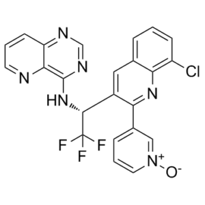
| SMILES | ClC1=CC=CC2=C1N=C(C1=CC=C[N+](=C1)[O-])C(=C2)[C@H](C(F)(F)F)NC1C2C(=CC=CN=2)N=CN=1 |
|
| InChi Key | LNLJHGXOFYUARS-OAQYLSRUSA-N | |
| InChi Code | InChI=1S/C23H14ClF3N6O/c24-16-6-1-4-13-10-15(18(31-19(13)16)14-5-3-9-33(34)11-14)21(23(25,26)27)32-22-20-17(29-12-30-22)7-2-8-28-20/h1-12,21H,(H,29,30,32)/t21-/m1/s1 | |
| Chemical Name |
|
|
| Synonyms |
|
|
| HS Tariff Code | 2934.99.9001 | |
| Storage |
Powder-20°C 3 years 4°C 2 years In solvent -80°C 6 months -20°C 1 month |
|
| Shipping Condition | Room temperature (This product is stable at ambient temperature for a few days during ordinary shipping and time spent in Customs) |
Biological Activity
| Targets |
PI3Kδ (IC50 = 12 nM); GSK-3α (IC50 = 1.5 nM) A potent, ATP-competitive, and highly selective PI3K inhibitor called seletalisib can prevent AKT phosphorylation after the BCR is activated in a B-cell line. Seletalisib inhibits superoxide release from human neutrophils when N-formyl peptides (fMLP) are stimulated, but not when phorbol myristate acetate (PMA), suggesting that it has a PI3Kδ-specific activity. Neither PBMCs nor other cell types treated with seletalisib show any signs of cytotoxicity. Seletalisib prevents human T cells from producing a number of cytokines after they become activated. The differentiation of T cells into Th1, Th2, and Th17 subtypes is inhibited by seletalisib. Seletalisib also prevents B-cell growth and cytokine production. Seletalisib blocks basophil degranulation induced by anti-IgE and B-cell activation in human whole blood assays[1]. |
| ln Vitro |
A potent, ATP-competitive, and highly selective PI3K inhibitor called seletalisib can prevent AKT phosphorylation after the BCR is activated in a B-cell line. Seletalisib inhibits superoxide release from human neutrophils when N-formyl peptides (fMLP) are stimulated, but not when phorbol myristate acetate (PMA), suggesting that it has a PI3Kδ-specific activity. Neither PBMCs nor other cell types treated with seletalisib show any signs of cytotoxicity. Seletalisib prevents human T cells from producing a number of cytokines after they become activated. The differentiation of T cells into Th1, Th2, and Th17 subtypes is inhibited by seletalisib. Seletalisib also prevents B-cell growth and cytokine production. Seletalisib blocks basophil degranulation induced by anti-IgE and B-cell activation in human whole blood assays[1]. Seletalisib potently inhibited anti-IgM-induced phosphorylation of AKT in Ramos human B-cell line with an IC50 of 15 nM (geomean; 95% CI: 9.3–23.5). [1] Seletalisib inhibited fMLP-stimulated superoxide release from human neutrophils with an IC50 of 16 nM (geomean; 95% CI: 11–24) but did not inhibit PMA-stimulated superoxide release, indicating PI3Kδ-specific activity. [1] In anti-CD3 stimulated human PBMCs, seletalisib inhibited secretion of IFNγ (IC50 = 54 nM), IL-17 (IC50 = 21 nM), and TNFα (IC50 = 31 nM). In rat PBMCs, it inhibited anti-CD3-induced TNFα release with an IC50 of 22 nM. [1] Seletalisib inhibited house dust mite (HDM)-induced IL-5 and IL-13 secretion from PBMCs of allergic donors with mean IC50 values of 15 nM and 2 nM, respectively. [1] Seletalisib inhibited anti-IgM-induced proliferation of human B-cells with an IC50 of 16 nM (range: 10–19 nM). [1] Seletalisib inhibited CpG ODN 2006-induced IL-6 and IL-10 release from purified human B-cells with IC50 values of 57 nM and 18.1 nM, respectively. [1] In human whole blood, seletalisib inhibited anti-IgM-induced CD69 expression on B-cells with an IC50 of 57 nM (geomean; 95% CI: 22–143). [1] In human whole blood, seletalisib inhibited anti-IgE-induced basophil degranulation (measured by CD63 expression) with an IC50 of 35 nM (geomean; 95% CI: 27–45). [1] BioMAP profiling showed that seletalisib selectively inhibited immune cell responses (e.g., B-cell proliferation, cytokine and IgG production in BT co-culture system) without showing cytotoxicity in PBMC assays or activity in non-leukocyte containing systems, in contrast to a pan-PI3K inhibitor. [1] |
| ln Vivo |
Seletalisib significantly reduces the amount of IL-2 released in rats after TCR stimulation. The inhibition of seletalisib is seen at all tested doses, reaching nearly complete inhibition at dose levels ≥1 mg/kg. The in vivo effects of seletalisib are strong, with an estimated IC50 value of <10 nM[1]. In a Lewis rat model of anti-CD3 antibody-induced IL-2 release, oral seletalisib (0.1–10 mg/kg) administered 30 minutes prior to anti-CD3 challenge significantly and dose-dependently inhibited IL-2 release. Nearly complete inhibition was achieved at doses ≥1 mg/kg. The estimated in vivo IC50 for IL-2 inhibition was <10 nM based on blood concentration-response relationship. [1] |
| Enzyme Assay |
Seletalisib is dissolved 1 mM solution in DMSO and tested in a concentration response (seletalisib), to explore the effects of PI3Kδ-specific inhibition compared with complete inhibition of class I PI3K signaling. Additionally, the effects of seletalisib at 1000, 100, 10, and 1 nM are examined in the BioMap BT cell system. Based on how the compounds affect cellular readout levels like cytokines, growth factors, adhesion molecules, and proliferation endpoints, an activity profile is created[1]. The inhibitory activity of seletalisib against PI3K isoforms was assessed using a competitive Time-Resolved Fluorescence Resonance Energy Transfer (TR-FRET) assay. In this assay, PI3K enzyme activity converts phosphatidylinositol bisphosphate (PIP2) to phosphatidylinositol triphosphate (PIP3). The generated PIP3 competes with a biotinylated PIP3 tracer for binding to a GST-tagged pleckstrin homology domain protein. The complex is detected using streptavidin-allophycocyanin and a europium-labeled anti-GST antibody, with proximity generating a TR-FRET signal. Inhibition of PI3K reduces PIP3 production, thereby increasing the TR-FRET signal. The assay was performed by pre-incubating compound with PIP2 and ATP, followed by addition of PI3K enzyme. After incubation, the reaction was stopped, detection reagents were added, and the TR-FRET signal was measured. The ATP-competitive nature of seletalisib was confirmed by running the assay at varying ATP concentrations (2, 40, 200, and 1000 µM). [1] Kinase selectivity profiling was performed using Z’-LYTE FRET and Adapta Universal Kinase assays at a screening concentration of 10 µM. [1] |
| Cell Assay |
Serially diluted seletalisib and goat anti-human F(ab)2 IgM in serum-free RPMI 1640 were added to Ramos cells plated in serum-free RPMI 1640. The plate was incubated for 10 minutes, placed on ice, and the cells were centrifuged to pellet them. The MSD assay was used to identify cellularly phosphorylated AKT. Ramos pAKT Assay: Ramos cells were plated in serum-free medium and treated with serially diluted seletalisib followed by stimulation with anti-IgM antibody. After a brief incubation, cells were lysed, and phosphorylated AKT (pAKT) levels were quantified using a mesoscale discovery (MSD) immunoassay. [1] Human Neutrophil Superoxide Release Assay: Neutrophils enriched from human blood were incubated with seletalisib in PBS containing a superoxide detection mix (dihydrocytochalasin B, horseradish peroxidase, sodium azide, Amplex Red) and stimulated with either fMLP or PMA. After incubation, fluorescence intensity was measured to quantify superoxide release. [1] T-cell Activation Assay (Human/Rat): PBMCs were isolated from human or rat blood. For human assays, plates were coated with anti-CD3 antibody. PBMCs and seletalisib were added to the plates and incubated for 48 hours. Culture supernatants were then harvested for cytokine analysis (IFNγ, IL-17, TNFα for human; TNFα for rat) using MSD or ELISA kits. [1] House Dust Mite (HDM) Assay: PBMCs from HDM-allergic donors were incubated with seletalisib and HDM extract for 6 days. Supernatants were harvested, and IL-5 and IL-13 levels were measured by ELISA. [1] B-cell Proliferation Assay: B-cells were isolated from PBMCs by CD19-positive selection, stained with CFSE, and co-cultured with mitomycin C-treated antigen-presenting cells. Cells were treated with seletalisib and stimulated with anti-IgM, IL-2, and IL-10. After 6 days, CFSE dilution in CD19+ cells was analyzed by flow cytometry to measure proliferation. [1] CpG-induced B-cell Cytokine Assay: Purified CD19+ B-cells were treated with seletalisib and stimulated with CpG ODN 2006 for 48 hours. Supernatants were collected, and IL-6 and IL-10 levels were measured by ELISA. [1] Whole Blood CD69 B-cell Activation Assay: Human whole blood aliquots were incubated with anti-IgM and seletalisib for 20 hours. Cells were then stained with anti-CD19 and anti-CD69 antibodies, lysed, and analyzed by flow cytometry to measure CD69 mean fluorescence intensity on CD19+ B-cells. [1] Whole Blood Basophil Degranulation Assay: Human whole blood was incubated with seletalisib, followed by stimulation with anti-IgE. After incubation, cells were stained with an antibody cocktail (CD123, HLA-DR, CD63), lysed, and analyzed by flow cytometry. Basophils were gated as SSC^lo, CD123+, HLA-DR- cells, and CD63 surface expression was measured as a marker of degranulation. [1] |
| Animal Protocol |
Rats: Rats receive seletalisib (0.1-10 mg/kg in 500 μL volume) or vehicle via oral gavage 30 min before receiving a 200 μL dose volume of anti-CD3 antibody intravenously. For oral and intravenous administration, the vehicle is methylcellulose or saline, respectively. Levels of IL-2 and seletalisib are measured[1]. Anti-CD3-induced IL-2 Release in Lewis Rats: Adult male Lewis rats (6-8 weeks old) were administered seletalisib (0.1, 1, 3, or 10 mg/kg) or vehicle (0.5% methylcellulose) via oral gavage in a volume of 500 µL. Thirty minutes later, rats received an intravenous injection of anti-CD3 antibody (100 µg/kg in 200 µL saline). A negative control group received both oral and i.v. vehicle. A positive control group received oral vehicle and i.v. anti-CD3 antibody. Ninety minutes post anti-CD3 administration, rats were anesthetized, blood was collected via terminal cardiac puncture into EDTA or heparin tubes, and plasma was prepared for IL-2 measurement by ELISA. Blood samples were also processed for seletalisib concentration analysis by LC-MS/MS. [1] |
| References |
[1]. Seletalisib: Characterization of a Novel, Potent, and Selective Inhibitor of PI3Kδ. J Pharmacol Exp Ther. 2017 Apr 25. pii: jpet.116.237347. |
| Additional Infomation |
Seletalisib has been used in trials studying the treatment and basic science of Primary Sjogren's Syndrome. Seletalisib (UCB-...) is a novel, potent, selective, and ATP-competitive small-molecule inhibitor of the PI3Kδ isoform. [1] Its selective inhibition of PI3Kδ, which is primarily expressed in leukocytes and signals downstream of immune receptors like the B-cell receptor (BCR), T-cell receptor (TCR), and Fc epsilon receptor I (FcεRI), suggests potential as a therapeutic for B-cell malignancies and autoimmune/inflammatory diseases (e.g., rheumatoid arthritis, systemic lupus erythematosus, psoriasis). [1] It has entered clinical testing. At the time of publication, it was being evaluated in healthy volunteers and patients with psoriasis (NCT02303509) and in patients with primary Sjögren’s Syndrome (NCT02610543). [1] |
Solubility Data
| Solubility (In Vitro) |
|
|||
| Solubility (In Vivo) |
Solubility in Formulation 1: ≥ 2.5 mg/mL (5.18 mM) (saturation unknown) in 10% DMSO + 90% Corn Oil (add these co-solvents sequentially from left to right, and one by one), clear solution. For example, if 1 mL of working solution is to be prepared, you can add 100 μL of 25.0 mg/mL clear DMSO stock solution to 900 μL of corn oil and mix evenly. (Please use freshly prepared in vivo formulations for optimal results.) |
| Preparing Stock Solutions | 1 mg | 5 mg | 10 mg | |
| 1 mM | 2.0710 mL | 10.3552 mL | 20.7104 mL | |
| 5 mM | 0.4142 mL | 2.0710 mL | 4.1421 mL | |
| 10 mM | 0.2071 mL | 1.0355 mL | 2.0710 mL |