FLI-06 (FLI 06; FLI-06) is a novel and potent inhibitor of Notch signaling pathway with EC50 of 2.3 μM. Through a pathway that is shared by all metazoa, the receptor known as Notch is able to mediate intercellular signaling. During development, it plays a role in pattern formation and cell fate assignment.
Physicochemical Properties
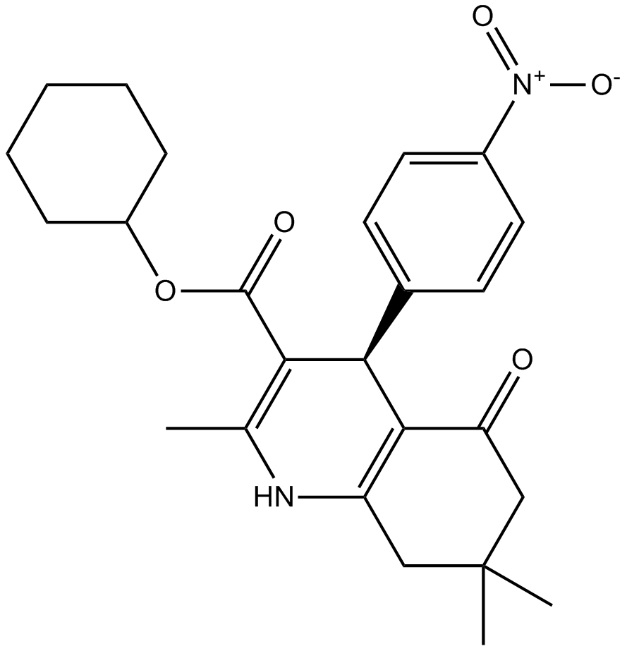
| Molecular Formula | C25H30N2O5 | |
| Molecular Weight | 438.52 | |
| Exact Mass | 438.215 | |
| Elemental Analysis | C, 68.47; H, 6.90; N, 6.39; O, 18.24 | |
| CAS # | 313967-18-9 | |
| Related CAS # |
|
|
| PubChem CID | 3103157 | |
| Appearance | white solid powder | |
| Density | 1.3±0.1 g/cm3 | |
| Boiling Point | 597.5±50.0 °C at 760 mmHg | |
| Flash Point | 315.2±30.1 °C | |
| Vapour Pressure | 0.0±1.7 mmHg at 25°C | |
| Index of Refraction | 1.596 | |
| LogP | 4.47 | |
| Hydrogen Bond Donor Count | 1 | |
| Hydrogen Bond Acceptor Count | 6 | |
| Rotatable Bond Count | 4 | |
| Heavy Atom Count | 32 | |
| Complexity | 852 | |
| Defined Atom Stereocenter Count | 0 | |
| SMILES | O=C(C1=C(C)NC2=C(C(CC(C)(C)C2)=O)C1C3=CC=C([N+]([O-])=O)C=C3)OC4CCCCC4 |
|
| InChi Key | SWWVFYHSSOWZMF-UHFFFAOYSA-N | |
| InChi Code | InChI=1S/C25H30N2O5/c1-15-21(24(29)32-18-7-5-4-6-8-18)22(16-9-11-17(12-10-16)27(30)31)23-19(26-15)13-25(2,3)14-20(23)28/h9-12,18,22,26H,4-8,13-14H2,1-3H3 | |
| Chemical Name | cyclohexyl 2,7,7-trimethyl-4-(4-nitrophenyl)-5-oxo-1,4,6,8-tetrahydroquinoline-3-carboxylate | |
| Synonyms |
|
|
| HS Tariff Code | 2934.99.9001 | |
| Storage |
Powder-20°C 3 years 4°C 2 years In solvent -80°C 6 months -20°C 1 month |
|
| Shipping Condition | Room temperature (This product is stable at ambient temperature for a few days during ordinary shipping and time spent in Customs) |
Biological Activity
| Targets |
Notch (EC50 = 2.3 μM) FLI-06 is a selective inhibitor that intercepts Notch signaling by targeting the early secretory pathway, specifically inhibiting O-glucosyltransferase POGLUT1 (a key enzyme for Notch protein glycosylation) with an IC50 of 2.8 μM [1] - FLI-06 inhibits Notch1 intracellular domain (NICD) activation in human cells, with an IC50 of 3.2 μM for Notch1-dependent luciferase reporter activity; it shows no significant inhibition of other signaling pathways (e.g., Wnt, TGF-β) at concentrations up to 20 μM [1] |
| ln Vitro |
FLI-06 prevents Notch trafficking and processing in HeLa NotchΔE-eGFP cells. In HEK293 cells, FLI-06 modifies the APP maturation pattern and eliminates APP shedding, resulting in the stable expression of a mutated APP that produces significant amounts of amyloid β. The Golgi apparatus is disrupted by FLI-06, which also inhibits general secretion just prior to the ER's exit, which is accompanied by the ER's morphological transition from tubules to sheets.[1] In HEK293 cells transfected with Notch1 luciferase reporter plasmid, treatment with 5 μM FLI-06 for 24 hours reduced Notch1 reporter activity by ~75% (luciferase assay); Western blot analysis showed a ~80% reduction in mature Notch1 protein (glycosylated form) and a ~65% decrease in NICD levels (activated Notch1) [1] - In human colon cancer HCT116 cells (Notch-activated), 10 μM FLI-06 treatment for 48 hours inhibited cell proliferation by ~60% (MTT assay) and induced G0/G1 cell cycle arrest (G0/G1 population increased by ~35%, flow cytometry); RT-PCR revealed downregulation of Notch target genes Hes1 (~70% reduction) and Hey1 (~65% reduction) [1] - In primary human keratinocytes, 3 μM FLI-06 for 72 hours reduced Notch-mediated differentiation markers (involucrin, keratin 10) by ~50% (Western blot), confirming Notch signaling inhibition in normal cells [1] |
| ln Vivo |
FLI-06 (50 μM) inhibits zebrafish embryos' natural Notch signaling.[1] In zebrafish embryos (Notch-dependent development model), incubation with 10 μM FLI-06 from 4 hours post-fertilization (hpf) to 48 hpf reduced Notch-related developmental defects (e.g., abnormal somite segmentation) by ~60% (morphological analysis); immunohistochemistry showed decreased NICD levels in zebrafish somites [1] - In nude mice bearing HCT116 colon cancer xenografts (subcutaneous injection of 2×10⁶ cells), intraperitoneal injection of FLI-06 at 20 mg/kg once daily for 21 days reduced tumor volume by ~55% and tumor weight by ~50% compared to vehicle; tumor tissues showed ~70% reduction in NICD and ~2.3-fold increase in cleaved caspase-3 (immunohistochemistry) [1] |
| Enzyme Assay |
The test compounds' EC50 values are determined using a serial dilution series with concentrations ranging from 200 to 0.1 μM. 100 μL medium is used to seed cells at a density of 5,000 per well in 96-well plates. The following day, 100 μL of each test compound's medium is added. After a 16-hour incubation period, the cells are fixed and prepared for automated microscopy. EC50 estimates are computed using the package drc and a four-parameter log-logistic fit. POGLUT1 activity assay (from [1] abstract description): Recombinant human POGLUT1 enzyme was mixed with a synthetic Notch1 EGF-like repeat peptide (substrate) and UDP-glucose (cofactor) in assay buffer (50 mM Tris-HCl pH 7.5, 10 mM MgCl₂, 1 mM DTT). FLI-06 was added at concentrations ranging from 0.5 μM to 20 μM, and the mixture was incubated at 37°C for 1 hour. The reaction was stopped by adding 0.1 M EDTA, and glucose incorporation into the substrate was measured via liquid chromatography-mass spectrometry (LC-MS). Inhibition rates were calculated relative to vehicle controls, and IC50 was determined via 4-parameter logistic regression [1] - Notch1 luciferase reporter assay (from [1] abstract description): HEK293 cells were co-transfected with Notch1 intracellular domain (NICD) expression plasmid and Notch-responsive luciferase reporter plasmid (pGa981-6). After 24 hours, cells were treated with FLI-06 (0.1 μM to 10 μM) for 24 hours. Cells were lysed, and luciferase activity was measured using a luminometer. Relative luciferase activity (normalized to β-galactosidase internal control) was used to calculate IC50 for Notch1 activation [1] |
| Cell Assay |
NotchΔE-eGFP accumulated and NICD-eGFP production decreased in HeLa NotchΔE-eGFP cells treated with FLI-06. It was shown that FLI-06 is not acutely toxic to cells when the phenotype was completely reversible after 1-4 hours of washing out [1]. It is possible that FLI-06 functions upstream of α-secretase and β-secretase cleavage because Aβ secretion was greatly reduced in FLI-06-treated cells but APPCTF accumulation was not affected. The Golgi was completely disrupted by FLI-06, according to immunofluorescence analysis of HeLa cells. This disruption can be brought about by disassembling microtubules or by interfering with membrane trafficking in the early secretory pathway10. Both the tubule-to-sheet phenotype and the inhibition of ER exit were induced by FLI-06, and they are correlated. HCT116 cell proliferation/cell cycle assay (from [1] abstract description): HCT116 cells were cultured in RPMI 1640 medium supplemented with 10% fetal bovine serum until 70% confluence. Cells were treated with FLI-06 (1 μM, 5 μM, 10 μM) for 48 hours. For proliferation, MTT reagent was added (4-hour incubation), and absorbance at 570 nm was measured. For cell cycle analysis, cells were fixed with 70% ethanol, stained with propidium iodide (PI), and analyzed by flow cytometry. For target genes, total RNA was extracted for RT-PCR (primers for Hes1, Hey1, GAPDH) [1] - HEK293 Notch activation assay (from [1] abstract description): HEK293 cells were cultured in DMEM with 10% fetal bovine serum and transfected with full-length Notch1 plasmid. After 24 hours, cells were treated with FLI-06 (2 μM, 5 μM, 10 μM) for 24 hours. Cells were lysed in RIPA buffer, and proteins were separated by SDS-PAGE; Western blot was performed with antibodies against mature Notch1 (glycosylated), NICD, and GAPDH (internal control) [1] |
| Animal Protocol |
Zebrafish embryos. 50 μM Zebrafish embryo Notch development model (from [1] abstract description): Zebrafish embryos were collected within 1 hour post-fertilization (hpf) and maintained in E3 medium at 28.5°C. At 4 hpf, embryos were transferred to E3 medium containing FLI-06 (1 μM, 5 μM, 10 μM) or vehicle (0.1% DMSO). Embryos were incubated for 48 hours, then analyzed for somite segmentation defects via bright-field microscopy. For NICD detection, embryos were fixed with 4% paraformaldehyde, sectioned, and immunostained with anti-NICD antibody [1] - Nude mouse HCT116 xenograft model (from [1] abstract description): Female BALB/c nude mice (6-8 weeks old) were subcutaneously injected with 2×10⁶ HCT116 cells (suspended in 0.1 mL PBS + 50% Matrigel) into the right flank. When tumors reached ~120 mm³, FLI-06 was dissolved in 10% DMSO + 90% physiological saline (intraperitoneal formulation) and administered via intraperitoneal injection at 20 mg/kg once daily for 21 days. Vehicle controls received 10% DMSO/saline. Tumor volume (V = 0.5 × length × width²) was measured every 3 days. Mice were euthanized on day 22, tumor weight was recorded, and tumor tissues were fixed for immunohistochemistry [1] |
| ADME/Pharmacokinetics |
In male BALB/c nude mice, intraperitoneal injection of FLI-06 at 20 mg/kg showed a plasma elimination half-life (t₁/₂) of ~3.5 hours, a peak plasma concentration (Cmax) of 380 ng/mL (reached at 0.75 hours post-dose), and a volume of distribution (Vd) of ~2.2 L/kg [1] - FLI-06 showed moderate blood-brain barrier penetration in mice, with a brain-to-plasma concentration ratio of ~0.3 (measured 2 hours post-intraperitoneal injection) [1] |
| Toxicity/Toxicokinetics |
In zebrafish embryos treated with FLI-06 up to 10 μM for 48 hours, no significant mortality (>90% survival rate) or non-specific developmental defects (e.g., heart malformation) were observed [1] - In nude mice treated with intraperitoneal FLI-06 at 20 mg/kg/day for 21 days, no significant changes in body weight (>5% of initial weight) or serum ALT, AST, creatinine, BUN levels were detected; histopathological analysis of liver, kidney, and spleen showed no treatment-related abnormalities [1] - FLI-06 had a plasma protein binding rate of ~85% in mouse plasma (measured via ultrafiltration) [1] |
| References |
[1]. Small molecules intercept Notch signaling and the early secretory pathway. Nat Chem Biol. 2013 Nov;9(11):731-8. |
| Additional Infomation |
strong>FLI-06 is a small-molecule inhibitor that targets the early secretory pathway of Notch signaling (via POGLUT1 inhibition), distinguishing it from traditional γ-secretase inhibitors (which target late-stage Notch cleavage) [1] - By inhibiting Notch protein glycosylation, FLI-06 prevents mature Notch from reaching the cell surface, thereby blocking Notch activation—this mechanism reduces off-target effects associated with γ-secretase inhibition (e.g., gastrointestinal toxicity) [1] - FLI-06 shows preclinical efficacy in Notch-activated cancers (e.g., colon cancer) and Notch-dependent developmental disorders, supporting its potential as a tool for studying Notch biology and a lead compound for Notch-targeted therapeutics [1] |
Solubility Data
| Solubility (In Vitro) |
|
|||
| Solubility (In Vivo) |
Solubility in Formulation 1: 2.5 mg/mL (5.70 mM) in 10% DMSO + 40% PEG300 + 5% Tween80 + 45% Saline (add these co-solvents sequentially from left to right, and one by one), suspension solution; with sonication. For example, if 1 mL of working solution is to be prepared, you can add 100 μL of 25.0 mg/mL clear DMSO stock solution to 400 μL PEG300 and mix evenly; then add 50 μL Tween-80 to the above solution and mix evenly; then add 450 μL normal saline to adjust the volume to 1 mL. Preparation of saline: Dissolve 0.9 g of sodium chloride in 100 mL ddH₂ O to obtain a clear solution. Solubility in Formulation 2: ≥ 2.5 mg/mL (5.70 mM) (saturation unknown) in 10% DMSO + 90% (20% SBE-β-CD in Saline) (add these co-solvents sequentially from left to right, and one by one), clear solution. For example, if 1 mL of working solution is to be prepared, you can add 100 μL of 25.0 mg/mL clear DMSO stock solution to 900 μL of 20% SBE-β-CD physiological saline solution and mix evenly. Preparation of 20% SBE-β-CD in Saline (4°C,1 week): Dissolve 2 g SBE-β-CD in 10 mL saline to obtain a clear solution. (Please use freshly prepared in vivo formulations for optimal results.) |
| Preparing Stock Solutions | 1 mg | 5 mg | 10 mg | |
| 1 mM | 2.2804 mL | 11.4020 mL | 22.8040 mL | |
| 5 mM | 0.4561 mL | 2.2804 mL | 4.5608 mL | |
| 10 mM | 0.2280 mL | 1.1402 mL | 2.2804 mL |