Physicochemical Properties
| Molecular Formula | C31H46O5 |
| Molecular Weight | 498.6940 |
| Exact Mass | 498.334 |
| CAS # | 137551-38-3 |
| PubChem CID | 5471851 |
| Appearance | White to off-white solid powder |
| Density | 1.1±0.1 g/cm3 |
| Boiling Point | 664.0±55.0 °C at 760 mmHg |
| Flash Point | 369.3±28.0 °C |
| Vapour Pressure | 0.0±4.6 mmHg at 25°C |
| Index of Refraction | 1.554 |
| LogP | 7.26 |
| Hydrogen Bond Donor Count | 3 |
| Hydrogen Bond Acceptor Count | 5 |
| Rotatable Bond Count | 10 |
| Heavy Atom Count | 36 |
| Complexity | 1000 |
| Defined Atom Stereocenter Count | 7 |
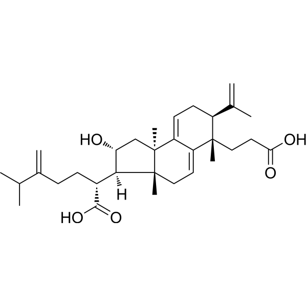
| SMILES | CC(C)C(=C)CC[C@H]([C@H]1[C@@H](C[C@@]2([C@@]1(CC=C3C2=CC[C@H]([C@]3(C)CCC(=O)O)C(=C)C)C)C)O)C(=O)O |
| InChi Key | KVAQLXUMUVEKGR-SMFZDKLCSA-N |
| InChi Code | InChI=1S/C31H46O5/c1-18(2)20(5)9-10-21(28(35)36)27-25(32)17-31(8)24-12-11-22(19(3)4)29(6,15-14-26(33)34)23(24)13-16-30(27,31)7/h12-13,18,21-22,25,27,32H,3,5,9-11,14-17H2,1-2,4,6-8H3,(H,33,34)(H,35,36)/t21-,22+,25-,27+,29+,30-,31+/m1/s1 |
| Chemical Name | (2R)-2-[(2R,3R,3aR,6S,7S,9bR)-6-(2-carboxyethyl)-2-hydroxy-3a,6,9b-trimethyl-7-prop-1-en-2-yl-1,2,3,4,7,8-hexahydrocyclopenta[a]naphthalen-3-yl]-6-methyl-5-methylideneheptanoic acid |
| HS Tariff Code | 2934.99.9001 |
| Storage |
Powder-20°C 3 years 4°C 2 years In solvent -80°C 6 months -20°C 1 month Note: This product requires protection from light (avoid light exposure) during transportation and storage. |
| Shipping Condition | Room temperature (This product is stable at ambient temperature for a few days during ordinary shipping and time spent in Customs) |
Biological Activity
| Targets |
Poricoic acid A exerts its effects by regulating the Gas6/Axl–NF-κB/Nrf2 signaling axis.[2] |
| ln Vitro |
Poricoic acid A demonstrated inhibitory activity against human lung adenocarcinoma A549 cells. The half maximal inhibitory concentration (IC₅₀) was determined to be 34.6 µg/mL.[1] The mixture of seco-lanostane triterpene acids (which contains Poricoic acid A) showed concentration-dependent inhibitory effects on the proliferation of A549 and Hela cells, with IC₅₀ values of 65.30 ± 0.33 µg/mL and 48.09 ± 6.75 µg/mL, respectively.[1] In normal rat kidney proximal tubular epithelial cells (NRK-52E) subjected to hypoxia/reoxygenation (H/R) injury, treatment with Poricoic acid A (10 µM) alone or in combination with melatonin (10 µM) reduced H/R-induced injury. It helped reverse the H/R-induced decrease in the epithelial cell marker E-cadherin. The compound alone or in combination attenuated the H/R-induced activation of the NF-κB pathway (reducing p-IκBα and nuclear p65 levels) and upregulated the antioxidant Nrf2 pathway (increasing Nrf2 and its downstream targets HO-1, catalase, GCLC, and NQO-1). The combination therapy showed stronger effects than Poricoic acid A alone.[2] A cell viability assay using a cell counting kit showed that Poricoic acid A at concentrations of 1, 10, 50, and 100 µM did not cause significant proliferative or cytotoxic effects on NRK-52E cells after 24 hours of treatment.[2] |
| ln Vivo |
In a rat model of renal ischemia-reperfusion injury (IRI), treatment with Poricoic acid A (10 mg/kg/day, oral administration from day 2 to day 13 after reperfusion) significantly reduced the elevated levels of serum creatinine and urea at days 3 and 14 post-IRI, indicating improved renal function. It also ameliorated renal histological damage, including tubular dilation, interstitial inflammation, and collagen deposition at day 14. Mechanistically, Poricoic acid A treatment regulated the Gas6/Axl pathway in a time-dependent manner: it upregulated the anti-inflammatory Gas6/Axl signaling at the acute kidney injury (AKI) stage (day 3) and downregulated the pro-fibrotic Gas6/Axl signaling at the chronic kidney disease (CKD) stage (day 14). At the AKI stage, Poricoic acid A attenuated inflammation by inhibiting NF-κB pathway activation (reducing p-IκBα, nuclear p65, and inflammatory markers MCP-1, COX-2, iNOS, 12-LO) and enhancing the Nrf2 antioxidant pathway (increasing Nrf2, HO-1, catalase, GCLC, NQO-1). It also reduced macrophage (ED-1 positive) and T-cell (CD3 positive) infiltration in the kidney. At the CKD stage, Poricoic acid A inhibited renal fibrosis, as evidenced by reduced collagen I and III deposition, lower protein levels of fibrotic markers (collagen I, fibronectin, FSP1, α-SMA, vimentin, TGF-β1), and increased expression of the epithelial marker E-cadherin. It also protected against podocyte injury by increasing podocyte markers (nephrin, podocin, podocalyxin, synaptopodin, WT1) and decreasing desmin expression. The combination of Poricoic acid A and melatonin showed superior renoprotective effects compared to Poricoic acid A alone.[2] |
| Cell Assay |
The article describes a general cytotoxicity assay for the evaluation of compounds (including the total acid extract). Human cancer cell lines (A549, HepG2, 2MIA, Hela, MGC803, MCF-7) were seeded in 96-well plates at a density of 4×10³ cells per well. After treatment with the test compound or serum-free medium for 48 hours, cell viability was assessed by adding MTT reagent. The absorbance was measured at 570 nm to determine cell growth inhibition.[1] The study employed a hypoxia/reoxygenation model using normal rat kidney proximal tubular epithelial cells (NRK-52E). Cells were cultured and then placed in a hypoxic chamber (1% O2) at 37°C for 9 hours, followed by reoxygenation under normal conditions for 6 or 12 hours. During the reoxygenation period, cells were treated with Poricoic acid A (10 µM) or vehicle. After treatment, cells were harvested for various analyses. Cell viability was assessed using a cell counting kit-8. Cells were seeded in 96-well plates, treated with different concentrations of Poricoic acid A for 24 hours, followed by the addition of the kit reagent. Absorbance was measured at 450 nm. Gene expression analysis was performed by quantitative real-time PCR. Total RNA was isolated, reverse transcribed into cDNA, and amplified using specific primers and a SYBR Green system. Protein expression was analyzed by western blot. Cells were lysed, proteins were separated by gel electrophoresis, transferred to membranes, and probed with specific primary and secondary antibodies. Bands were visualized using a chemiluminescence detection system. Immunofluorescence staining was performed. Cells grown on coverslips were fixed, permeabilized, blocked, and incubated with primary antibodies overnight, followed by fluorescent secondary antibodies and DAPI for nuclear staining. Images were captured using a confocal microscope. For gene knockdown, Axl-specific siRNA was transfected into NRK-52E cells using a lipid-based transfection reagent. After 72 hours, cells were collected for subsequent experiments to validate the role of Axl.[2] |
| Animal Protocol |
The study used a rat model of renal ischemia-reperfusion injury. Male Sprague-Dawley rats were anesthetized. The renal pedicles were clamped for 1 hour to induce ischemia, followed by reperfusion. Sham-operated rats underwent laparotomy without clamping. Rats were randomly divided into groups, including an IRI model group and an IRI group treated with Poricoic acid A. Poricoic acid A was administered by intragastric gavage at a dose of 10 mg/kg per day, starting from day 2 after reperfusion and continuing until day 13. Renal function was assessed by measuring serum creatinine and urea levels at days 3 and 14 post-surgery. At the endpoint (day 14), kidney tissues were collected for histological analysis (H&E, Masson's trichrome, picrosirius red staining), immunohistochemistry, immunofluorescence, and molecular biology analyses (western blot, qPCR).[2] |
| References |
[1]. Enrichment and separation of antitumor triterpene acids from the epidermis of Poria cocos by pH-zone-refining counter-current chromatography and conventional high-speed counter-current chromatography. J Sep Sci. 2015 Jun;38(11):1977-82. [2]. Poricoic acid A enhances melatonin inhibition of AKI-to-CKD transition by regulating Gas6/AxlNFκB/Nrf2 axis. Free Radic Biol Med. 2019 Apr;134:484-497. |
| Additional Infomation |
Poricoic acid A is a tricyclic triterpenoid isolated from Poria cocos. It has a role as a fungal metabolite. It is a tricyclic triterpenoid, a dicarboxylic acid and a secondary alcohol. Poricoic acid A has been reported in Phellodendron amurense and Wolfiporia cocos with data available. Poricoic acid A is a seco-lanostane type triterpene acid and one of the major antitumor components found in the epidermis of Poria cocos (Schw.) Wolf.[1] Poricoic acid A was successfully isolated from an enriched triterpene acid mixture using conventional high-speed counter-current chromatography (HSCCC). From 120 mg of the enriched mixture, 50 mg of Poricoic acid A was obtained with a purity of 98%.[1] The chemical structure of Poricoic acid A was confirmed by comparison of its ¹H NMR and ¹³C NMR spectral data with literature values.[1] The study concluded that Poricoic acid A was a significant anti-lung cancer constituent within the mixture of seco-lanostane triterpene acids from P. cocos epidermis.[1] Poricoic acid A is a major triterpenoid component isolated from the surface layer of Poria cocos Wolf. In this study, Poricoic acid A was extracted and purified from the surface layer of Poria cocos. The plant material was extracted with ethanol, partitioned, and then subjected to multiple column chromatography steps (MCI gel, silica gel, RP-18) and finally semi-preparative HPLC to obtain the pure compound. The chemical structure of Poricoic acid A is provided in the article (Fig. 1A). The study identifies Axl, a receptor tyrosine kinase, as a promising therapeutic target for preventing AKI-to-CKD transition, and Poricoic acid A acts in part through modulating this pathway. Poricoic acid A enhanced the inhibitory effects of melatonin on the AKI-to-CKD transition, suggesting a potential synergistic combination therapy.[2] |
Solubility Data
| Solubility (In Vitro) | DMSO : ~100 mg/mL (~200.53 mM) |
| Solubility (In Vivo) |
Solubility in Formulation 1: ≥ 2.5 mg/mL (5.01 mM) (saturation unknown) in 10% DMSO + 90% Corn Oil (add these co-solvents sequentially from left to right, and one by one), clear solution. For example, if 1 mL of working solution is to be prepared, you can add 100 μL of 25.0 mg/mL clear DMSO stock solution to 900 μL of corn oil and mix evenly. (Please use freshly prepared in vivo formulations for optimal results.) |
| Preparing Stock Solutions | 1 mg | 5 mg | 10 mg | |
| 1 mM | 2.0053 mL | 10.0263 mL | 20.0525 mL | |
| 5 mM | 0.4011 mL | 2.0053 mL | 4.0105 mL | |
| 10 mM | 0.2005 mL | 1.0026 mL | 2.0053 mL |