Physicochemical Properties
| Molecular Formula | C56H98N16O13 |
| Molecular Weight | 1203.4767 |
| Exact Mass | 1202.749 |
| CAS # | 4135-11-9 |
| Related CAS # | 1405-20-5 |
| PubChem CID | 5702105 |
| Appearance | White to off-white solid powder |
| Density | 1.3±0.1 g/cm3 |
| Boiling Point | 1571.6±65.0 °C at 760 mmHg |
| Flash Point | 904.2±34.3 °C |
| Vapour Pressure | 0.0±0.3 mmHg at 25°C |
| Index of Refraction | 1.592 |
| LogP | -2.87 |
| Hydrogen Bond Donor Count | 20 |
| Hydrogen Bond Acceptor Count | 22 |
| Rotatable Bond Count | 29 |
| Heavy Atom Count | 90 |
| Complexity | 2240 |
| Defined Atom Stereocenter Count | 0 |
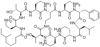
| SMILES | CC[C@H](C)CCCCC(=O)N[C@@H](CCN)C(=O)N[C@@H]([C@@H](C)O)C(=O)N[C@@H](CCN)C(=O)N[C@H]1CCNC(=O)[C@@H](NC(=O)[C@@H](NC(=O)[C@@H](NC(=O)[C@@H](NC(=O)[C@H](NC(=O)[C@@H](NC1=O)CCN)CC2=CC=CC=C2)CC(C)C)CCN)CCN)[C@@H](C)O |
| InChi Key | HFMDLUQUEXNBOP-UHFFFAOYSA-N |
| InChi Code | InChI=1S/C56H98N16O13.H2O4S/c1-7-32(4)13-11-12-16-44(75)63-36(17-23-57)51(80)72-46(34(6)74)56(85)68-39(20-26-60)48(77)67-41-22-28-62-55(84)45(33(5)73)71-52(81)40(21-27-61)65-47(76)37(18-24-58)66-53(82)42(29-31(2)3)69-54(83)43(30-35-14-9-8-10-15-35)70-49(78)38(19-25-59)64-50(41)79;1-5(2,3)4/h8-10,14-15,31-34,36-43,45-46,73-74H,7,11-13,16-30,57-61H2,1-6H3,(H,62,84)(H,63,75)(H,64,79)(H,65,76)(H,66,82)(H,67,77)(H,68,85)(H,69,83)(H,70,78)(H,71,81)(H,72,80);(H2,1,2,3,4) |
| Chemical Name | N-[4-amino-1-[[1-[[4-amino-1-oxo-1-[[6,9,18-tris(2-aminoethyl)-15-benzyl-3-(1-hydroxyethyl)-12-(2-methylpropyl)-2,5,8,11,14,17,20-heptaoxo-1,4,7,10,13,16,19-heptazacyclotricos-21-yl]amino]butan-2-yl]amino]-3-hydroxy-1-oxobutan-2-yl]amino]-1-oxobutan-2-yl]-6-methyloctanamide;sulfuric acid |
| Synonyms | 4135-11-9; UNII-B35S89JSRU; B35S89JSRU; (6S)-N-[(2S)-4-amino-1-[[(2S,3R)-1-[[(2S)-4-amino-1-oxo-1-[[(3S,6S,9S,12S,15R,18S,21S)-6,9,18-tris(2-aminoethyl)-15-benzyl-3-[(1R)-1-hydroxyethyl]-12-(2-methylpropyl)-2,5,8,11,14,17,20-heptaoxo-1,4,7,10,13,16,19-heptazacyclotricos-21-yl]amino]butan-2-yl]amino]-3-hydroxy-1-oxobutan-2-yl]amino]-1-oxobutan-2-yl]-6-methyloctanamide; CHEMBL2397513; BRN 0505906; L-THREONINE, N2-(6-METHYL-1-OXOOCTYL)-L-2,4-DIAMINOBUTANOYL-L-THREONYL-L-2,4-DIAMINOBUTANOYL-L-2,4-DIAMINOBUTANOYL-L-2,4-DIAMINOBUTANOYL-D-PHENYLALANYL-L-LEUCYL-L-2,4-DIAMINOBUTANOYL-L-2,4-DIAMINOBUTANOYL-, CYCLIC (10->4)-PEPTIDE; N2-(6-METHYL-1-OXOOCTYL)-L-2,4-DIAMINOBUTANOYL-L-THREONYL-L-2,4-DIAMINOBUTANOYL-L-2,4-DIAMINOBUTANOYL-L-2,4-DIAMINOBUTANOYL-D-PHENYLALANYL-L-LEUCYL-L-2,4-DIAMINOBUTANOYL-L-2,4-DIAMINOBUTANOYL-L-THREONINE CYCLIC (10->4)-PEPTIDE; |
| HS Tariff Code | 2934.99.9001 |
| Storage |
Powder-20°C 3 years 4°C 2 years In solvent -80°C 6 months -20°C 1 month Note: Please store this product in a sealed and protected environment (e.g. under nitrogen), avoid exposure to moisture and light. |
| Shipping Condition | Room temperature (This product is stable at ambient temperature for a few days during ordinary shipping and time spent in Customs) |
Biological Activity
| Targets | Polypeptide antibacterial |
| ln Vitro | Polymyxin B1 exhibits antibacterial activity against isolates of Pseudomonas aeruginosa ATCC 27853, Acinetobacter baumannii ATCC BAA 747, Klebsiella pneumoniae ATCC 13883, Pseudomonas aeruginosa 9019, Acinetobacter baumannii 1261, and Klebsiella pneumoniae VM9, with MIC values of 4 μg/mL, 2 μg/mL, 2 μg/mL, and 2 μg/mL, respectively [3]. Staphylococcus aureus, Escherichia coli, and yeast all exhibit significant inhibitions of protein synthesis when exposed to polymyxin B1 [4]. |
| ln Vivo | In an arterial model, the pharmacokinetics of Polymyxin B1 were investigated intravenously at a dose of 0.8 mg/kg. Compared to colistin A and colistin B, polymyxin B1 had a greater area under the concentration-time curve. Colistin A Colistin B: Its peptide binding rate is 82.3%, its elimination half-life is 79.5 minutes, and its AUC0-∞ is 365 mg. The clearance rate of polymyxin B1 is 2.39 mL/min/kg.min/L [5]. |
| Enzyme Assay |
Plasma Protein Binding [5] The plasma protein binding of the individual polymyxins was determined by ultracentrifugation with healthy drug-free rat plasma. Aliquots (200 μL) of each plasma sample were spiked with 5 mg/L of each individual polymyxin component. Plasma samples were incubated for 30 min at 37 °C. Samples were then ultracentrifuged at 215000g for 4 h at 37 °C. From the triplicate spiked plasma samples, two samples were subjected to buffer fraction analysis in which 50 μL of protein-free supernatant was removed. The final sample from the triplicate was subjected to complete plasma analysis in which 50 μL of the total resuspended content was removed. The polymyxin concentration in the supernatant and the resuspended samples was determined by LC-MS/MS. |
| Animal Protocol |
Pharmacokinetics [5] Sixteen rats were cannulated in the jugular vein for polymyxin administration and in the carotid artery for blood collection. Following cannulation, animals were allowed to recover for a day. Rats were divided into four groups (n = 4, in each group) and received a 0.8 mg/kg bolus dose of a polymyxin via the jugular vein cannula. Blood (200 μL) was collected prior to the administration of the individual polymyxin components and at 10, 20, 30, 60, 90, 120, 180, 240, and 360 min thereafter and centrifuged immediately (10000g, 10 min) at 4 °C. Urine was collected in chilling chambers from the metabolic cage prior to administration of the individual components and over the intervals of 0–6 h and 6–24 h thereafter. For the plasma and urine assay, calibration curves were constructed with concentrations of individual polymyxin components ranging from 0.1 to 8.0 mg/L. The intraday accuracy and reproducibility of individual polymyxin components were <15% at 0.3 mg/L and <10% at 6 mg/L. The pharmacokinetic data were analyzed using WinNonlin (the noncompartmental model). Urinary recovery was calculated via the following equation: Total polymyxin is calculated as follows: Concentration of polymyxin (μg/mL) × urine volume (mL, 0–24 h). Differences between the values of the pharmacokinetic parameters for the polymyxin components were evaluated using one-way ANOVA Kruskal–Wallis test. A P-value of <0.05 was considered significant. Random blood samples (specifically timed in relation to the dose administered) were obtained, which were allowed to clot, and serum was obtained by centrifugation. Timing and the number of sample obtained per patient were primarily determined by logistic convenience and as permitted by the clinical conditions of the patients. Serum samples were stored in −70 °C until drug assay. Polymyxin B1 (the major component in commercial polymyxin B) (Orwa et al., 2000) concentrations in the serum samples were determined by a validated methodology using liquid chromatography mass spectroscopy. The serum concentration profiles of all the patients (including the number of doses into therapy if blood samples were taken before the 14th dose of therapy, in case steady state had not been achieved) were analyzed by a single population pharmacokinetic analysis using the nonparametric adaptive grid (NPAG) program (Leary et al., 2001). A 1- or 2-compartment model with zero-order time-limited infusion into the central compartment and 1st-order elimination from the central compartment (and 1st-order transfer rates between central and peripheral compartments in the 2-compartment model) was fit to the data. The interday variation of the assay was used to determine the variance structural model by a quadratic relationship between the SDs and mean polymyxin B1 concentrations. Total observation variability was assumed to be proportional to the assay variance, and the proportionality constant was optimized after each iteration. The best-fit models were discriminated using the likelihood ratio test, adjusted for the difference in degrees of freedom. Protein binding of polymyxin B1 in human serum was determined in triplicate by comparing the peak area ratio of a known spiked sample of pooled human serum with and without ultrafiltration (Ultrafree-MC low-binding Durapore membrane 0.1-μm filter unit; Millipore, Bedford, MA) at 21 000 × g for 30 min at 20 and 37 °C (preincubated for 2 h). Nonspecific drug binding to the ultrafiltration membrane was investigated in triplicate using spiked samples in preincubated deproteinized serum matrix (at 37 °C for 2 h) and in water at 20 °C. [1] |
| ADME/Pharmacokinetics | Polymyxin B is increasingly used clinically for the treatment of multidrug-resistant Gram-negative infections, despite very limited understanding of its disposition in humans. The disposition of intravenous polymyxin B1 in 9 adult patients was characterized. Random blood samples (specifically timed in relation to the dose administered) were obtained, and the serum concentrations of polymyxin B1 were assayed using a validated methodology by liquid chromatography mass spectroscopy. The serum concentration profiles of all the patients were analyzed by a population pharmacokinetic analysis using the nonparametric adaptive grid program. The mean volume of distribution and elimination half-life were found to be 47.2 L and 13.6 h, respectively. This is the 1st case series to date in which the pharmacokinetics of polymyxin B1 after intravenous administration are described. The results of the series in conjunction with pharmacodynamic and susceptibility surveillance studies could facilitate an approach to the design of optimal dosing regimens. [1] |
| Toxicity/Toxicokinetics |
11228650 mouse LD50 intraperitoneal 19 mg/kg CRC Handbook of Antibiotic Compounds, Vols.1- , Berdy, J., Boca Raton, FL, CRC Press, 1980, 4(1)(334), 1980 11228650 mouse LD50 subcutaneous 80 mg/kg CRC Handbook of Antibiotic Compounds, Vols.1- , Berdy, J., Boca Raton, FL, CRC Press, 1980, 4(1)(334), 1980 11228650 mouse LD50 intravenous 1500 ug/kg CRC Handbook of Antibiotic Compounds, Vols.1- , Berdy, J., Boca Raton, FL, CRC Press, 1980, 4(1)(334), 1980 |
| References |
[1]. Pharmacokinetics of polymyxin B1 in patients with multidrug-resistant Gram-negative bacterial infections. Diagn Microbiol Infect Dis. 2008 Feb;60(2):163-7. [2]. Interaction of the antimicrobial peptide polymyxin B1 with both membranes of E. coli: a molecular dynamics study. PLoS Comput Biol. 2015 Apr 17;11(4):e1004180. [3]. In vitro potency of various polymyxin B components. Antimicrob Agents Chemother. 2011 Sep;55(9):4490-1. [4]. Compounds affecting membranes that inhibit protein synthesis in yeast. Antimicrob Agents Chemother. 1979 Dec;16(6):750-6. [5]. Pharmacokinetics of the Individual Major Components of Polymyxin B and Colistin in Rats. J Nat Prod. 2017 Jan 27;80(1):225-229. |
| Additional Infomation |
Polymyxin B sulphate is a cyclic peptide. A mixture of polymyxins B1 and B2, obtained from BACILLUS POLYMYXA strains. They are basic polypeptides of about eight amino acids and have cationic detergent action on cell membranes. Polymyxin B is used for treatment of infections with gram-negative bacteria, but may be neurotoxic and nephrotoxic. See also: Polymyxin B Sulfate (annotation moved to). Antimicrobial peptides are small, cationic proteins that can induce lysis of bacterial cells through interaction with their membranes. Different mechanisms for cell lysis have been proposed, but these models tend to neglect the role of the chemical composition of the membrane, which differs between bacterial species and can be heterogeneous even within a single cell. Moreover, the cell envelope of Gram-negative bacteria such as E. coli contains two membranes with differing compositions. To this end, we report the first molecular dynamics simulation study of the interaction of the antimicrobial peptide, polymyxin B1 with complex models of both the inner and outer membranes of E. coli. The results of >16 microseconds of simulation predict that polymyxin B1 is likely to interact with the membranes via distinct mechanisms. The lipopeptides aggregate in the lipopolysaccharide headgroup region of the outer membrane with limited tendency for insertion within the lipid A tails. In contrast, the lipopeptides readily insert into the inner membrane core, and the concomitant increased hydration may be responsible for bilayer destabilization and antimicrobial function. Given the urgent need to develop novel, potent antibiotics, the results presented here reveal key mechanistic details that may be exploited for future rational drug development. [2] The pharmacokinetics of polymyxin B1, polymyxin B2, colistin A, and colistin B were investigated in a rat model following intravenous administration (0.8 mg/kg) of each individual component. Plasma and urine concentrations were determined by LC-MS/MS, and plasma protein binding was measured by ultracentrifugation. Total and unbound pharmacokinetic parameters for each component were calculated using noncompartmental analysis. All of the polymyxin components had a similar clearance, volume of distribution, elimination half-life, and urinary recovery. The area under the concentration–time curve for polymyxins B1 and B2 was greater than those of colistins A and B. Colistin A (56.6 ± 9.25%) and colistin B (41.7 ± 12.4%) displayed lower plasma protein binding in rat plasma compared to polymyxin B1 (82.3 ± 4.30%) and polymyxin B2 (68.4 ± 3.50%). These differences in plasma protein binding potentially equate to significant differences in unbound pharmacokinetics, highlighting the need for more stringent standardization of the composition of commercial products currently available for clinical use. [5] |
Solubility Data
| Solubility (In Vitro) | May dissolve in DMSO (in most cases), if not, try other solvents such as H2O, Ethanol, or DMF with a minute amount of products to avoid loss of samples |
| Solubility (In Vivo) |
Note: Listed below are some common formulations that may be used to formulate products with low water solubility (e.g. < 1 mg/mL), you may test these formulations using a minute amount of products to avoid loss of samples. Injection Formulations (e.g. IP/IV/IM/SC) Injection Formulation 1: DMSO : Tween 80: Saline = 10 : 5 : 85 (i.e. 100 μL DMSO stock solution → 50 μL Tween 80 → 850 μL Saline) *Preparation of saline: Dissolve 0.9 g of sodium chloride in 100 mL ddH ₂ O to obtain a clear solution. Injection Formulation 2: DMSO : PEG300 :Tween 80 : Saline = 10 : 40 : 5 : 45 (i.e. 100 μL DMSO → 400 μLPEG300 → 50 μL Tween 80 → 450 μL Saline) Injection Formulation 3: DMSO : Corn oil = 10 : 90 (i.e. 100 μL DMSO → 900 μL Corn oil) Example: Take the Injection Formulation 3 (DMSO : Corn oil = 10 : 90) as an example, if 1 mL of 2.5 mg/mL working solution is to be prepared, you can take 100 μL 25 mg/mL DMSO stock solution and add to 900 μL corn oil, mix well to obtain a clear or suspension solution (2.5 mg/mL, ready for use in animals). Injection Formulation 4: DMSO : 20% SBE-β-CD in saline = 10 : 90 [i.e. 100 μL DMSO → 900 μL (20% SBE-β-CD in saline)] *Preparation of 20% SBE-β-CD in Saline (4°C,1 week): Dissolve 2 g SBE-β-CD in 10 mL saline to obtain a clear solution. Injection Formulation 5: 2-Hydroxypropyl-β-cyclodextrin : Saline = 50 : 50 (i.e. 500 μL 2-Hydroxypropyl-β-cyclodextrin → 500 μL Saline) Injection Formulation 6: DMSO : PEG300 : castor oil : Saline = 5 : 10 : 20 : 65 (i.e. 50 μL DMSO → 100 μLPEG300 → 200 μL castor oil → 650 μL Saline) Injection Formulation 7: Ethanol : Cremophor : Saline = 10: 10 : 80 (i.e. 100 μL Ethanol → 100 μL Cremophor → 800 μL Saline) Injection Formulation 8: Dissolve in Cremophor/Ethanol (50 : 50), then diluted by Saline Injection Formulation 9: EtOH : Corn oil = 10 : 90 (i.e. 100 μL EtOH → 900 μL Corn oil) Injection Formulation 10: EtOH : PEG300:Tween 80 : Saline = 10 : 40 : 5 : 45 (i.e. 100 μL EtOH → 400 μLPEG300 → 50 μL Tween 80 → 450 μL Saline) Oral Formulations Oral Formulation 1: Suspend in 0.5% CMC Na (carboxymethylcellulose sodium) Oral Formulation 2: Suspend in 0.5% Carboxymethyl cellulose Example: Take the Oral Formulation 1 (Suspend in 0.5% CMC Na) as an example, if 100 mL of 2.5 mg/mL working solution is to be prepared, you can first prepare 0.5% CMC Na solution by measuring 0.5 g CMC Na and dissolve it in 100 mL ddH2O to obtain a clear solution; then add 250 mg of the product to 100 mL 0.5% CMC Na solution, to make the suspension solution (2.5 mg/mL, ready for use in animals). Oral Formulation 3: Dissolved in PEG400 Oral Formulation 4: Suspend in 0.2% Carboxymethyl cellulose Oral Formulation 5: Dissolve in 0.25% Tween 80 and 0.5% Carboxymethyl cellulose Oral Formulation 6: Mixing with food powders Note: Please be aware that the above formulations are for reference only. InvivoChem strongly recommends customers to read literature methods/protocols carefully before determining which formulation you should use for in vivo studies, as different compounds have different solubility properties and have to be formulated differently. (Please use freshly prepared in vivo formulations for optimal results.) |
| Preparing Stock Solutions | 1 mg | 5 mg | 10 mg | |
| 1 mM | 0.8309 mL | 4.1546 mL | 8.3092 mL | |
| 5 mM | 0.1662 mL | 0.8309 mL | 1.6618 mL | |
| 10 mM | 0.0831 mL | 0.4155 mL | 0.8309 mL |