Pentixafor is a ligand/intermediate used for the preparation of gallium Ga 68-pentixafor, which is a ligand for chemokine receptor C-X-C chemokine receptor type 4 (CXCR4), and a radioconjugate consisting a cyclic pentapeptide analog of stromal-cell derived factor-1 (SDF-1 or CXCL12).
Physicochemical Properties
| Molecular Formula | C60H80N14O14 |
| Molecular Weight | 1221.36261367798 |
| Exact Mass | 1220.597 |
| Elemental Analysis | C, 59.00; H, 6.60; N, 16.06; O, 18.34 |
| CAS # | 1341207-62-2 |
| Related CAS # | 1341207-62-2;1342253-77-3 (Gallium);1345698-96-5 (Ga-68); |
| PubChem CID | 54575322 |
| Appearance | White to off-white solid powder |
| LogP | -6.3 |
| Hydrogen Bond Donor Count | 12 |
| Hydrogen Bond Acceptor Count | 19 |
| Rotatable Bond Count | 23 |
| Heavy Atom Count | 88 |
| Complexity | 2330 |
| Defined Atom Stereocenter Count | 4 |
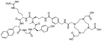
| SMILES | CN1[C@@H](C(=O)N[C@H](C(=O)N[C@H](C(=O)NCC(=O)N[C@@H](C1=O)CC2=CC=C(C=C2)O)CC3=CC4=CC=CC=C4C=C3)CCCN=C(N)N)CCCNC(=O)C5=CC=C(C=C5)CNC(=O)CN6CCN(CCN(CCN(CC6)CC(=O)O)CC(=O)O)CC(=O)O |
| InChi Key | OSUJVKAXNLHVRB-HUMWUIFSSA-N |
| InChi Code | nChI=1S/C60H80N14O14/c1-70-49(9-5-20-63-55(84)43-16-10-40(11-17-43)33-65-51(77)35-71-22-24-72(36-52(78)79)26-28-74(38-54(82)83)29-27-73(25-23-71)37-53(80)81)58(87)68-46(8-4-21-64-60(61)62)57(86)69-47(32-41-12-15-42-6-2-3-7-44(42)30-41)56(85)66-34-50(76)67-48(59(70)88)31-39-13-18-45(75)19-14-39/h2-3,6-7,10-19,30,46-49,75H,4-5,8-9,20-29,31-38H2,1H3,(H,63,84)(H,65,77)(H,66,85)(H,67,76)(H,68,87)(H,69,86)(H,78,79)(H,80,81)(H,82,83)(H4,61,62,64)/t46-,47-,48+,49+/m0/s1 SMILES Code: OC1=CC=C(C[C@@H](NC(CNC([C@H](CC2=CC3=C(C=CC=C3)C=C2)NC4=O)=O)=O)C(N(C)[C@H](CCCNC(C5=CC=C(CNC(CN6CCN(CC(O)=O)CCN(CC(O)=O)CCN(CC(O)=O)CC6)=O)C=C5)=O)C(N[C@H]4CCCNC(N)=N)=O)=O)C=C1 |
| Chemical Name | 2,2',2''-(10-(2-((4-((3-((2R,5S,8S,14R)-5-(3-guanidinopropyl)-14-(4-hydroxybenzyl)-1-methyl-8-(naphthalen-2-ylmethyl)-3,6,9,12,15-pentaoxo-1,4,7,10,13-pentaazacyclopentadecan-2-yl)propyl)carbamoyl)benzyl)amino)-2-oxoethyl)-1,4,7,10-tetraazacyclododecane-1,4,7-triyl)triacetic acid |
| Synonyms | CPCR 4-2; CPCR4-2; CPCR-4-2; CPCR42; CPCR4-2; TOZ93UY3AX; UNII-TOZ93UY3AX; 2-[4,7-bis(carboxymethyl)-10-[2-[[4-[3-[(2R,5S,8S,14R)-5-[3-(diaminomethylideneamino)propyl]-14-[(4-hydroxyphenyl)methyl]-1-methyl-8-(naphthalen-2-ylmethyl)-3,6,9,12,15-pentaoxo-1,4,7,10,13-pentazacyclopentadec-2-yl]propylcarbamoyl]phenyl]methylamino]-2-oxoethyl]-1,4,7,10-tetrazacyclododec-1-yl]acetic acid; (68GA)PENTIXAFOR; BOCLATIXAFORTIDE; Ligand of gallium Ga 68-pentixafor; CPCR-42; CPCR 42; Pentixafor |
| HS Tariff Code | 2934.99.9001 |
| Storage |
Powder-20°C 3 years 4°C 2 years In solvent -80°C 6 months -20°C 1 month Note: Please store this product in a sealed and protected environment, away from moisture and light. |
| Shipping Condition | Room temperature (This product is stable at ambient temperature for a few days during ordinary shipping and time spent in Customs) |
Biological Activity
| Targets | CXCR4/chemokine receptor C-X-C chemokine receptor type 4 |
| ln Vitro | A growing body of literature reports on the upregulation of C-X-C motif chemokine receptor 4 (CXCR4) in a variety of cancer entities, rendering this receptor as suitable target for molecular imaging and endoradiotherapy in a theranostic setting. For instance, the CXCR4-targeting positron emission tomography (PET) agent [68 Ga]PentixaFor has been proven useful for a comprehensive assessment of the current status quo of solid tumors, including adrenocortical carcinoma or small-cell lung cancer. In addition, [68 Ga]PentixaFor has also provided an excellent readout for hematological malignancies, such as multiple myeloma, marginal zone lymphoma, or mantle cell lymphoma. PET-based quantification of the CXCR4 capacities in vivo allows for selecting candidates that would be suitable for treatment using the theranostic equivalent [177Lu]/[90Y]PentixaTher. This CXCR4-directed theranostic concept has been used as a conditioning regimen prior to hematopoietic stem cell transplantation and to achieve sufficient anti-lymphoma/-tumor activity in particular for malignant tissues that are highly sensitive to radiation, such as the hematological system. Increasing the safety margin, pretherapeutic dosimetry is routinely performed to determine the optimal activity to enhance therapeutic efficacy and to reduce off-target adverse events[1]. |
| ln Vivo |
In mice affected with either CXCR4( −) or CXCR4( +) leukemia xenografts, an increased [68Ga]PentixaFor signal was observed in the latter animals [1]. After intravenous administration, [177Lu]PentixaTher binds to plasma proteins with high metabolic stability, and only a small fraction of about 4% is attached to leukocytes and platelets via CXCR4 binding. Scintigraphically detectable activity accumulations are found in kidney, liver, spleen, and bone marrow, as well as in CXCR4-expressing malignant tissues. An example of measured time functions of activity retention in organs and tissues in a patient with MM is shown in Fig. 5. The figure, like the results summarized below unless otherwise specified, is taken from a recently published study on [177Lu]PentixaTher biokinetics and dosimetry.[1] |
| Enzyme Assay |
Biokinetics and pretherapeutic dosimetry[1] After intravenous administration, [177Lu]PentixaTher binds to plasma proteins with high metabolic stability, and only a small fraction of about 4% is attached to leukocytes and platelets via CXCR4 binding. Scintigraphically detectable activity accumulations are found in kidney, liver, spleen, and bone marrow, as well as in CXCR4-expressing malignant tissues. An example of measured time functions of activity retention in organs and tissues in a patient with MM is shown in Fig. 5. The figure, like the results summarized below unless otherwise specified, is taken from a recently published study on [177Lu]PentixaTher biokinetics and dosimetry. |
| Animal Protocol | Aiming to provide a roadmap among a broad spectrum of neoplasms, a recent bicentric study assessed [68Ga]PentixaFor uptake and image contrast among the largest cohort of subjects imaged with CXCR4-directed PET to date, thereby determining the most relevant clinical applications. Investigating 690 patients affected with various solid tumors and hematological neoplasms scheduled for 777 scans, 68.9% demonstrated uptake in sites of disease The highest tracer uptake was recorded in MM (maximum SUV > 12). The second highest uptake was then found in ACC, MCL, adrenocortical adenoma, and SCL. Osteosarcoma, bladder cancer, head and neck cancer, and Ewing sarcoma, on the other hand, exhibited the lowest average SUV (< 6; Fig. 4A). Comparable findings were recorded for target-to-background ratio (TBR), thereby reflecting image contrast. Again, the highest TBR was found in advanced blood cancers, including MM, MCL, and acute lymphoblastoid leukemia (Fig. 4B). Moreover, lower specific activity is characterized by higher amounts of cold mass, thereby having a relevant impact on image interpretation. The authors did not record any relevant significant associations with semiquantitative parameters and specific activity, supporting the hypothesis that read-out capabilities are not hampered, regardless of the amount of specific activities[1]. |
| ADME/Pharmacokinetics |
The total body 177Lu activity typically decays bi-exponentially. About half of the activity is eliminated with a median effective half-life of about 10 h mainly by renal excretion; the remainder decays with a mean effective half-life of about 4 days. Activity concentration in blood typically shows three components with about 10%, 2.5%, and 0.2% of the administered activity per liter of blood decaying with half-lives of 0.23 h, 7 h, and 40 h, respectively. [177Lu]PentixaTher accumulates in the bone marrow and remains there with a half-life of several days, making the bone marrow the critical organ where acute toxicity is foremost expected. The calculated specific bone marrow doses were heterogeneous, ranging from 0.14 to 2.3 (median value, 0.5) Gy/GBq 177Lu. Given high individual variability and the uncertainties of bone marrow dosimetry, therapeutic use of PentixaTher may be confined to myeloablative therapies. However, it must be considered in myeloablative treatment that the long residence time of the activity in the bone marrow requires a long decay time before a stem cell transplantation can be safely performed. Therefore, in order to reduce the duration of the phase of aplasia and the associated risk of threatening complications, therapy is usually performed with the nuclide 90Y instead of 177Lu[1]. |
| Toxicity/Toxicokinetics |
Toxicity profile[1] Investigating the safety profile, 22 patients with advanced blood cancer treated with [177Lu] or [90Y]PentixaTher and subsequent chemotherapy followed by HSCT were investigated. As expected, all patients developed cytopenia (including hemoglobin, leukocytes, granulocytes, and platelets; Fig. 7A). One patient developed tumor lysis syndrome, followed by grade 3 acute kidney failure, while all other adverse effects were manageable and did not cause any delay for further treatment. In this regard, time interval between CXCR4 ERT and conditioning therapy was significantly longer with [177Lu]PentixaTher, which can be explained by the longer half-life of 6.7 days when compared to [90Y]PentixaTher (2.7 days; Fig. 7B). The ongoing COLPRIT trial is a prospective phase I/II study which will further elucidate the therapeutic efficacy and safety of this theranostic strategy in patients with advanced blood cancer (Eudra‐CT 2015‐001817‐28). |
| References |
[1]. Eur J Nucl Med Mol Imaging. 2022 Oct;49(12):4133-4144. [2]. J Nucl Cardiol. 2022 Apr;29(2):503-505. |
| Additional Infomation | CXCR4 is upregulated on various cancer cells, rendering this receptor as a potential target for tumor read-out and treatment strategies. The CXCR4-targeted PET agent [68Ga]PentixaFor has been successfully applied to patients with solid and advanced blood cancers, demonstrating substantially increased radiotracer accumulation in ACC, SCLC, MM, MZL, MCL, or gastric MALT. In addition to assessment of widespread disease, such a functional imaging approach allows to assess the capacities of the target in-vivo. Thus, quantification of [68Ga]PentixaFor accumulation may then allow to estimate the efficacy of non-radioactive CXCR4 inhibitory treatments (e.g., with anti-human CXCR4 IgG monoclonal antibodies for MM patients) or to identify patients that would be eligible for treatment with hot CXCR4-directed theranostic radiotracers, such as [177Lu]/[90Y]PentixaTher. The latter concept has already been applied to hematological malignancies known to be sensitive to radiation, e.g., in advanced MM, ALL, or diffuse large B cell lymphoma. In this context, pretherapeutic dosimetry can determine the appropriate amount of activity to achieve anti-tumor effects and to minimize off-target effects. CXCR4 ERT also caused desired bone marrow ablation and has therefore been incorporated in the therapeutic algorithm of advanced blood cancer patients (allogenic/autologous HSCT following CXCR4 ERT along with successful engraftment). Therapeutic efficacy of those treatment regimens led to remarkable outcome benefits in those heavily pretreated patients. Given substantial high doses in the tumor, some patients experienced tumor lysis syndrome and thus, those individuals should be closely monitored[1]. |
Solubility Data
| Solubility (In Vitro) |
DMSO: ~100 mg/mL (81.9 mM) Methanol: ≥ 125 mg/mL (102.3 mM) |
| Solubility (In Vivo) |
Note: Listed below are some common formulations that may be used to formulate products with low water solubility (e.g. < 1 mg/mL), you may test these formulations using a minute amount of products to avoid loss of samples. Injection Formulations (e.g. IP/IV/IM/SC) Injection Formulation 1: DMSO : Tween 80: Saline = 10 : 5 : 85 (i.e. 100 μL DMSO stock solution → 50 μL Tween 80 → 850 μL Saline) *Preparation of saline: Dissolve 0.9 g of sodium chloride in 100 mL ddH ₂ O to obtain a clear solution. Injection Formulation 2: DMSO : PEG300 :Tween 80 : Saline = 10 : 40 : 5 : 45 (i.e. 100 μL DMSO → 400 μLPEG300 → 50 μL Tween 80 → 450 μL Saline) Injection Formulation 3: DMSO : Corn oil = 10 : 90 (i.e. 100 μL DMSO → 900 μL Corn oil) Example: Take the Injection Formulation 3 (DMSO : Corn oil = 10 : 90) as an example, if 1 mL of 2.5 mg/mL working solution is to be prepared, you can take 100 μL 25 mg/mL DMSO stock solution and add to 900 μL corn oil, mix well to obtain a clear or suspension solution (2.5 mg/mL, ready for use in animals). Injection Formulation 4: DMSO : 20% SBE-β-CD in saline = 10 : 90 [i.e. 100 μL DMSO → 900 μL (20% SBE-β-CD in saline)] *Preparation of 20% SBE-β-CD in Saline (4°C,1 week): Dissolve 2 g SBE-β-CD in 10 mL saline to obtain a clear solution. Injection Formulation 5: 2-Hydroxypropyl-β-cyclodextrin : Saline = 50 : 50 (i.e. 500 μL 2-Hydroxypropyl-β-cyclodextrin → 500 μL Saline) Injection Formulation 6: DMSO : PEG300 : castor oil : Saline = 5 : 10 : 20 : 65 (i.e. 50 μL DMSO → 100 μLPEG300 → 200 μL castor oil → 650 μL Saline) Injection Formulation 7: Ethanol : Cremophor : Saline = 10: 10 : 80 (i.e. 100 μL Ethanol → 100 μL Cremophor → 800 μL Saline) Injection Formulation 8: Dissolve in Cremophor/Ethanol (50 : 50), then diluted by Saline Injection Formulation 9: EtOH : Corn oil = 10 : 90 (i.e. 100 μL EtOH → 900 μL Corn oil) Injection Formulation 10: EtOH : PEG300:Tween 80 : Saline = 10 : 40 : 5 : 45 (i.e. 100 μL EtOH → 400 μLPEG300 → 50 μL Tween 80 → 450 μL Saline) Oral Formulations Oral Formulation 1: Suspend in 0.5% CMC Na (carboxymethylcellulose sodium) Oral Formulation 2: Suspend in 0.5% Carboxymethyl cellulose Example: Take the Oral Formulation 1 (Suspend in 0.5% CMC Na) as an example, if 100 mL of 2.5 mg/mL working solution is to be prepared, you can first prepare 0.5% CMC Na solution by measuring 0.5 g CMC Na and dissolve it in 100 mL ddH2O to obtain a clear solution; then add 250 mg of the product to 100 mL 0.5% CMC Na solution, to make the suspension solution (2.5 mg/mL, ready for use in animals). Oral Formulation 3: Dissolved in PEG400 Oral Formulation 4: Suspend in 0.2% Carboxymethyl cellulose Oral Formulation 5: Dissolve in 0.25% Tween 80 and 0.5% Carboxymethyl cellulose Oral Formulation 6: Mixing with food powders Note: Please be aware that the above formulations are for reference only. InvivoChem strongly recommends customers to read literature methods/protocols carefully before determining which formulation you should use for in vivo studies, as different compounds have different solubility properties and have to be formulated differently. (Please use freshly prepared in vivo formulations for optimal results.) |
| Preparing Stock Solutions | 1 mg | 5 mg | 10 mg | |
| 1 mM | 0.8188 mL | 4.0938 mL | 8.1876 mL | |
| 5 mM | 0.1638 mL | 0.8188 mL | 1.6375 mL | |
| 10 mM | 0.0819 mL | 0.4094 mL | 0.8188 mL |