Physicochemical Properties
| Molecular Formula | C16H15N3O2S |
| Molecular Weight | 313.37 |
| Exact Mass | 313.09 |
| Elemental Analysis | C, 61.32; H, 4.82; N, 13.41; O, 10.21; S, 10.23 |
| CAS # | 2765218-56-0 |
| PubChem CID | 155387479 |
| Appearance | White to off-white solid |
| LogP | 3 |
| Hydrogen Bond Donor Count | 1 |
| Hydrogen Bond Acceptor Count | 5 |
| Rotatable Bond Count | 6 |
| Heavy Atom Count | 22 |
| Complexity | 363 |
| Defined Atom Stereocenter Count | 0 |
| InChi Key | UFASECSYKSMYET-UHFFFAOYSA-N |
| InChi Code | InChI=1S/C16H15N3O2S/c20-16(18-8-4-7-15-17-9-10-22-15)13-11-14(21-19-13)12-5-2-1-3-6-12/h1-3,5-6,9-11H,4,7-8H2,(H,18,20) |
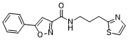
| Chemical Name | 5-phenyl-N-(3-(thiazol-2-yl)propyl)isoxazole-3-carboxamide |
| Synonyms | PY-60; PY 60; YAP activator PY-60; PY60; SV956D8WK8; PY-60?; PY60; |
| HS Tariff Code | 2934.99.9001 |
| Storage |
Powder-20°C 3 years 4°C 2 years In solvent -80°C 6 months -20°C 1 month |
| Shipping Condition | Room temperature (This product is stable at ambient temperature for a few days during ordinary shipping and time spent in Customs) |
Biological Activity
| Targets | ANXA2 (Kd = 1.4 µM) |
| ln Vitro | PY-60 activates YAP by targeting ANXA2[1]. PY-60 is a thiazole-substituted gel that activated 293A-TEAD-LUC cells' luciferase activity at a dosage. When cells were injected at high densities (EC50= 1.5 and 1.6 μM, respectively), serum was not present. Additionally, PY-60 treatment caused the nuclear localization of YAP in response to increasing cell density and promoted the binding of TEAD and YAP proteins in cells. PY-60 activates proliferative YAP in animals to regulate cell contraction program and can induce apoptosis through proliferation[1]. PY-60 also significantly increases the levels of YAP-controlled calibrators (i.e., ANRKD1, CYR61, and CTGF) in 293A cells and other human cells (i.e., MCF10A, HEK293T, H69 levels, and HaCaT). PY-60 competes with ANXA2 for the binding of phosphoinositides by releasing the ANXA2-YAP complex from the membrane [1]. |
| ln Vivo | Hematoxylin and eosin and anti-K14 histology staining demonstrated a considerable growth of duct cells and K14 precursors when PY-60 (10 μM) was applied topically to the skin surface of wild-type puncta C57BL/6 mice at 10 days. Because PY-60 production increases the number of epidermal cells per unit length of skin, tip thickness is approximately doubled [1]. |
| Enzyme Assay |
In vitro binding experiments [1] Recombinant ANXA2 was generated by first subcloning a codon-optimized gBlock (IDT) encoding a C-terminally hexahistidine-tagged ANXA2 (NM_001002858) into the pET-22b expression vector. After transformation into BL21 bacteria, protein was allowed to express for 24 h at 30 °C with shaking. Recombinant ANXA2 was first purified by standard batchwise nickel-nitrilotriacetic acid-based purification according to the supplier’s recommendations (Qiagen), and then separated by sizing column using standard techniques into storage buffer (20 mM Tris, 137 mM NaCl and 10% glycerol pH 8.0). Crosslinking reactions were performed by the addition of 1 µg of recombinant ANXA2 to 100 µl of 1× PBS (250 nM ANXA2) and PY-PAP such that its final concentration was as indicated and that the final concentration of DMSO was 1% (vol/vol). Samples were incubated at 37 °C for 1 h in PCR strips and then subjected to UV-B-based crosslinking (365 nm) using a Stratalinker UV instrument for 10 min. Next, 10 µl of concentrated click reaction master mixes with rhodamine azide was added as above and reactions were incubated for 1 h at room temperature. Reactions were then separated using SDS–PAGE (100 ng of recombinant protein per well, 15-well gels, 4–12% Bis-tris BOLT gels and visualized using a ChemiDoc instrument. Parallel SDS–PAGE gels of the above reactions were silver stained using the Pierce Silver Stain Kit to confirm equal recombinant protein loading. |
| Cell Assay | Primary mouse keratinocytes were isolated from C57BL/6 neonatal animals at 2 days of age and then cultured on a mitomycin-C-treated 3T3 feeder layer in FAD complete medium. DMSO vehicle control or compound PY-60 (10 µM) was added to the medium during initial plating. For secondary replating assays, primary keratinocytes were passaged onto a new 3T3 cell feeder layer. After 9 days of growth, keratinocytes were fixed with paraformaldehyde and then stained with 1% rhodamine B [1]. |
| Animal Protocol | C57BL/6 WT mice (8 weeks of age) were treated once per day (topically, on the skin) for ten consecutive days with either vehicle only (acetone) or PY-60 (10 mg ml–1). The epidermis was assessed for thickness using H&E staining. Anti-K14 and anti-K167 immunofluorescent staining were used to assess cell expansion. In all models, animals were randomly assorted into groups of equal mean body weight before topical dosing. No animals were excluded from the analysis. The investigators were not blinded during group allocation or dosing. All animal studies were perfomed in compliance with protocols approved by the Boston Children’s Hospital Institutional Animal Care and Use Committee [1]. |
| References |
[1]. YAP-dependent proliferation by a small molecule targeting annexin A2. Nat Chem Biol. 2021;17(7):767-775. |
| Additional Infomation | The transcriptional coactivator Yes-associated protein 1 (YAP) orchestrates a proproliferative transcriptional program that controls the fate of somatic stem cells and the regenerative responses of certain tissues. As such, agents that activate YAP may hold therapeutic potential in disease states exacerbated by insufficient proliferative repair. Here we report the discovery of a small molecule, termed PY-60, which robustly activates YAP transcriptional activity in vitro and promotes YAP-dependent expansion of epidermal keratinocytes in mouse following topical drug administration. Chemical proteomics revealed the relevant target of PY-60 to be annexin A2 (ANXA2), a protein that directly associates with YAP at the cell membrane in response to increased cell density. PY-60 treatment liberates ANXA2 from the membrane, ultimately promoting a phosphatase-bound, nonphosphorylated and transcriptionally active form of YAP. This work reveals ANXA2 as a previously undescribed, druggable component of the Hippo pathway and suggests a mechanistic rationale to promote regenerative repair in disease.[1] |
Solubility Data
| Solubility (In Vitro) | DMSO : ~100 mg/mL (~319.11 mM) |
| Solubility (In Vivo) |
Solubility in Formulation 1: ≥ 2.5 mg/mL (7.98 mM) (saturation unknown) in 10% DMSO + 40% PEG300 +5% Tween-80 + 45% Saline (add these co-solvents sequentially from left to right, and one by one), clear solution. For example, if 1 mL of working solution is to be prepared, you can add 100 μL of 25.0 mg/mL clear DMSO stock solution to 400 μL PEG300 and mix evenly; then add 50 μL Tween-80 + to the above solution and mix evenly; then add 450 μL normal saline to adjust the volume to 1 mL. Preparation of saline: Dissolve 0.9 g of sodium chloride in 100 mL ddH₂ O to obtain a clear solution. (Please use freshly prepared in vivo formulations for optimal results.) |
| Preparing Stock Solutions | 1 mg | 5 mg | 10 mg | |
| 1 mM | 3.1911 mL | 15.9556 mL | 31.9112 mL | |
| 5 mM | 0.6382 mL | 3.1911 mL | 6.3822 mL | |
| 10 mM | 0.3191 mL | 1.5956 mL | 3.1911 mL |