PI3Kδ-IN-1 is a novel, potent, selective, and efficacious PI3Kδ inhibitor with an IC50 of 1.7 nM. PI3Kδ plays an important role controlling immune cell function and has therefore been identified as a potential target for the treatment of immunological disorders. PI3Kδ-IN-1 improved Caco-2 permeability, reduced Caco-2 efflux, reduced hERG PC activity, and increased selectivity profile while maintaining potency in the CD69 hWB assay. The optimization of the aryl substitution then identified a 4'-CN group that improved the human/rodent correlation in microsomal metabolic stability. PI3Kδ-IN-1 is very potent in PK/PD assays and highly efficacious in a mouse collagen-induced arthritis model.
Physicochemical Properties
| Molecular Formula | C22H20F3N7O2 |
| Molecular Weight | 471.43511390686 |
| Exact Mass | 471.16 |
| Elemental Analysis | C, 56.05; H, 4.28; F, 12.09; N, 20.80; O, 6.79 |
| CAS # | 1911564-39-0 |
| PubChem CID | 132220479 |
| Appearance | White to off-white solid powder |
| LogP | 1.6 |
| Hydrogen Bond Donor Count | 1 |
| Hydrogen Bond Acceptor Count | 9 |
| Rotatable Bond Count | 2 |
| Heavy Atom Count | 34 |
| Complexity | 872 |
| Defined Atom Stereocenter Count | 0 |
| InChi Key | ZDXIDBRGTYDHBQ-UHFFFAOYSA-N |
| InChi Code | InChI=1S/C22H20F3N7O2/c1-12(33)31-7-6-30(20(34)21(31,2)3)16-8-13(4-5-14(16)10-26)17-9-15(22(23,24)25)18-19(27)28-11-29-32(17)18/h4-5,8-9,11H,6-7H2,1-3H3,(H2,27,28,29) |
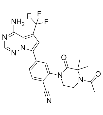
| Chemical Name | 2-(4-acetyl-3,3-dimethyl-2-oxopiperazin-1-yl)-4-[4-amino-5-(trifluoromethyl)pyrrolo[2,1-f][1,2,4]triazin-7-yl]benzonitrile |
| Synonyms | PI3Kδ-IN-1 |
| HS Tariff Code | 2934.99.9001 |
| Storage |
Powder-20°C 3 years 4°C 2 years In solvent -80°C 6 months -20°C 1 month |
| Shipping Condition | Room temperature (This product is stable at ambient temperature for a few days during ordinary shipping and time spent in Customs) |
Biological Activity
| Targets |
Phosphatidylinositol 3-kinase δ (PI3Kδ) (ADP-Glo IC50 = 1.7 ± 0.9 nM; HTRF IC50 < 0.1 nM)[1] PI3Kγ (ADP-Glo IC50 = 170 ± 70 nM; ~100-fold selectivity over PI3Kδ)[1] PI3Kα (ADP-Glo IC50 = 1200 ± 500 nM; ~700-fold selectivity over PI3Kδ)[1] PI3Kβ (ADP-Glo IC50 > 5000 nM)[1] |
| ln Vitro |
Comparing PI3Kδ-IN-1 (compound 52) to other PI3K isoforms, it is more than 100 times more selective. With a selectivity over 660 times that of MNK1 and other reagents in the HTRF experiment, kinome selectivity is likewise quite good. Notably, PI3Kδ-IN-1 exhibits good permeability features and enhanced human/rodent in vitro stability correlations [1]. PI3Kδ-IN-1 shows potent inhibitory activity in human whole blood (hWB) functional assays. In the CD69 hWB assay, the IC50 is 35 ± 20 nM, and in the IFNγ hWB assay, the IC50 is 35 ± 15 nM, demonstrating its ability to inhibit PI3Kδ-mediated immune cell activation ex vivo.[1] In a mouse basophil ex vivo pharmacodynamic assay, PI3Kδ-IN-1 demonstrated dose-dependent inhibition of anti-mIgE-induced CD63+ cell expression, with an EC50 of 2.0 ± 0.8 nM and an EC90 of 9 ± 5 nM. This activity is consistent with its potency in a mouse whole blood (mWB) assay (IC50 = 5.1 ± 1.8 nM).[1] The compound exhibits excellent kinase selectivity. In an HTRF binding assay against 235 kinases, including phosphatidylinositol 3-kinase-related kinases, it showed >100-fold selectivity over other PI3K isoforms and >660-fold selectivity over kinases like MNK1 (IC50 = 66 ± 24 nM).[1] |
| ln Vivo |
PI3Kδ-IN-1 (2, 5 mg/kg, orally administered for 42 days) significantly reduced mouse paw edema. After 24 hours in mice, PI3Kδ-IN-1 has an EC50 of 10 nM (ED50 ∼1.25 mg/kg) [1]. PI3Kδ-IN-1 demonstrated significant efficacy in a mouse KLH-induced serum IgG production model. When administered orally twice daily beginning on the day of immunization, it dose-dependently suppressed anti-KLH IgG antibody titers measured on day 13.[1] In a mouse collagen-induced arthritis (CIA) model, oral administration of PI3Kδ-IN-1 (compound 52) twice daily from day 1 post-immunization significantly reduced the clinical arthritis score (sum of scores for all four paws) compared to the vehicle-treated group, indicating potent in vivo efficacy against autoimmune inflammation.[1] |
| Enzyme Assay |
The inhibitory activity against PI3K isoforms (α, β, γ, δ) was determined using an ADP-Glo functional assay. The assay measures the consumption of ATP by the kinase. Reactions were performed in a buffer system containing specific concentrations of each PI3K enzyme, ATP, and lipid substrate (PIP2). The final enzyme concentrations were: PI3Kα (0.5 nM), PI3Kβ (2 nM), PI3Kγ (20 nM), PI3Kδ (0.5 nM). ATP concentrations varied by isoform. Test compounds were serially diluted in DMSO and added to the reaction. After incubation, ADP-Glo reagent was added to stop the reaction and convert ADP to ATP, followed by a detection reagent to generate luminescence. Luminescence signals were compared to no-enzyme (100% inhibition) and vehicle-only (0% inhibition) controls. Dose-response curves were generated, and IC50 values were calculated by nonlinear regression analysis.[1] Kinase selectivity profiling was performed using a Homogeneous Time-Resolved Fluorescence (HTRF) binding competition assay. The assay mixture contained the target kinase, a terbium-labeled anti-His tag antibody, an ATP-competitive fluorescein-labeled kinase tracer at its Kd concentration, and the test compound in assay buffer. After incubation, fluorescence intensities at emission wavelengths for fluorescein and terbium were measured. Inhibition data were calculated from the fluorescence ratio, comparing signals from no-protein controls and DMSO-only controls. Compounds were tested at 11 concentrations with 3-fold serial dilution.[1] |
| Cell Assay |
The CD69 Human Whole Blood (hWB) Assay was used to evaluate functional inhibition of PI3Kδ in a physiologically relevant context. Human ACD-treated whole blood was aliquoted into assay plates. Test compounds, pre-diluted in DMSO and transferred via acoustic dispensing, were added to the blood and incubated. B-cell activation was then stimulated by adding dextran-conjugated anti-human IgD overnight. The following day, cells were stained with fluorescently labeled anti-human CD19 and anti-human CD69 antibodies. After red blood cell lysis and fixation, samples were analyzed by flow cytometry. Activation was measured as CD69 expression on CD19+ B cells. Inhibition data were plotted as percentage activation, and IC50 values were determined after background correction.[1] The IFNγ Human Whole Blood (hWB) Assay assessed T-cell cytokine production. Human heparinized whole blood was incubated with serially diluted compound for 1 hour. T-cells were then stimulated by adding a combination of anti-human CD3 and anti-human CD28 antibodies cross-linked with a secondary antibody, followed by overnight incubation. Cell culture supernatant was collected the next day, and IFNγ levels were quantified using an AlphaLISA immunoassay kit according to the manufacturer's protocol.[1] The Mouse Basophil Ex Vivo Assay evaluated pharmacodynamic response. Heparin-treated whole blood was collected from mice 1 hour after oral compound administration. The blood was diluted and stimulated with anti-mouse IgE. After stimulation, cells were stained with fluorescent antibodies against IgE and CD63. Red blood cells were lysed, and remaining cells were analyzed by flow cytometry to determine the percentage of CD63+ cells within the IgE+ basophil population. Response was compared between compound-treated and vehicle-treated samples.[1] |
| Animal Protocol |
Animal/Disease Models: Male DBA/1 mice (20−25g) [1] Doses: 0.5, 2, 5 mg/kg Route of Administration: Orally bid for 42 days. Experimental Results: A dose-dependent reduction in clinical scores was observed. Doses of 2 and 5 mg/kg demonstrated more than 50% inhibition of paw swelling [1]. For the Mouse Basophil Ex Vivo Pharmacodynamic Study, BALB/c mice were administered PI3Kδ-IN-1 orally. Blood was drawn 1 hour post-dose for the ex vivo stimulation assay described in the Cell Assay section.[1] For the KLH-Induced Serum IgG Production study, female BALB/c mice were immunized intraperitoneally with KLH. PI3Kδ-IN-1, dissolved in a vehicle of 85% PEG300, 5% Pluronic L44, and 10% 400 mM citric acid, was administered by oral gavage twice daily starting on the day of immunization. Blood samples were collected on day 13 under anesthesia, and serum anti-KLH IgG titers were determined by ELISA.[1] For the Collagen-Induced Arthritis (CIA) efficacy study, male DBA/1 mice were immunized with bovine type II collagen on day 0 and day 21. Vehicle or PI3Kδ-IN-1 in vehicle was administered by oral gavage twice daily beginning on day 1. Mice were monitored for paw inflammation after the second immunization. Each paw was scored for swelling and joint involvement (0-4 scale), and scores from all four paws were summed for each mouse.[1] For pharmacokinetic (PK) studies, PI3Kδ-IN-1 was administered to BALB/c mice (10 mg/kg, P.O.) and other species (rat, dog, monkey) both intravenously (IV) and orally (P.O.) to determine parameters like Cmax, AUC, clearance, volume of distribution, half-life, and bioavailability.[1] An in vivo rabbit electrophysiology (EP) study was conducted to assess cardiovascular safety. Anesthetized New Zealand white rabbits received cumulative IV doses of PI3Kδ-IN-1 (3, 10, and 20 mg/kg) or vehicle. Hemodynamic parameters (heart rate, blood pressure) and ECG intervals (QTc, PR, QRS) were continuously monitored. Blood samples were collected for plasma concentration analysis.[1] |
| ADME/Pharmacokinetics |
PI3Kδ-IN-1 showed good metabolic stability in vitro in liver microsomes from human (94% remaining), rat (91% remaining), and mouse (81% remaining) after a 6-minute incubation.[1] The in vitro half-life (t1/2) in microsomes was >120 min for human, 67 min for rat, and 72 min for mouse.[1] Plasma protein binding was relatively high across species: human (84% bound, 16% free), rat (89% bound, 11% free), mouse (86% bound, 14% free), rabbit (91% bound, 9% free).[1] It exhibited good passive membrane permeability in a PAMPA assay (590 nm/s at pH 7.4) and moderate permeability in a Caco-2 assay (110 nm/s, A-to-B) with an efflux ratio of 4.[1] No significant inhibition (>20 µM IC50) was observed against major human cytochrome P450 enzymes (1A2, 2B6, 2C8, 2C9, 2C19, 2D6, 3A4).[1] In vivo PK in BALB/c mice (10 mg/kg, P.O.) showed a Cmax of 8.2 ± 1.2 µM and an AUC24h of 17 ± 3 µM·h, representing a significant improvement over earlier analogs.[1] It demonstrated good oral bioavailability across species: mouse (94%), rat (71%), dog (75%).[1] Clearance was low in dog, resulting in a long IV half-life of 29 ± 8 h. IV half-lives were shorter in rat (2.2 h) and monkey (4.6 h).[1] |
| Toxicity/Toxicokinetics |
PI3Kδ-IN-1 showed inhibitory activity against the hERG potassium channel in a patch clamp (PC) assay, with 25 ± 5% inhibition at 3 µM and 40 ± 10% inhibition at 10 µM.[1] In an anesthetized rabbit electrophysiology study, IV administration caused dose-dependent QTc interval prolongation (+2.2 ms at 3 mg/kg, +13 ms at 10 mg/kg, +25 ms at 20 mg/kg). Significant increases in heart rate and decreases in blood pressure were also observed at the 10 and 20 mg/kg doses.[1] The free plasma concentration at the 3 mg/kg dose, which did not produce significant QTc changes, provided an approximately 10-fold safety margin over the predicted efficacious free Cmax.[1] |
| References |
[1]. Identification of a Potent, Selective, and Efficacious Phosphatidylinositol 3-Kinase δ (PI3Kδ) Inhibitor for the Treatment of Immunological Disorders. J Med Chem. 2017 Jun 22;60(12):5193-5208. |
| Additional Infomation |
PI3Kδ-IN-1 (compound 52) is a highly potent and selective inhibitor of PI3Kδ, identified through structure-activity relationship (SAR) optimization of a pyrrolotriazine scaffold. Key optimizations included replacing a polar C-5 heterocycle with a trifluoromethyl group to improve properties and introducing a 4'-cyano substituent on the phenyl ring to improve metabolic stability correlation between humans and rodents.[1] The compound's primary therapeutic target is immunological disorders, supported by its efficacy in preclinical models of immune activation (KLH) and autoimmune arthritis (CIA).[1] An X-ray co-crystal structure of an earlier analog (pyrazole 22) bound to PI3Kδ (PDB: 5EM) guided the molecular design, showing key interactions with hinge residues V828 and E826.[1] |
Solubility Data
| Solubility (In Vitro) | DMSO : ~62.5 mg/mL (~132.57 mM) |
| Solubility (In Vivo) |
Solubility in Formulation 1: 2.08 mg/mL (4.41 mM) in 10% DMSO + 40% PEG300 + 5% Tween80 + 45% Saline (add these co-solvents sequentially from left to right, and one by one), suspension solution; with sonication. For example, if 1 mL of working solution is to be prepared, you can add 100 μL of 20.8 mg/mL clear DMSO stock solution to 400 μL PEG300 and mix evenly; then add 50 μL Tween-80 to the above solution and mix evenly; then add 450 μL normal saline to adjust the volume to 1 mL. Preparation of saline: Dissolve 0.9 g of sodium chloride in 100 mL ddH₂ O to obtain a clear solution. Solubility in Formulation 2: ≥ 2.08 mg/mL (4.41 mM) (saturation unknown) in 10% DMSO + 90% Corn Oil (add these co-solvents sequentially from left to right, and one by one), clear solution. For example, if 1 mL of working solution is to be prepared, you can add 100 μL of 20.8 mg/mL clear DMSO stock solution to 900 μL of corn oil and mix evenly. (Please use freshly prepared in vivo formulations for optimal results.) |
| Preparing Stock Solutions | 1 mg | 5 mg | 10 mg | |
| 1 mM | 2.1212 mL | 10.6058 mL | 21.2116 mL | |
| 5 mM | 0.4242 mL | 2.1212 mL | 4.2423 mL | |
| 10 mM | 0.2121 mL | 1.0606 mL | 2.1212 mL |