Physicochemical Properties
| Molecular Formula | C25H30N8O |
| Molecular Weight | 458.558703899384 |
| Exact Mass | 458.254 |
| CAS # | 2644673-07-2 |
| PubChem CID | 156704693 |
| Appearance | Light yellow to brown solid powder |
| LogP | 4.2 |
| Hydrogen Bond Donor Count | 3 |
| Hydrogen Bond Acceptor Count | 8 |
| Rotatable Bond Count | 7 |
| Heavy Atom Count | 34 |
| Complexity | 632 |
| Defined Atom Stereocenter Count | 0 |
| InChi Key | PQIKUMYNSRASMV-UHFFFAOYSA-N |
| InChi Code | InChI=1S/C25H30N8O/c1-4-32-9-11-33(12-10-32)22-8-7-20(14-23(22)34-3)29-25-26-15-17(2)24(30-25)28-19-6-5-18-16-27-31-21(18)13-19/h5-8,13-16H,4,9-12H2,1-3H3,(H,27,31)(H2,26,28,29,30) |
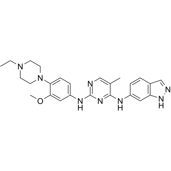
| Chemical Name | 2-N-[4-(4-ethylpiperazin-1-yl)-3-methoxyphenyl]-4-N-(1H-indazol-6-yl)-5-methylpyrimidine-2,4-diamine |
| HS Tariff Code | 2934.99.9001 |
| Storage |
Powder-20°C 3 years 4°C 2 years In solvent -80°C 6 months -20°C 1 month |
| Shipping Condition | Room temperature (This product is stable at ambient temperature for a few days during ordinary shipping and time spent in Customs) |
Biological Activity
| ln Vitro | Osteosarcoma cancer cells (U2OS, MG63, MNNG/HOS, and SAOS-2) are inhibited in their proliferation and colony formation by PDGFR-IN-1 (Compound 7m) (0-0.4 μM, 48 hours) [1]. A dose-dependent mechanism of cell cycle arrest is induced by PDGFR-IN-1 (0-0.4 μM, 48 hours) [1]. In MNNG/HOS and MG63 cells, PDGFR-IN-1 (0-1.6 μM, 48 h) causes apoptosis in a dose-dependent manner [1]. In MNNG/HOS and MG63 cells, PDGFR-IN-1 (0-0.4 μM, 15 minutes) suppresses the expression of α-tubulin [1]. PDGFR-IN-1 (0-0.4 μM, 48 hours) suppresses downstream signaling (p-AKT, p-ERK, and p-STAT3) and PDGFRβ phosphorylation [1]. By suppressing FAK expression and cell front distribution, PDGFR-IN-1 (0-0.4 μM, 48 h) dramatically reduces the migration and invasion of osteosarcoma cancer cells [1]. |
| ln Vivo | PDGFR-IN-1 (BALB/c mice, MNNG/HOS xenograft mouse, 15, 30 mg/kg, orally, daily, 14 days) shows less toxicity and a greater antitumor efficaciousness that considerably reduces tumor growth[1]. It is safe to use PDGFR-IN-1 (C57/BL6 mice, 40–80 mg/kg, orally, daily for 10 days) for in vivo research[1]. PDGFR-IN-1 (Sprague-Dawley rats, oral bioavailability: good, maximum concentration and exposure, acceptable half-life, and 4 mg/kg IP, once) exhibits a decent profile[1]. |
| Cell Assay |
Cell proliferation assay Cell Types: Human osteosarcoma cancer cell lines (U2OS, MG63, MNNG/HOS and SAOS-2) [1] Tested Concentrations: 0.1, 0.2 and 0.4 μM. Incubation Duration: 48 h Experimental Results: It demonstrated strong anti-proliferative activity against MG63, U2OS, MNNG/HOS and SAOS-2, with IC50 values of 0.44, 0.42, 1.03 and 0.37 μM respectively. Exhibits dose-dependent inhibition of colony formation. Cell cycle analysis Cell Types: MG63 and MNNG/HOS cells [1] Tested Concentrations: 0, 0.1, 0.2, 0.4 μM Incubation Duration: 48 hrs (hours) Experimental Results: Induction of G2/M cell cycle arrest in MNNG/HOS and G0/G1 cells - Cycle arrest in MG63 cells was dose-dependent. Apoptosis analysis Cell Types: MG63 and MNNG/HOS cells [1] Tested Concentrations: 0, 0.4, 0.8, 1.6 μM Incubation Duration: 48 h Experimental Results: Apoptosis of MNNG/HOS and MG63 cells was induced in a dose-dependent manner. Immunofluorescence Cell Types: MG63 and MNNG/HOS[1] Tested Concentrations: 0, 0.1, 0.2, 0.4 μM Incubation Duration: 15 min Experimental Results: Inhibited the expression of α-tubulin in MNNG/HOS |
| Animal Protocol |
Animal/Disease Models: balb/c (Bagg ALBino) mouse (18-20 g, MNNG/HOS xenograft mice, eight groups) [1] Doses: 15, 30 mg/kg Route of Administration: Orally, one time/day for 14 days Experimental Results: Dramatically inhibits tumor growth, exhibits stronger anti-tumor efficacy, does not cause significant changes in body weight or organ weight (heart, lungs, liver, spleen, kidney), strongly inhibits tumor cell proliferation and induces tumor tissue apoptosis. Animal/Disease Models: C57/BL6 black mouse [1] Doses: 40, 80 mg/kg Route of Administration: Orally, one time/day for 10 days. Experimental Results: No obvious morphological distortion of organs and tissues was found. |
| References |
[1]. Discovery of Potent and Orally Bioavailable Platelet-Derived Growth Factor Receptor (PDGFR) Inhibitors for the Treatment of Osteosarcoma. J Med Chem. 2022;65(7):5374-5391. |
Solubility Data
| Solubility (In Vitro) | May dissolve in DMSO (in most cases), if not, try other solvents such as H2O, Ethanol, or DMF with a minute amount of products to avoid loss of samples |
| Solubility (In Vivo) |
Note: Listed below are some common formulations that may be used to formulate products with low water solubility (e.g. < 1 mg/mL), you may test these formulations using a minute amount of products to avoid loss of samples. Injection Formulations (e.g. IP/IV/IM/SC) Injection Formulation 1: DMSO : Tween 80: Saline = 10 : 5 : 85 (i.e. 100 μL DMSO stock solution → 50 μL Tween 80 → 850 μL Saline) *Preparation of saline: Dissolve 0.9 g of sodium chloride in 100 mL ddH ₂ O to obtain a clear solution. Injection Formulation 2: DMSO : PEG300 :Tween 80 : Saline = 10 : 40 : 5 : 45 (i.e. 100 μL DMSO → 400 μLPEG300 → 50 μL Tween 80 → 450 μL Saline) Injection Formulation 3: DMSO : Corn oil = 10 : 90 (i.e. 100 μL DMSO → 900 μL Corn oil) Example: Take the Injection Formulation 3 (DMSO : Corn oil = 10 : 90) as an example, if 1 mL of 2.5 mg/mL working solution is to be prepared, you can take 100 μL 25 mg/mL DMSO stock solution and add to 900 μL corn oil, mix well to obtain a clear or suspension solution (2.5 mg/mL, ready for use in animals). Injection Formulation 4: DMSO : 20% SBE-β-CD in saline = 10 : 90 [i.e. 100 μL DMSO → 900 μL (20% SBE-β-CD in saline)] *Preparation of 20% SBE-β-CD in Saline (4°C,1 week): Dissolve 2 g SBE-β-CD in 10 mL saline to obtain a clear solution. Injection Formulation 5: 2-Hydroxypropyl-β-cyclodextrin : Saline = 50 : 50 (i.e. 500 μL 2-Hydroxypropyl-β-cyclodextrin → 500 μL Saline) Injection Formulation 6: DMSO : PEG300 : castor oil : Saline = 5 : 10 : 20 : 65 (i.e. 50 μL DMSO → 100 μLPEG300 → 200 μL castor oil → 650 μL Saline) Injection Formulation 7: Ethanol : Cremophor : Saline = 10: 10 : 80 (i.e. 100 μL Ethanol → 100 μL Cremophor → 800 μL Saline) Injection Formulation 8: Dissolve in Cremophor/Ethanol (50 : 50), then diluted by Saline Injection Formulation 9: EtOH : Corn oil = 10 : 90 (i.e. 100 μL EtOH → 900 μL Corn oil) Injection Formulation 10: EtOH : PEG300:Tween 80 : Saline = 10 : 40 : 5 : 45 (i.e. 100 μL EtOH → 400 μLPEG300 → 50 μL Tween 80 → 450 μL Saline) Oral Formulations Oral Formulation 1: Suspend in 0.5% CMC Na (carboxymethylcellulose sodium) Oral Formulation 2: Suspend in 0.5% Carboxymethyl cellulose Example: Take the Oral Formulation 1 (Suspend in 0.5% CMC Na) as an example, if 100 mL of 2.5 mg/mL working solution is to be prepared, you can first prepare 0.5% CMC Na solution by measuring 0.5 g CMC Na and dissolve it in 100 mL ddH2O to obtain a clear solution; then add 250 mg of the product to 100 mL 0.5% CMC Na solution, to make the suspension solution (2.5 mg/mL, ready for use in animals). Oral Formulation 3: Dissolved in PEG400 Oral Formulation 4: Suspend in 0.2% Carboxymethyl cellulose Oral Formulation 5: Dissolve in 0.25% Tween 80 and 0.5% Carboxymethyl cellulose Oral Formulation 6: Mixing with food powders Note: Please be aware that the above formulations are for reference only. InvivoChem strongly recommends customers to read literature methods/protocols carefully before determining which formulation you should use for in vivo studies, as different compounds have different solubility properties and have to be formulated differently. (Please use freshly prepared in vivo formulations for optimal results.) |
| Preparing Stock Solutions | 1 mg | 5 mg | 10 mg | |
| 1 mM | 2.1807 mL | 10.9037 mL | 21.8074 mL | |
| 5 mM | 0.4361 mL | 2.1807 mL | 4.3615 mL | |
| 10 mM | 0.2181 mL | 1.0904 mL | 2.1807 mL |