Physicochemical Properties
| Molecular Formula | C47H44N6NA4O17P4.13.5H2O |
| Molecular Weight | 1424.01 |
| Related CAS # | 907547-55-1 (free acid); 1006028-37-0 (sodium) |
| Appearance | White to yellow solid powder |
| InChi Key | GSMUPMANKNAMAS-UHFFFAOYSA-J |
| InChi Code | InChI=1S/C47H48N6O17P4.4Na/c1-27-9-13-33(45(56)50-39-15-11-29(23-71(59,60)61)17-35(39)25-73(65,66)67)21-41(27)52-43(54)31-5-3-7-37(19-31)48-47(58)49-38-8-4-6-32(20-38)44(55)53-42-22-34(14-10-28(42)2)46(57)51-40-16-12-30(24-72(62,63)64)18-36(40)26-74(68,69)70;;;;/h3-22H,23-26H2,1-2H3,(H,50,56)(H,51,57)(H,52,54)(H,53,55)(H2,48,49,58)(H2,59,60,61)(H2,62,63,64)(H2,65,66,67)(H2,68,69,70);;;;/q;4*+1/p-4 |
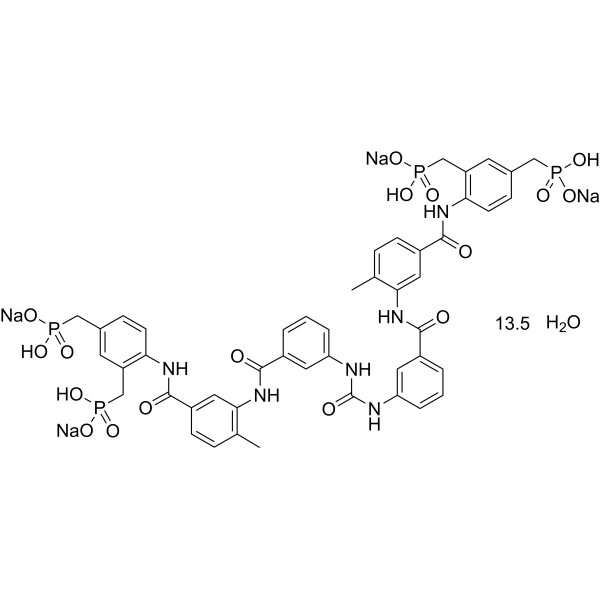
| Chemical Name | tetrasodium;[2-[[3-[[3-[[3-[[5-[[2,4-bis[[hydroxy(oxido)phosphoryl]methyl]phenyl]carbamoyl]-2-methylphenyl]carbamoyl]phenyl]carbamoylamino]benzoyl]amino]-4-methylbenzoyl]amino]-5-[[hydroxy(oxido)phosphoryl]methyl]phenyl]methyl-hydroxyphosphinate hydrate |
| HS Tariff Code | 2934.99.9001 |
| Storage |
Powder-20°C 3 years 4°C 2 years In solvent -80°C 6 months -20°C 1 month Note: Please store this product in a sealed and protected environment, avoid exposure to moisture. |
| Shipping Condition | Room temperature (This product is stable at ambient temperature for a few days during ordinary shipping and time spent in Customs) |
Biological Activity
| Targets | P2Y11 (pEC50 = 6.27) |
| ln Vitro |
NF546(hydrate) is comparatively more selective for P2Y11 than P2Y1, P2Y2, P2Y4, P2Y6, P2Y12, P2X1, P2X2, and P2X2-X3. In human monocyte-derived dendritic cells, NF546(hydrate)(100 μM; 24 hours) is equally effective in inducing TSP-1 release and preventing the release of IL-12p70 triggered by LPS[1].
The G protein-coupled P2Y(11) receptor is involved in immune system modulation. In-depth physiological evaluation is hampered, however, by a lack of selective and potent ligands. By screening a library of sulfonic and phosphonic acid derivatives at P2Y(11) receptors recombinantly expressed in human 1321N1 astrocytoma cells (calcium and cAMP assays), the selective non-nucleotide P2Y(11) agonist NF546 [4,4'-(carbonylbis(imino-3,1-phenylene-carbonylimino-3,1-(4-methyl-phenylene)carbonylimino))-bis(1,3-xylene-alpha,alpha'-diphosphonic acid) tetrasodium salt] was identified. NF546 had a pEC(50) of 6.27 and is relatively selective for P2Y(11) over P2Y(1), P2Y(2), P2Y(4), P2Y(6), P2Y(12), P2X(1), P2X(2), and P2X(2)-X(3). Adenosine-5'-O-(3-thio)triphosphate (ATPgammaS), a nonhydrolyzable analog of the physiological P2Y(11) agonist ATP, and NF546 use a common binding site as suggested by molecular modeling studies and their competitive behavior toward the nanomolar potency antagonist NF340 [4,4'-(carbonylbis(imino-3,1-(4-methyl-phenylene)carbonylimino))bis(naphthalene-2,6-disulfonic acid) tetrasodium salt] in Schild analysis. The pA(2) of NF340 was 8.02 against ATPgammaS and 8.04 against NF546 (calcium assays). NF546 was further tested for P2Y(11)-mediated effects in monocyte-derived dendritic cells. Similarly to ATPgammaS, NF546 led to thrombospondin-1 secretion and inhibition of lipopolysaccharide-stimulated interleukin-12 release, whereas NF340 inhibited these effects. Further, for the first time, it was shown that ATPgammaS or NF546 stimulation promotes interleukin 8 (IL-8) release from dendritic cells, which could be inhibited by NF340. In conclusion, we have described the first selective, non-nucleotide agonist NF546 for P2Y(11) receptors in both recombinant and physiological expression systems and could show a P2Y(11)-stimulated IL-8 release, further supporting the immunomodulatory role of P2Y(11) receptors [1]. In this study, we investigated the role of P2Y11R signaling in vascular dysfunction. P2Y11R activity was modulated using its pharmacological agonist NF546 and antagonist NF340. Rat aortic rings were exposed to angiotensin II (AngII) and evaluated for their vasomotor response. The P2Y11R agonist NF546 reduced AngII-induced vascular dysfunction by promoting EC-dependent vasorelaxation, through an increased nitric oxide (NO) bioavailability and reduced AngII-induced H2O2 release; these effects were prevented by the use of the P2Y11R antagonist NF340. Human vascular SMCs and ECs were subjected to AngII or H/R simulation in vitro. P2Y11R agonist modulated vasoactive factors in human ECs, that is, endothelial nitric oxide synthase (eNOS) and endothelin-1, reduced SMC proliferation and prevented the switch towards a synthetic phenotype. H/R and AngII increased ECs secretome-induced SMC proliferation, an effect prevented by P2Y11R activation. Thus, our data suggest that P2Y11R activation may protect blood vessels from HR-/AngII-induced injury and reduce vascular dysfunctions. These results open the way for new vasculoprotective interventions.[2] Here, researchers find that cAMP synthesis in response to elevated glucose and the selective P2Y11 agonist NF546 is blocked by disruption of A-kinase anchoring protein 5 (AKAP5) function in arterial myocytes. Glucose and NF546-induced potentiation of L-type Ca2+ channels, vasoconstriction and decreased blood flow are prevented in AKAP5 null arterial myocytes/arteries. These responses are nucleated via the AKAP5-dependent clustering of P2Y11/ P2Y11-like receptors, AC5, PKA and CaV1.2 into nanocomplexes at the plasma membrane of human and mouse arterial myocytes. Hence, data reveal an AKAP5 signaling module that regulates L-type Ca2+ channel activity and vascular reactivity upon elevated glucose. This AKAP5-anchored nanocomplex may contribute to vascular complications during diabetic hyperglycemia [3]. |
| Enzyme Assay |
ROS Measurement [2] H2O2-released by aortic rings was evaluated by Ferrous iron xylenol orange Oxidation (FOX) assay using the PeroxiDetect Kit according to the manufacturer’s instructions. A 30-min incubation was performed in the absence or presence of AngII. The two groups were also incubated at the same time with NF546, NF340 or both. |
| Cell Assay |
Cell Culture and Reagents [2] Human umbilical vein endothelial cells (HUVECs) and human coronary artery smooth muscle cells (HCASMC) and their corresponding culture media (endothelial cell growth medium 2 and smooth muscle cell growth medium 2) were purchased from Promocell and cultured according to the manufacturer’s instructions. Cells were used from passage 3 to 6 for experimental procedures. For H/R experiments, HUVEC and HCASMC were submitted to 5 h hypoxia (1% O2, 5% CO2, DPBS with CaMg) in a hypoxia chamber. DPBS was equilibrated overnight before the experiment. Several durations of reoxygenation (DMEM/F12, 5% CO2, 21% O2) were used depending on the experiment and the type of cell. Hypoxia and reoxygenation durations were based on previous observations from our group. All modulating agents were added at the onset of the reoxygenation. Concomitantly, a simulation of stress environment was induced by AngII (100 nM). Modulation of P2Y11R was performed with its agonist NF546 (10 µM) and antagonist NF340 (10 µM); the maximal concentration effect was chosen according to Meis et al [2]. |
| Animal Protocol |
Vascular Function Analysis [2] Rat aortic rings were mounted on an isometric force transducer (Myograph DMT 620M) in an organ chamber. This chamber contained 5 mL of Krebs solution equilibrated 30 min at 37 °C with O2, and 10 µM diclofenac to block the generation of prostaglandins, then the major vasodilator remaining active is the NO. Aortic rings were then stretched and equilibrated at a tension of 2 g (isometric tension). The maximal contraction level was evaluated in response to a 80 mM KCl solution. After washing steps and equilibration, aortic rings were exposed to angiotensin II (AngII) (100 nM) or vehicle (control group) during 30 min. Both the control group and the AngII-treated group were incubated simultaneously with or without NF546 (10 µM), NF340 (10 µM), or both. These are respectively the specific agonist and antagonist of P2Y11R. Contraction was evaluated in response to phenylephrine, and was adjusted to reach a preconstriction level of 80% of the contraction elicited by KCl. Relaxation was evaluated in response to cumulative doses of acetylcholine. |
| References |
[1]. NF546 [4,4'-(carbonylbis(imino-3,1-phenylene-carbonylimino-3,1-(4-methyl-phenylene)-carbonylimino))-bis(1,3-xylene-alpha,alpha'-diphosphonic acid) tetrasodium salt] is a non-nucleotide P2Y11 agonist and stimulates release of interleukin-8 from human monocyte-derived dendritic cells. J Pharmacol Exp Ther. 2010 Jan;332(1):238-47. [2]. P2Y11 Agonism Prevents Hypoxia/Reoxygenation- and Angiotensin II-Induced Vascular Dysfunction and Intimal Hyperplasia Development. Int J Mol Sci. 2021 Jan 16;22(2):855. [3]. AKAP5 complex facilitates purinergic modulation of vascular L-type Ca2+ channel CaV1.2. Nat Commun. 2020 Oct 20;11(1):5303. |
| Additional Infomation |
The L-type Ca2+ channel CaV1.2 is essential for arterial myocyte excitability, gene expression and contraction. Elevations in extracellular glucose (hyperglycemia) potentiate vascular L-type Ca2+ channel via PKA, but the underlying mechanisms are unclear. Here, we find that cAMP synthesis in response to elevated glucose and the selective P2Y11 agonist NF546 is blocked by disruption of A-kinase anchoring protein 5 (AKAP5) function in arterial myocytes. Glucose and NF546-induced potentiation of L-type Ca2+ channels, vasoconstriction and decreased blood flow are prevented in AKAP5 null arterial myocytes/arteries. These responses are nucleated via the AKAP5-dependent clustering of P2Y11/ P2Y11-like receptors, AC5, PKA and CaV1.2 into nanocomplexes at the plasma membrane of human and mouse arterial myocytes. Hence, data reveal an AKAP5 signaling module that regulates L-type Ca2+ channel activity and vascular reactivity upon elevated glucose. This AKAP5-anchored nanocomplex may contribute to vascular complications during diabetic hyperglycemia.[3] Vascular dysfunction in cardiovascular diseases includes vasomotor response impairments, endothelial cells (ECs) activation, and smooth muscle cells (SMCs) proliferation and migration to the intima. This results in intimal hyperplasia and vessel failure. We previously reported that activation of the P2Y11 receptor (P2Y11R) in human dendritic cells, cardiofibroblasts and cardiomyocytes was protective against hypoxia/reoxygenation (HR) lesions. In this study, we investigated the role of P2Y11R signaling in vascular dysfunction. P2Y11R activity was modulated using its pharmacological agonist NF546 and antagonist NF340. Rat aortic rings were exposed to angiotensin II (AngII) and evaluated for their vasomotor response. The P2Y11R agonist NF546 reduced AngII-induced vascular dysfunction by promoting EC-dependent vasorelaxation, through an increased nitric oxide (NO) bioavailability and reduced AngII-induced H2O2 release; these effects were prevented by the use of the P2Y11R antagonist NF340. Human vascular SMCs and ECs were subjected to AngII or H/R simulation in vitro. P2Y11R agonist modulated vasoactive factors in human ECs, that is, endothelial nitric oxide synthase (eNOS) and endothelin-1, reduced SMC proliferation and prevented the switch towards a synthetic phenotype. H/R and AngII increased ECs secretome-induced SMC proliferation, an effect prevented by P2Y11R activation. Thus, our data suggest that P2Y11R activation may protect blood vessels from HR-/AngII-induced injury and reduce vascular dysfunctions. These results open the way for new vasculoprotective interventions. [2] |
Solubility Data
| Solubility (In Vitro) | May dissolve in DMSO (in most cases), if not, try other solvents such as H2O, Ethanol, or DMF with a minute amount of products to avoid loss of samples |
| Solubility (In Vivo) |
Note: Listed below are some common formulations that may be used to formulate products with low water solubility (e.g. < 1 mg/mL), you may test these formulations using a minute amount of products to avoid loss of samples. Injection Formulations (e.g. IP/IV/IM/SC) Injection Formulation 1: DMSO : Tween 80: Saline = 10 : 5 : 85 (i.e. 100 μL DMSO stock solution → 50 μL Tween 80 → 850 μL Saline) *Preparation of saline: Dissolve 0.9 g of sodium chloride in 100 mL ddH ₂ O to obtain a clear solution. Injection Formulation 2: DMSO : PEG300 :Tween 80 : Saline = 10 : 40 : 5 : 45 (i.e. 100 μL DMSO → 400 μLPEG300 → 50 μL Tween 80 → 450 μL Saline) Injection Formulation 3: DMSO : Corn oil = 10 : 90 (i.e. 100 μL DMSO → 900 μL Corn oil) Example: Take the Injection Formulation 3 (DMSO : Corn oil = 10 : 90) as an example, if 1 mL of 2.5 mg/mL working solution is to be prepared, you can take 100 μL 25 mg/mL DMSO stock solution and add to 900 μL corn oil, mix well to obtain a clear or suspension solution (2.5 mg/mL, ready for use in animals). Injection Formulation 4: DMSO : 20% SBE-β-CD in saline = 10 : 90 [i.e. 100 μL DMSO → 900 μL (20% SBE-β-CD in saline)] *Preparation of 20% SBE-β-CD in Saline (4°C,1 week): Dissolve 2 g SBE-β-CD in 10 mL saline to obtain a clear solution. Injection Formulation 5: 2-Hydroxypropyl-β-cyclodextrin : Saline = 50 : 50 (i.e. 500 μL 2-Hydroxypropyl-β-cyclodextrin → 500 μL Saline) Injection Formulation 6: DMSO : PEG300 : castor oil : Saline = 5 : 10 : 20 : 65 (i.e. 50 μL DMSO → 100 μLPEG300 → 200 μL castor oil → 650 μL Saline) Injection Formulation 7: Ethanol : Cremophor : Saline = 10: 10 : 80 (i.e. 100 μL Ethanol → 100 μL Cremophor → 800 μL Saline) Injection Formulation 8: Dissolve in Cremophor/Ethanol (50 : 50), then diluted by Saline Injection Formulation 9: EtOH : Corn oil = 10 : 90 (i.e. 100 μL EtOH → 900 μL Corn oil) Injection Formulation 10: EtOH : PEG300:Tween 80 : Saline = 10 : 40 : 5 : 45 (i.e. 100 μL EtOH → 400 μLPEG300 → 50 μL Tween 80 → 450 μL Saline) Oral Formulations Oral Formulation 1: Suspend in 0.5% CMC Na (carboxymethylcellulose sodium) Oral Formulation 2: Suspend in 0.5% Carboxymethyl cellulose Example: Take the Oral Formulation 1 (Suspend in 0.5% CMC Na) as an example, if 100 mL of 2.5 mg/mL working solution is to be prepared, you can first prepare 0.5% CMC Na solution by measuring 0.5 g CMC Na and dissolve it in 100 mL ddH2O to obtain a clear solution; then add 250 mg of the product to 100 mL 0.5% CMC Na solution, to make the suspension solution (2.5 mg/mL, ready for use in animals). Oral Formulation 3: Dissolved in PEG400 Oral Formulation 4: Suspend in 0.2% Carboxymethyl cellulose Oral Formulation 5: Dissolve in 0.25% Tween 80 and 0.5% Carboxymethyl cellulose Oral Formulation 6: Mixing with food powders Note: Please be aware that the above formulations are for reference only. InvivoChem strongly recommends customers to read literature methods/protocols carefully before determining which formulation you should use for in vivo studies, as different compounds have different solubility properties and have to be formulated differently. (Please use freshly prepared in vivo formulations for optimal results.) |
| Preparing Stock Solutions | 1 mg | 5 mg | 10 mg | |
| 1 mM | 0.7022 mL | 3.5112 mL | 7.0224 mL | |
| 5 mM | 0.1404 mL | 0.7022 mL | 1.4045 mL | |
| 10 mM | 0.0702 mL | 0.3511 mL | 0.7022 mL |