Neratinib maleate, the maleate salt of neratinib (HKI-272; PB-272; Nerlynx), is an orally bioavailable HER2 and EGFR inhibitor approved by FDA in 2017 to reduce the risk of breast cancer returning. In cell-free assays, it inhibits HER2 and EGFR with IC50s of 59 nM and 92 nM, respectively.
Physicochemical Properties
| Molecular Formula | C34H33CLN6O7 |
| Molecular Weight | 673.11 |
| Exact Mass | 672.209 |
| Elemental Analysis | C, 60.67; H, 4.94; Cl, 5.27; N, 12.49; O, 16.64 |
| CAS # | 915942-22-2 |
| Related CAS # | Neratinib;698387-09-6 |
| PubChem CID | 67307512 |
| Appearance | White to off-white solid powder |
| Hydrogen Bond Donor Count | 4 |
| Hydrogen Bond Acceptor Count | 12 |
| Rotatable Bond Count | 13 |
| Heavy Atom Count | 48 |
| Complexity | 1000 |
| Defined Atom Stereocenter Count | 0 |
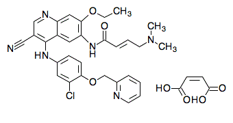
| SMILES | C(/C(=O)O)=C/C(=O)O.N(C1C=CC(OCC2N=CC=CC=2)=C(Cl)C=1)C1=C(C#N)C=NC2=CC(=C(C=C12)NC(=O)/C=C/CN(C)C)OCC |
| InChi Key | VXZCUHNJXSIJIM-MEBGWEOYSA-N |
| InChi Code | InChI=1S/C30H29ClN6O3.C4H4O4/c1-4-39-28-16-25-23(15-26(28)36-29(38)9-7-13-37(2)3)30(20(17-32)18-34-25)35-21-10-11-27(24(31)14-21)40-19-22-8-5-6-12-33-22;5-3(6)1-2-4(7)8/h5-12,14-16,18H,4,13,19H2,1-3H3,(H,34,35)(H,36,38);1-2H,(H,5,6)(H,7,8)/b9-7+;2-1- |
| Chemical Name | (Z)-but-2-enedioic acid;(E)-N-[4-[3-chloro-4-(pyridin-2-ylmethoxy)anilino]-3-cyano-7-ethoxyquinolin-6-yl]-4-(dimethylamino)but-2-enamide |
| Synonyms | HKI-272 maleate or PB272; HKI272; 915942-22-2; hki-272 maleate; Nerlynx; Neratinib maleate [MI]; UNII-9RM7XY23ZS; 9RM7XY23ZS; Neratinib (maleate); HKI 272; PB 272; PB-272 maleate |
| HS Tariff Code | 2934.99.9001 |
| Storage |
Powder-20°C 3 years 4°C 2 years In solvent -80°C 6 months -20°C 1 month Note: Please store this product in a sealed and protected environment, avoid exposure to moisture. |
| Shipping Condition | Room temperature (This product is stable at ambient temperature for a few days during ordinary shipping and time spent in Customs) |
Biological Activity
| Targets |
HER2 (IC50 = 59 nM); EGFR (IC50 = 92 nM) Neratinib shows no action against tyrosine kinase c-Met[1], Akt, cyclin D1/cdk4, cyclin E/cdk2, cyclin B1/cdk1, IKK-2, MK-2, PDK1, c-Raf, and Tpl-2, among other serine-threonine kinases[1]. Neratinib is much less active in cell lines that express neither EGFR nor HER-2 (3T3, MDA-MB-435, and SW620), and it inhibits the proliferation of cell lines that exhibit high levels of HER-2 (3T3/neu, SK-Br-3, and BT474)[1]. Neratinib (0-2 nM, 12-16 h) arrests BT474 cell cycle at G1-S phase[1]. Neratinib causes cyclin D1 levels to be down-regulated, Akt and MAPK phosphorylation to be inhibited, and p27 to be induced[1]. |
| ln Vitro |
Neratinib shows no action against tyrosine kinase c-Met[1], Akt, cyclin D1/cdk4, cyclin E/cdk2, cyclin B1/cdk1, IKK-2, MK-2, PDK1, c-Raf, and Tpl-2, among other serine-threonine kinases[1]. Neratinib is much less active in cell lines that express neither EGFR nor HER-2 (3T3, MDA-MB-435, and SW620), and it inhibits the proliferation of cell lines that exhibit high levels of HER-2 (3T3/neu, SK-Br-3, and BT474)[1]. Neratinib (0-2 nM, 12-16 h) arrests BT474 cell cycle at G1-S phase[1]. Neratinib causes cyclin D1 levels to be down-regulated, Akt and MAPK phosphorylation to be inhibited, and p27 to be induced[1]. Neratinib Maleate (HKI-272) potently inhibited the proliferation of HER-2-overexpressing human breast cancer cell lines BT474 and SK-Br-3 with IC50 values of 2 nM. It also inhibited the proliferation of HER-2-transfected mouse fibroblast 3T3/neu cells (IC50 = 3 nM), showing >200-fold selectivity over the parental 3T3 line (IC50 = 700 nM). The compound inhibited proliferation of EGFR-overexpressing A431 cells with an IC50 of 81 nM, but was much less active against MDA-MB-435 and SW620 cells that lack HER-2 and EGFR expression (IC50 ~690-960 nM). Neratinib Maleate (HKI-272) inhibited ligand-independent HER-2 autophosphorylation in BT474 cells and EGF-stimulated EGFR phosphorylation in A431 cells with IC50 values of 5 nM and 3 nM, respectively. Treatment of BT474 cells with Neratinib Maleate (HKI-272) resulted in inhibition of downstream signaling pathways, including phosphorylation of MAPK and Akt (IC50 = 2 nM for both), down-regulation of cyclin D1 levels (IC50 = 9 nM), reduction of retinoblastoma protein (Rb) phosphorylation, and induction of p27. This led to a G1-S phase cell cycle arrest (IC50 for reduction of S-phase cells = 2 nM) and, at higher concentrations, an increase in sub-G1 population indicative of apoptosis. Neratinib Maleate (HKI-272) functioned as an irreversible inhibitor. Inhibition of receptor phosphorylation persisted after compound washout. Direct covalent binding to the HER-2 kinase domain was demonstrated using [14C]HKI-272. In contrast, trastuzumab (up to 30 µg/ml) only partially inhibited HER-2 phosphorylation and downstream signaling in BT474 cells. [1] |
| ln Vivo |
Neratinib (HKI-272) exhibits anticancer properties against cancer cells that express high levels of EGFR or HER-2 (0-80 mg/kg/day; i.e., 42 days)[1]. In vivo, HKI-272 is active in HER-2- and EGFR-dependent tumor xenograft models when dosed orally on a once daily schedule. On the basis of its favorable preclinical pharmacological profile, HKI-272 has been selected as a candidate for additional development as an antitumor agent in breast and other HER-2-dependent cancers.[1] Oral administration of Neratinib Maleate (HKI-272) once daily inhibited the growth of HER-2-dependent tumor xenografts in athymic mice. In the 3T3/neu model, doses of 20-80 mg/kg/day produced 53-98% inhibition of tumor growth. In BT474 breast cancer xenografts, doses of 5-40 mg/kg/day produced 70-93% inhibition of tumor growth. A single oral dose (40 mg/kg) inhibited HER-2 phosphorylation in BT474 tumors by 84% at 1 hour and 97% at 6 hours, with inhibition sustained at 43% after 24 hours. In SK-OV-3 ovarian cancer xenografts, doses of 5-60 mg/kg/day produced 31-85% inhibition. In EGFR-dependent A431 xenografts, Neratinib Maleate (HKI-272) was less potent, with 40 mg/kg/day producing 76% inhibition, compared to the more robust effects seen in HER-2 models. The compound showed minimal activity (28% inhibition at 80 mg/kg/day) in MCF-7 xenografts, a breast cancer line with low HER-2/EGFR expression. [1] |
| Enzyme Assay |
Prepared as 10 mg/mL stocks in DMSO, neratinib is diluted in 25 mM HEPES (pH 7.5; 0.002 ng/mL–20 μg/mL). In 96-well ELISA plates, purified recombinant COOH-terminal fragments of HER2 (amino acids 676-1255) or epidermal growth factor receptor (EGFR) (amino acids 645-1186) are diluted in 100 mM HEPES (pH 7.5) and 50% glycerol. The mixture is then incubated with increasing concentrations of Neratinib in 4 mM HEPES (pH 7.5), 0.4 mM MnCl2, 20 μM sodium vanadate, and 0.2 mM DTT for 15 minutes at room temperature. 40 μM ATP and 20 mM MgCl2 are added to start the kinase reaction, which is then left to run at room temperature for an hour. Wash the plates, then use anti-phospho-tyrosine antibodies (15 ng/well) labeled with europium to detect phosphorylation. Using a Victor2 fluorescence reader (excitation wavelength of 340 nm and emission wavelength of 615 nm), the signal is detected following the washing and enhancement stages. An inhibition curve is used to determine the concentration of Neratinib (IC50) at which receptor phosphorylation is inhibited by 50%. Activity of HER-2 and EGFR cytoplasmic domains was measured by an autophosphorylation assay using time-resolved fluorometry. Compounds were prepared as 10 mg/ml stocks in DMSO and diluted in 25 mm HEPES (pH 7.5; 0.002 ng/ml–20 μg/ml). Enzyme [diluted in 100 mm HEPES (pH 7.5) and 50% glycerol] was incubated with inhibitor in 4 mm HEPES (pH 7.5), 0.4 mm MnCl2, 20 μm sodium vanadate, and 0.2 mm DTT for 15 min at room temperature in 96-well ELISA plates. The kinase reaction was initiated by the addition of 40 μm ATP and 20 mm MgCl2 and allowed to proceed for 1 h at room temperature. Plates were washed, and phosphorylation was detected using Europium-labeled anti-phospho-tyrosine antibodies (15 ng/well; Wallac). After washing and enhancement steps according to the manufacturer’s recommendations, signal was detected using a Victor2 fluorescence reader (excitation wavelength 340 nm, emission wavelength 615 nm). The concentration of compound that inhibited receptor phosphorylation by 50% (IC50) was calculated from inhibition curves.[1] Assays for other kinases were performed using recombinant enzymes expressed in bacterial, insect, or human cell lines. All enzymes used were serine-threonine kinases, except c-Met, KDR, src (tyrosine kinases), and MEK1 (dual specificity). Substrates used were peptides (Akt, IKK-2, MK2, PDK1, src, and Tpl2), proteins (cyclin D1/CDK4, cyclin E/CDK2, cyclin B1/CDK1, and c-Raf), poly(glutamic acid4-tyrosine) (KDR), or the kinase itself (autophosphorylation; c-met). Phosphorylation was measured using TMB peroxidase substrate for cyclin/cyclin-dependent kinase (cdk), LabChip for MK-2, or DELPHIA/LANCE for all others.[1] The cytoplasmic domains of HER-2 and EGFR, expressed as recombinant His-tagged proteins in insect cells, were purified using nickel affinity chromatography. Purity was estimated to be >80% by SDS-PAGE. For the kinase activity assay, the enzyme was incubated with the inhibitor in a buffer containing HEPES, MnCl2, sodium vanadate, and DTT for 15 minutes at room temperature. The kinase reaction was initiated by adding ATP and MgCl2 and proceeded for 1 hour at room temperature. Receptor autophosphorylation was detected using europium-labeled anti-phosphotyrosine antibodies in a time-resolved fluorescence assay (DELFIA). The IC50 was calculated from the inhibition curves. [1] |
| Cell Assay |
Different concentrations of Neratinib are applied to cells (3T3, 3T3/neu, A431, BT474, SK-Br-3, MDA-MB-435, and SW480) for a period of either two or six days. A protein-binding dye called sulforhodamine B is used to measure cell proliferation. In short, cells are thoroughly cleaned with water after being fixed with 10% trichloroacetic acid. After staining the cells with 0.1% sulforhodamine B, they are rinsed in 5% acetic acid. After solubilizing the protein-associated dye in 10 mM Tris, the absorbance is calculated at 450 nM. Inhibition curves are used to calculate the concentration of neratinib (IC50) that inhibits cell proliferation by 50%. For cell proliferation assays, cells were plated in 96-well plates. The next day, serial dilutions of the compound were added. After 2 days (6 days for BT474), cell proliferation was assessed using the sulforhodamine B (SRB) protein-binding dye. Cells were fixed with trichloroacetic acid, stained with SRB, and the solubilized dye was measured spectrophotometrically. IC50 values were determined from inhibition curves. For analysis of receptor and signaling protein phosphorylation or expression, cells were treated with the compound for specified durations (e.g., 3 hours for receptor phosphorylation, overnight for downstream markers). Cells were then lysed, and proteins were separated by SDS-PAGE, transferred to nitrocellulose, and detected by immunoblotting with specific antibodies and enhanced chemiluminescence. For binding studies, recombinant HER-2 cytoplasmic domain or intact BT474 cells were incubated with [14C]HKI-272 in the presence or absence of excess unlabeled compound. Samples were denatured, separated by SDS-PAGE, and analyzed by fluorography. For cell cycle analysis, BT474 cells treated with the compound overnight were pulse-labeled with bromodeoxyuridine (BrdUrd). Cells were fixed, denatured, stained with anti-BrdUrd-FITC antibody and propidium iodide, and analyzed by flow cytometry to determine the percentage of cells in different cell cycle phases. [1] |
| Animal Protocol |
Female athymic (nude) mice, tumor xenograft[1] 10, 20, 40, 60 or 80 mg/kg/day Gavage, 42 days Tumor Xenograft Studies.[1] Tumor cells (maintained in tissue culture) or tumor fragments were implanted s.c. in the flanks of female athymic (nude) mice. For estrogen-dependent cell lines (BT474, MCF-7, and SK-OV-3), animals were implanted with hormone pellets (0.72 mg of 17-β estradiol, 60-day release) 1 week before implantation of tumors. Additionally, SK-OV-3 cells were suspended in Matrigel basement membrane matrix for implantation. Treatment was initiated after tumors had reached a size of 90–200 mg, following random assignment of the animals to different treatment groups (staging, day 0). For 3T3/neu xenografts, treatment was initiated the day after tumor implantation (day 0). HKI-272 was formulated in 0.5% methocellulose-0.4% polysorbate-80 (Tween 80) and administered daily, p.o., by gavage. Tumor mass [(length × width2)/2] was determined every 7 days. Tumor outgrowth in all xenograft studies, except 3T3/neu, was expressed as relative tumor growth: the ratio of the mean tumor mass to the mean tumor mass on day 0. Inhibition of tumor growth was calculated relative to vehicle-treated controls. Statistical significance of inhibition was demonstrated using one-tailed Student’s t test (equal variance) after log transformation of the data.[1] HER-2 Phosphorylation in Xenografts.[1] Athymic female nude mice (5 animals/group) were implanted s.c. with BT474 tumor fragments (∼30 mm3). When tumors reached 200–300 mg, animals were given a single oral dose (40 mg/kg) of HKI-272 in pH 2.0 water. Tumors from control and treated animals were excised at 1, 3, 6, and 24 h and minced. Tumor fragments were suspended in 10 mm Tris (pH 7.5), 5 mm EDTA, 150 mm NaCl, 1% Triton X-100, 1% sodium deoxycholate, 0.1% SDS, 1 mm phenylmethylsulfonyl fluoride, 10 μg/ml pepstatin, 10 μg/ml leupeptin, 10 μg/ml aprotinin, 2 mm sodium vanadate, and 100 mm sodium fluoride and lysed by homogenization on ice with a polytron. After clarification by centrifugation, protein concentration in lysates was estimated using the Bio-Rad DC protein assay. Sixty μg of lysate pooled from each group were analyzed by SDS-PAGE and immunoblotting with phospho-tyrosine-specific antibodies. Pooled extracts were also immunoprecipitated using 4 μg of anti-HER-2 antibodies for 1 h at 4°C. Immune complexes were collected on protein A-agarose, washed, and analyzed by immunoblotting using phospho-tyrosine-specific antibodies. Extracts from individual tumors were analyzed to determine variability between animals. Female athymic (nude) mice were implanted subcutaneously with tumor cells or tumor fragments. For estrogen-dependent cell lines (BT474, MCF-7, SK-OV-3), animals were implanted with estradiol pellets one week prior to tumor implantation. Treatment began when tumors reached a predetermined size (90-200 mg), except for rapidly growing 3T3/neu tumors where treatment started the day after implantation. Neratinib Maleate (HKI-272) was formulated in 0.5% methocellulose and 0.4% Tween 80 in water and administered orally by gavage once daily for 10-20 days depending on the study. Tumor dimensions were measured periodically, and tumor mass was calculated. Tumor growth inhibition was calculated relative to vehicle-treated controls. For the assessment of HER-2 phosphorylation in xenografts, mice bearing BT474 tumors were given a single oral dose of the compound. Tumors were excised at various time points, homogenized, and lysates were analyzed by immunoblotting for phospho-tyrosine and HER-2. [1] |
| ADME/Pharmacokinetics |
The terminal half-life of Neratinib Maleate (HKI-272) after a single oral dose (20 mg/kg) in nude mice was approximately 4 hours. [1] |
| Toxicity/Toxicokinetics |
Effects During Pregnancy and Lactation ◉ Summary of Use during Lactation No information is available on the clinical use of neratanib during breastfeeding. Because neratanib and its metabolite are over 99% bound to plasma proteins, the amount in milk is likely to be low. The manufacturer recommends that breastfeeding be discontinued during neratanib therapy. ◉ Effects in Breastfed Infants Relevant published information was not found as of the revision date. ◉ Effects on Lactation and Breastmilk Relevant published information was not found as of the revision date. In the xenograft studies conducted, Neratinib Maleate (HKI-272) was well tolerated at the doses tested (up to 80 mg/kg/day). No weight loss or other compound-related toxicity was observed. [1] |
| References |
[1]. Antitumor activity of HKI-272, an orally active, irreversible inhibitor of the HER-2 tyrosine kinase. Cancer Res, 2004, 64(11), 3958-3965. |
| Additional Infomation |
Neratinib Maleate is the maleate salt form of neratinib, an orally available, quinazoline-based, irreversible inhibitor of both the receptor tyrosine kinases (RTKs) human epidermal growth factor receptor 2 (HER2; ERBB2) and human epidermal growth factor receptor (EGFR), with potential antineoplastic activity. Upon administration, neratinib targets and covalently binds to the cysteine residue in the ATP-binding pockets of both HER2 and EGFR. This inhibits their activity and results in the inhibition of downstream signal transduction events, induces cell cycle arrest, apoptosis and ultimately decreases cellular proliferation in HER2- and EGFR-expressing tumor cells. EGFR and HER2, RTKs that are mutated or overactivated in many tumor cell types, play key roles in tumor cell proliferation and tumor vascularization. See also: Neratinib (has active moiety). Drug Indication Nerlynx is indicated for the extended adjuvant treatment of adult patients with early stage hormone receptor positive HER2-overexpressed/amplified breast cancer and who are less than one year from the completion of prior adjuvant trastuzumab based therapy. Neratinib Maleate (HKI-272) is an orally active, irreversible inhibitor of the HER-2 tyrosine kinase, designed based on the chemical scaffold of the EGFR inhibitor EKB-569 but modified for improved HER-2 activity. It contains a Michael acceptor group that covalently binds to a conserved cysteine residue (Cys-805) in the ATP-binding pocket of HER-2, leading to sustained inhibition. Its mechanism of action involves direct inhibition of HER-2 and EGFR kinase activity, leading to blockade of downstream MAPK and Akt signaling pathways, downregulation of cell cycle regulators (cyclin D1, phospho-Rb), induction of p27, cell cycle arrest at G1-S phase, and ultimately decreased cell proliferation and induction of apoptosis. The study positions Neratinib Maleate (HKI-272) as a promising candidate for the treatment of HER-2-dependent cancers, such as breast and ovarian cancer, and potentially for tumors overexpressing both HER-2 and EGFR. [1] |
Solubility Data
| Solubility (In Vitro) |
|
|||
| Solubility (In Vivo) |
|
| Preparing Stock Solutions | 1 mg | 5 mg | 10 mg | |
| 1 mM | 1.4856 mL | 7.4282 mL | 14.8564 mL | |
| 5 mM | 0.2971 mL | 1.4856 mL | 2.9713 mL | |
| 10 mM | 0.1486 mL | 0.7428 mL | 1.4856 mL |