Methyl-β-cyclodextrin, the randomly methylated form of β-cyclodextrin, is a cyclic heptasaccharide used to improve the water solubility of non-polar compounds such as fatty acids, lipids, vitamins and cholesterol for use in cell culture applications and to extract cholesterol from lipid membranes. As a cyclic heptasaccharide, it can also be used to deliver hydrophobic drugs based on its property of solubilizing non-polar substances. Methyl-β-cyclodextrin is also extensively used as a cholesterol-depleting reagent. Methyl-β-cyclodextrin strongly reduces clathrin-dependent endocytosis.
Physicochemical Properties
| Molecular Formula | C54H94O35 |
| Molecular Weight | 1303.3032 |
| Exact Mass | 1302.557 |
| CAS # | 128446-36-6 |
| PubChem CID | 51051622 |
| Appearance | White to off-white solid powder |
| Density | 1.4±0.1 g/cm3 |
| Boiling Point | 1206.9±65.0 °C at 760 mmHg |
| Melting Point | 180-182ºC |
| Flash Point | 683.7±34.3 °C |
| Vapour Pressure | 0.0±0.6 mmHg at 25°C |
| Index of Refraction | 1.567 |
| LogP | 6.95 |
| Hydrogen Bond Donor Count | 9 |
| Hydrogen Bond Acceptor Count | 35 |
| Rotatable Bond Count | 19 |
| Heavy Atom Count | 89 |
| Complexity | 2050 |
| Defined Atom Stereocenter Count | 35 |
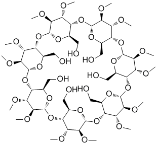
| SMILES | CO[C@H]1[C@@H]([C@H]2[C@H](O[C@@H]1O[C@@H]3[C@H](O[C@@H]([C@H]([C@@H]3OC)OC)O[C@@H]4[C@H](O[C@@H]([C@H]([C@@H]4OC)OC)O[C@@H]5[C@H](O[C@@H]([C@H]([C@@H]5OC)OC)O[C@@H]6[C@H](O[C@@H]([C@H]([C@@H]6OC)OC)O[C@@H]7[C@H](O[C@@H]([C@H]([C@@H]7OC)OC)O[C@@H]8[C@H](O[C@H](O2)[C@H]([C@@H]8O)OC)CO)CO)CO)CO)CO)CO)CO)O |
| InChi Key | YZOUYRAONFXZSI-SBHWVFSVSA-N |
| InChi Code | InChI=1S/C54H94O35/c1-64-36-28(63)30-21(14-56)76-48(36)83-29-20(13-55)77-49(37(65-2)27(29)62)85-31-22(15-57)79-51(44(72-9)38(31)66-3)87-33-24(17-59)81-53(46(74-11)40(33)68-5)89-35-26(19-61)82-54(47(75-12)42(35)70-7)88-34-25(18-60)80-52(45(73-10)41(34)69-6)86-32-23(16-58)78-50(84-30)43(71-8)39(32)67-4/h20-63H,13-19H2,1-12H3/t20-,21-,22-,23-,24-,25-,26-,27-,28-,29-,30-,31-,32-,33-,34-,35-,36+,37+,38-,39-,40-,41-,42-,43+,44+,45+,46+,47+,48-,49-,50-,51-,52-,53-,54-/m1/s1 |
| Chemical Name | (1S,3R,5R,6R,8R,10R,11R,13R,15R,16R,18R,20R,21R,23R,25R,26R,28R,30R,31S,33R,35R,36R,37S,38R,39S,40R,41S,42R,43S,44R,45S,46R,47S,48R,49S)-5,10,15,20,25,30,35-heptakis(hydroxymethyl)-37,39,40,41,42,43,44,45,46,47,48,49-dodecamethoxy-2,4,7,9,12,14,17,19,22,24,27,29,32,34-tetradecaoxaoctacyclo[31.2.2.23,6.28,11.213,16.218,21.223,26.228,31]nonatetracontane-36,38-diol |
| Synonyms | 128446-36-6; Methyl-b-cyclodextrin; Methyl-; A-cyclodextrin; MFCD00074980; |
| HS Tariff Code | 2934.99.9001 |
| Storage |
Powder-20°C 3 years 4°C 2 years In solvent -80°C 6 months -20°C 1 month |
| Shipping Condition | Room temperature (This product is stable at ambient temperature for a few days during ordinary shipping and time spent in Customs) |
Biological Activity
| Targets |
IC50: 3.33-4.23 mM (PEL cell growth)[1]; Solubility enhancer; Deodorizer Actin cytoskeleton. Treatment with 1 mM Methyl-β-cyclodextrin (MCD) for 4 hours significantly depolymerized the actin network in HeLa cells, reducing the fluorescence intensity of phalloidin-stained actin filaments by 49 ± 3% (p < 0.01). MCD did not perturb the microtubule network [1] Plasma membrane cholesterol. Methyl-β-cyclodextrin (MβCD) selectively extracts cholesterol from the plasma membrane of mammalian cells [2] |
| ln Vitro |
For the purpose of enhancing the intake of tiny molecules like glucose and nanoparticles, methyl-β-cyclodextrin is widely employed to raise the permeability of cells[4]. Cyclodextrins are a kind of cyclic oligosaccharides that have a lipophilic core chamber and a hydrophilic outside. Generally speaking, cyclodextrin molecules do not penetrate lipophilic membranes since they are rather big molecules with many hydrogen sources and acceptors. The principal application of cyclodextrins in the pharmaceutical industry has been as complexing agents to improve the aqueous solubility, bioavailability, and stability of poorly soluble medicines. Drugs' bioavailability is one of the many uses for cyclodextrins in medicinal applications[4]. By rapidly removing cholesterol from the plasma membrane, methyl-β-cyclodextrin causes PEL cells to undergo caspase-dependent death. All PEL cell lines are inhibited in their development by methyl-β-cyclodextrin in a way that is dose dependent. Every cell line has an IC50 of 3.33–4.23 mM[5]. Among the several agents that deplete cholesterol from cells, methyl-β-cyclodextrin—a highly soluble cyclic heptasaccharide with a β glucopyranose unit—has been found to be the most efficient[5]. Methyl-β-cyclodextrin (MCD) (1 mM, 4h) depolymerized the actin cytoskeleton in HeLa cells, reducing F-actin fluorescence intensity by 49%. This led to a 56% reduction in paxillin focal adhesion area and a 66% reduction in phosphorylated focal adhesion kinase (pFAK) area. Atomic force microscopy showed a 50% decrease in cell stiffness (from 4.3 ± 0.3 kPa to 2.1 ± 0.2 kPa). Traction force microscopy revealed a 65% reduction in cellular traction force (from 367 ± 15 Pa to 128 ± 11 Pa). MCD treatment increased the intracellular accumulation of microtubule-targeting agents (MTAs) like BODIPY-vinblastine, crocin, and curcumin by approximately 50% compared to untreated cells. Pretreatment with 1 mM MCD for 4 hours potentiated the antiproliferative effects of vinblastine, taxol, and crocin in HeLa cells, lowering their IC50 values. For example, the IC50 of vinblastine decreased from 4.2 ± 0.1 nM (alone) to 1.9 ± 0.2 nM (with MCD pretreatment). MCD pretreatment also augmented the effects of vinblastine on cell cycle arrest at G2/M phase and enhanced the disruption of interphase and mitotic microtubules by vinblastine, taxol, and crocin. Furthermore, the combination of low-dose MCD (0.25 or 1 mM) with vinblastine synergistically inhibited HeLa cell proliferation, with Combination Index (CI) values of 0.55 and 0.43, respectively. Similar sensitization effects of MCD pretreatment were observed in breast (MCF-7), liver (Huh7), prostate (PC3) cancer cells, and a multidrug-resistant breast cancer cell line (EMT6/AR1) [1] Pretreatment with 1 mM MCD for 4 hours alone did not affect HeLa cell proliferation up to 24 hours, but higher concentrations showed inhibitory effects [1] Pretreatment of various cell lines (HEp-2, NIH/3T3, MDCK II, A431) with 10 mM Methyl-β-cyclodextrin (MβCD) for 15 minutes at 37°C strongly inhibited clathrin-dependent endocytosis. Endocytosis of transferrin was reduced by approximately 50% across all tested cell lines. Endocytosis of epidermal growth factor (EGF) was also strongly reduced (by ~40%). In contrast, endocytosis of ricin, a general membrane marker that enters via both clathrin-dependent and -independent pathways, was less affected, showing only about 20% inhibition. This differential effect suggests that MβCD specifically perturbs clathrin-dependent endocytosis more than clathrin-independent pathways. The inhibition of transferrin endocytosis was concentration-dependent (50% reduction at ~10 mM MβCD) and was completely reversible upon removal of MβCD. Recovery of endocytosis occurred over time (fully by 3 hours) even in serum-free medium and was dependent on cholesterol synthesis, as it was inhibited by lovastatin (an inhibitor of cholesterol synthesis) unless water-soluble cholesterol was added concurrently. Electron microscopy revealed that MβCD treatment caused the disappearance of typically invaginated caveolae and strongly inhibited the invagination of clathrin-coated pits, leading to an accumulation of shallow, flattened pits. However, quantitative immunogold labeling showed that transferrin receptors (TfRs) were still concentrated in these shallow coated pits to the same degree (~7-fold) as in control cells. Surface binding of transferrin was increased nearly twofold after MβCD treatment, consistent with inhibited internalization. Treatment with 10 mM MβCD did not significantly affect cellular protein synthesis or intracellular potassium content, indicating no large-scale plasma membrane permeability changes under these conditions [2] The inhibitory effect on transferrin endocytosis was specific to cyclodextrins capable of binding cholesterol (MβCD and β-cyclodextrin), while α-cyclodextrin and γ-cyclodextrin had no significant effect [2] Treatment with Methyl-β-cyclodextrin blocked migrasome formation in cells, indicating that cholesterol is necessary for migrasome biogenesis[3] |
| ln Vivo | Methyl-β-cyclodextrin effectively suppresses PEL cell growth and invasion in a PEL xenograft mice model without causing any evident side effects. Mice treated with methyl-β-cyclodextrin seem healthy, whereas those not treated have enlarged abdomens. The mice treated with methyl-β-cyclodextrin had significantly lower body weights than the control group. Mice treated with methyl-β-cyclodextrin had a much smaller volume of ascites than mice not treated with it[4]. Cyclodextrins have been found in studies on humans and animals to be useful in enhancing the distribution of nearly any kind of medication formulation. Around 30 distinct pharmaceutical products with drug/cyclodextrin complexes are available on the market at the moment[6] throughout the world. |
| Cell Assay |
PEL cells are incubated in triplicate in a 96-well microculture plate in the presence of different concentrations of methyl-β-cyclodextrin (0-10 mM) in a final volume of 0.1 mL for 24 h at 37°C. Subsequently, MTT (0.5 mg/mL final concentration) is added to each well. After 3 h of additional incubation, 100 μL of a 0.04 N HCl is added to dissolve the crystals. Absorption values at 570 nm are determined[1]. Tetrazolium dye methylthiotetrazole (MTT) assay[5] The antiproliferative activities of M-β-CyD against PEL cell lines were measured by the methylthiotetrazole (MTT) method. Briefly, 2 × 10⁴ cells were incubated in triplicate in a 96-well microculture plate in the presence of different concentrations of M-β-CyD (0–10 mM) in a final volume of 0.1 ml for 24 h at 37 °C. Subsequently, MTT (0.5 mg/ml final concentration) was added to each well. After 3 h of additional incubation, 100 μl of a 0.04 N HCl was added to dissolve the crystals. Absorption values at 570 nm were determined with an automatic enzyme-linked immunosorbent assay (ELISA) plate reader. Values were normalized to untreated (control) samples. Cell viability assay[5] Cell viability was examined by the propidium iodide (PI) exclusion method as described previously. Briefly, BCBL-1 cells (2 × 10⁵ cells/ml) were cultured in the presence or absence of M-β-CyD for 1–6 h in 6-well culture plates. After being incubated, cells were stained with PI (final concentration; 2 μg/ml) and cell viability was analyzed by LSR II flow cytometry. 1) Immunofluorescence for Cytoskeleton and Focal Adhesions: Cells were seeded on coverslips, treated with 1 mM Methyl-β-cyclodextrin (MCD) for 4 hours, and subsequently with other drugs as specified. After incubation, cells were fixed, permeabilized, and blocked. They were incubated with primary antibodies (e.g., anti-β-tubulin, anti-paxillin, anti-pFAK) overnight at 4°C, followed by appropriate fluorescent secondary antibodies. Actin was stained using fluorescent phalloidin, and nuclei were stained with Hoechst. Images were captured using a confocal microscope, and fluorescence intensity or focal adhesion area was quantified using image analysis software [1] 2) Drug Accumulation Assay: Cells were treated with 1 mM MCD for 4 hours. The medium was replaced with fresh medium containing fluorescent drugs (e.g., 5 nM BODIPY-vinblastine for 6h; 1 µM crocin or 5 µM curcumin for 24h). Cells were then trypsinized, washed, counted, and lysed. The fluorescence (for BODIPY-vinblastine) or absorbance (for crocin and curcumin) of the supernatant (cytosolic extract) was measured to determine intracellular drug concentration relative to untreated controls [1] 3) Cell Proliferation Assay (Sulforhodamine B assay): Cells were seeded in a plate, allowed to attach, and then incubated with 1 mM MCD (or 200 nM Latrunculin B as a positive control) for 4 hours. The medium was replaced with fresh medium containing varying concentrations of the test drug (e.g., vinblastine, taxol, crocin). After incubation for one cell cycle, cells were fixed, stained with Sulforhodamine B dye, and the absorbance was measured at 520 nm to determine the inhibition of proliferation and calculate IC50 values [1] 4) Cell Cycle Analysis by Flow Cytometry: Cells were treated with 1 mM MCD for 4 hours, followed by incubation with a drug (e.g., 5 nM vinblastine) for 24 hours. Cells were then fixed, treated with RNase A and propidium iodide (PI), and analyzed by flow cytometry to determine DNA content and cell cycle distribution [1] 5) BrdU Assay: Cells on coverslips were treated with 1 mM MCD for 4 hours (or left untreated), followed by treatment with 5 nM vinblastine for 8 hours. BrdU reagent was added for the last 4 hours of incubation. Cells were then processed for immunofluorescence using an anti-BrdU antibody and a fluorescent secondary antibody to label cells in S-phase. Nuclei were counterstained with Hoechst. The percentage of BrdU-positive cells was manually counted [1] 6) Trypsin Deadhesion Assay: Cells were seeded on coverslips and treated with drugs. After treatment, trypsin was added, and time-lapse images were captured at intervals until cells completely detached. Cell area over time was quantified, and time constants (τ1 and τ2) describing detachment kinetics were calculated using curve-fitting software [1] 7) Traction Force Microscopy (TFM): Cells were seeded on soft polyacrylamide gels embedded with fluorescent beads and coated with collagen. After treatment with MCD and/or other drugs, phase-contrast images of cells and fluorescence images of the beads were taken. Cells were then lysed, and bead images were taken again. Bead displacement fields were calculated, and traction forces exerted by cells were computed using specialized analysis code [1] 8) Atomic Force Microscopy (AFM): Cells on coverslips were treated with 1 mM MCD for 4 hours. A pyramidal tip probe was used in contact mode to obtain force curves on the cell surface. The stiffness (Young's modulus) of the cells was calculated by fitting the force curves with the Hertz model [1] 9) Determination of Combination Index: Cells were incubated with different concentrations of vinblastine in combination with either 0.25 mM or 1 mM MCD for 24 hours. Cell proliferation was assessed. The median dose (Dm) for each agent alone was determined from dose-response curves. The Combination Index (CI) was calculated using the Chou and Talalay method, where CI < 1 indicates synergy [1] 1) Endocytosis Assays (Transferrin, EGF, Ricin): Cells were washed and preincubated with or without Methyl-β-cyclodextrin (MβCD) (e.g., 10 mM) in HEPES-buffered medium for 15 minutes at 37°C. Radiolabeled ligands (¹²⁵I-transferrin, ¹²⁵I-EGF, or ¹²⁵I-ricin) were then added to the cells. For transferrin and EGF, after a short incubation (5 or 10 minutes) at 37°C, surface-bound ligand was removed (using protease treatment for transferrin or low-pH buffer wash for EGF). The remaining cell-associated radioactivity (representing endocytosed ligand) was measured using a gamma counter. For ricin, after a 15-minute incubation, cells were washed with medium containing lactose to remove surface-bound toxin, and the internalized radioactivity was measured [2] 2) Cholesterol Extraction Assay: Cells were labeled with [³H]cholesterol either briefly (15 min) to label the cell surface or for 20 hours to equilibrate with intracellular pools. After washing, cells were incubated with or without 10 mM MβCD in medium for various times (15, 30, 60 min) at 37°C. Cells were then lysed, and the remaining radioactivity in the lysate was measured by scintillation counting to determine the percentage of cholesterol extracted [2] 3) Electron Microscopy and Morphometric Analysis: Cells treated with or without MβCD were fixed with formaldehyde/glutaraldehyde, post-fixed with osmium tetroxide, and processed for embedding in resin. Ultrathin sections were stained and examined by electron microscopy. The frequency of invaginated caveolae and the morphology of clathrin-coated pits (classified as shallow, invaginated, or nearly pinched-off) were quantified [2] 4) Immunogold Labeling for Transferrin Receptors: Cells were fixed, incubated with a primary anti-transferrin receptor antibody, followed by a secondary antibody conjugated to colloidal gold particles. After processing for electron microscopy, the distribution of gold particles on the cell surface (within coated pits vs. outside pits) was quantified to calculate the concentration efficiency of receptors in coated pits [2] 5) Protein Synthesis Assay: Cells were treated with or without MβCD, then incubated with [³H]leucine for 10 minutes. Cells were washed, precipitated with trichloroacetic acid, and the incorporated radioactivity in the precipitated protein was measured using a beta-counter [2] 6) Intracellular Potassium Measurement: Cells treated with or without MβCD were washed with magnesium chloride, air-dried, and dissolved in sodium hydroxide. The potassium content in the solution was determined using ion-selective electrodes [2] 7) Reversibility and Cholesterol Dependence Assay: Cells were treated with MβCD to inhibit endocytosis, then the medium was replaced with fresh medium without MβCD. Cells were further incubated for varying times (15 min to 3 hours) in the presence or absence of fetal calf serum, water-soluble cholesterol, and/or lovastatin. After this recovery period, transferrin endocytosis was measured as described above to assess recovery [2] Cells were treated with Methyl-β-cyclodextrin to deplete cholesterol. After treatment, migrasome formation was assessed by confocal microscopy. The treatment resulted in inhibition of migrasome formation, supporting the role of cholesterol in migrasome assembly[3] |
| Animal Protocol | NOD/Rag-2/Jak3-double deficient (NRJ) mice were housed and monitored in our animal research facility according to institutional guidelines. All experimental procedures and protocols were approved by the Institutional Animal Care and Use Committee at Kumamoto University. Eight- to ten-week-old female NRJ mice were intraperitoneally inoculated with 7 × 10⁶ BCBL-1 cells suspended in 200 μl PBS. The mice were then treated with intraperitoneal injections of PBS or M-β-CyD (500 mg/kg per day). Tumor burdens were evaluated by measuring body weights and ascites.[5] |
| Toxicity/Toxicokinetics |
Treatment of cells with 10 mM Methyl-β-cyclodextrin (MβCD) for 15 minutes did not cause significant membrane leakage, as evidenced by no reduction in protein synthesis and no change in intracellular potassium content in multiple cell lines (HEp-2, MDCK II, A431, NIH/3T3) [2] |
| References |
[1]. Methyl-β-cyclodextrin, an actin depolymerizer augments the antiproliferative potential of microtubule-targeting agents. Sci Rep. 2019 May 21;9(1):7638. [2]. Extraction of cholesterol with methyl-beta-cyclodextrin perturbs formation of clathrin-coated endocytic vesicles. Mol Biol Cell. 1999;10(4):961-974. [3]. Migrasome formation is mediated by assembly of micron-scale tetraspanin macrodomains. Nat Cell Biol. 2019 Aug;21(8):991-1002. [4]. Cholesterol depletion from the plasma membrane triggers ligand-independent activation of the epidermal growth factor receptor. J Biol Chem. 2002 Dec 20;277(51):49631-7. [5]. The antitumor effects of methyl-β-cyclodextrin against primary effusion lymphoma via the depletion of cholesterol from lipid rafts. Biochem Biophys Res Commun. 2014 Dec 12;455(3-4):285-9. [6]. Cyclodextrins in delivery systems: Applications. J Pharm Bioallied Sci. 2010 Apr;2(2):72-9. |
| Additional Infomation |
Cyclodextrins (CDs) are a family of cyclic oligosaccharides with a hydrophilic outer surface and a lipophilic central cavity. CD molecules are relatively large with a number of hydrogen donors and acceptors and, thus in general, they do not permeate lipophilic membranes. In the pharmaceutical industry, CDs have mainly been used as complexing agents to increase aqueous solubility of poorly soluble drugs and to increase their bioavailability and stability. CDs are used in pharmaceutical applications for numerous purposes, including improving the bioavailability of drugs. Current CD-based therapeutics is described and possible future applications are discussed. CD-containing polymers are reviewed and their use in drug delivery is presented. Of specific interest is the use of CD-containing polymers to provide unique capabilities for the delivery of nucleic acids. Studies in both humans and animals have shown that CDs can be used to improve drug delivery from almost any type of drug formulation. Currently, there are approximately 30 different pharmaceutical products worldwide containing drug/CD complexes in the market.[6] \n\nPrimary effusion lymphoma (PEL) is a subtype of aggressive and chemotherapy-resistant non-Hodgkin lymphoma that occurs predominantly in patients with advanced AIDS. In this study, we examined the antitumor activity of methyl-β-cyclodextrin (M-β-CyD) in vitro and in vivo. M-β-CyD quickly induced caspase-dependent apoptosis in PEL cells via cholesterol depletion from the plasma membrane. In a PEL xenograft mouse model, M-β-CyD significantly inhibited the growth and invasion of PEL cells without apparent adverse effects. These results strongly suggest that M-β-CyD has the potential to be an effective antitumor agent against PEL.[5] \n\nMethyl-β-cyclodextrin (MCD), an established pharmacological excipient, depolymerizes the actin cytoskeleton. In this work, we investigated the effect of MCD-mediated actin depolymerization on various cellular phenotypes including traction force, cell stiffness, focal adhesions, and intracellular drug accumulation. In addition to a reduction in the contractile cellular traction, MCD acutely inhibits the maturation of focal adhesions. Alteration of contractile forces and focal adhesions affects the trypsin-mediated detachment kinetics of cells. Moreover, MCD-mediated actin depolymerization increases the intracellular accumulation of microtubule-targeting agents (MTAs) by ~50% with respect to the untreated cells. As MCD treatment enhances the intracellular concentration of drugs, we hypothesized that the MCD-sensitized cancer cells could be effectively killed by low doses of MTAs. Our results in cervical, breast, hepatocellular, prostate cancer and multidrug-resistant breast cancer cells confirmed the above hypothesis. Further, the combined use of MCD and MTAs synergistically inhibits the proliferation of tumor cells. These results indicate the potential use of MCD in combination with MTAs for cancer chemotherapy and suggest that targeting both actin and microtubules simultaneously may be useful for cancer therapy. Importantly, the results provide significant insight into the crosstalk between actin and microtubules in regulating the traction force and dynamics of cell deadhesion.[1] \n\nThe importance of cholesterol for endocytosis has been investigated in HEp-2 and other cell lines by using methyl-beta-cyclodextrin (MbetaCD) to selectively extract cholesterol from the plasma membrane. MbetaCD treatment strongly inhibited endocytosis of transferrin and EGF, whereas endocytosis of ricin was less affected. The inhibition of transferrin endocytosis was completely reversible. On removal of MbetaCD it was restored by continued incubation of the cells even in serum-free medium. The recovery in serum-free medium was inhibited by addition of lovastatin, which prevents cholesterol synthesis, but endocytosis recovered when a water-soluble form of cholesterol was added together with lovastatin. Electron microscopical studies of MbetaCD-treated HEp-2 cells revealed that typical invaginated caveolae were no longer present. Moreover, the invagination of clathrin-coated pits was strongly inhibited, resulting in accumulation of shallow coated pits. Quantitative immunogold labeling showed that transferrin receptors were concentrated in coated pits to the same degree (approximately sevenfold) after MbetaCD treatment as in control cells. Our results therefore indicate that although clathrin-independent (and caveolae-independent) endocytosis still operates after removal of cholesterol, cholesterol is essential for the formation of clathrin-coated endocytic vesicles.[2] Methyl-β-cyclodextrin (MCD) is an established pharmacological excipient used to enhance drug solubility, stability, and bioavailability. It is biocompatible and some derivatives are FDA-approved. In this study, MCD is shown to depolymerize the actin cytoskeleton, which increases plasma membrane permeability. This property allows it to enhance the intracellular accumulation and efficacy of co-administered Microtubule-Targeting Agents (MTAs) like vinblastine and taxol. The work suggests that combining actin cytoskeleton perturbation (via MCD) with microtubule perturbation (via MTAs) represents a potential strategy to overcome drug resistance and improve cancer chemotherapy [1] Methyl-β-cyclodextrin (MβCD) is used in this study as a tool to selectively deplete cholesterol from the plasma membrane. The results demonstrate that cholesterol is essential for the invagination and vesicle formation stages of clathrin-dependent endocytosis, but not for the initial assembly of clathrin coats or the concentration of specific receptors (like the transferrin receptor) within these coated pits. The study also shows that a clathrin-independent (and caveolae-independent) endocytic pathway is less sensitive to cholesterol depletion [2] Methyl-β-cyclodextrin was used as a cholesterol-depleting reagent to study the role of cholesterol in migrasome formation. It was shown that cholesterol depletion impairs migrasome formation, indicating cholesterol is an essential component of migrasomal membrane domains[3] |
Solubility Data
| Solubility (In Vitro) |
DMSO : ≥ 100 mg/mL H2O : ≥ 50 mg/mL |
| Solubility (In Vivo) |
Note: Listed below are some common formulations that may be used to formulate products with low water solubility (e.g. < 1 mg/mL), you may test these formulations using a minute amount of products to avoid loss of samples. Injection Formulations (e.g. IP/IV/IM/SC) Injection Formulation 1: DMSO : Tween 80: Saline = 10 : 5 : 85 (i.e. 100 μL DMSO stock solution → 50 μL Tween 80 → 850 μL Saline) *Preparation of saline: Dissolve 0.9 g of sodium chloride in 100 mL ddH ₂ O to obtain a clear solution. Injection Formulation 2: DMSO : PEG300 :Tween 80 : Saline = 10 : 40 : 5 : 45 (i.e. 100 μL DMSO → 400 μLPEG300 → 50 μL Tween 80 → 450 μL Saline) Injection Formulation 3: DMSO : Corn oil = 10 : 90 (i.e. 100 μL DMSO → 900 μL Corn oil) Example: Take the Injection Formulation 3 (DMSO : Corn oil = 10 : 90) as an example, if 1 mL of 2.5 mg/mL working solution is to be prepared, you can take 100 μL 25 mg/mL DMSO stock solution and add to 900 μL corn oil, mix well to obtain a clear or suspension solution (2.5 mg/mL, ready for use in animals). Injection Formulation 4: DMSO : 20% SBE-β-CD in saline = 10 : 90 [i.e. 100 μL DMSO → 900 μL (20% SBE-β-CD in saline)] *Preparation of 20% SBE-β-CD in Saline (4°C,1 week): Dissolve 2 g SBE-β-CD in 10 mL saline to obtain a clear solution. Injection Formulation 5: 2-Hydroxypropyl-β-cyclodextrin : Saline = 50 : 50 (i.e. 500 μL 2-Hydroxypropyl-β-cyclodextrin → 500 μL Saline) Injection Formulation 6: DMSO : PEG300 : castor oil : Saline = 5 : 10 : 20 : 65 (i.e. 50 μL DMSO → 100 μLPEG300 → 200 μL castor oil → 650 μL Saline) Injection Formulation 7: Ethanol : Cremophor : Saline = 10: 10 : 80 (i.e. 100 μL Ethanol → 100 μL Cremophor → 800 μL Saline) Injection Formulation 8: Dissolve in Cremophor/Ethanol (50 : 50), then diluted by Saline Injection Formulation 9: EtOH : Corn oil = 10 : 90 (i.e. 100 μL EtOH → 900 μL Corn oil) Injection Formulation 10: EtOH : PEG300:Tween 80 : Saline = 10 : 40 : 5 : 45 (i.e. 100 μL EtOH → 400 μLPEG300 → 50 μL Tween 80 → 450 μL Saline) Oral Formulations Oral Formulation 1: Suspend in 0.5% CMC Na (carboxymethylcellulose sodium) Oral Formulation 2: Suspend in 0.5% Carboxymethyl cellulose Example: Take the Oral Formulation 1 (Suspend in 0.5% CMC Na) as an example, if 100 mL of 2.5 mg/mL working solution is to be prepared, you can first prepare 0.5% CMC Na solution by measuring 0.5 g CMC Na and dissolve it in 100 mL ddH2O to obtain a clear solution; then add 250 mg of the product to 100 mL 0.5% CMC Na solution, to make the suspension solution (2.5 mg/mL, ready for use in animals). Oral Formulation 3: Dissolved in PEG400 Oral Formulation 4: Suspend in 0.2% Carboxymethyl cellulose Oral Formulation 5: Dissolve in 0.25% Tween 80 and 0.5% Carboxymethyl cellulose Oral Formulation 6: Mixing with food powders Note: Please be aware that the above formulations are for reference only. InvivoChem strongly recommends customers to read literature methods/protocols carefully before determining which formulation you should use for in vivo studies, as different compounds have different solubility properties and have to be formulated differently. (Please use freshly prepared in vivo formulations for optimal results.) |
| Preparing Stock Solutions | 1 mg | 5 mg | 10 mg | |
| 1 mM | 0.7673 mL | 3.8364 mL | 7.6728 mL | |
| 5 mM | 0.1535 mL | 0.7673 mL | 1.5346 mL | |
| 10 mM | 0.0767 mL | 0.3836 mL | 0.7673 mL |