ML133 HCl, the hydrochloride salt of ML-133, is a novel, potent and selective inhibitor of the inward-rectifier potassium channel 2 (Kir2) identified from a high-throughput screening (HTS) of more than 300,000 small molecules. It inhibits Kir2.1 with an IC50 of 1.8 μM (at pH 7.4) and 290 nM (at pH 8.5), and shows no/little effect on Kir1.1, Kir4.1 and Kir7.1. The K(ir) inward rectifying potassium channels have a broad tissue distribution and are implicated in a variety of functional roles.
Physicochemical Properties
| Molecular Formula | C19H19NO.HCL | |
| Molecular Weight | 313.82 | |
| Exact Mass | 313.123 | |
| CAS # | 1222781-70-5 | |
| Related CAS # |
|
|
| PubChem CID | 44247466 | |
| Appearance | White to off-white solid powder | |
| LogP | 5.331 | |
| Hydrogen Bond Donor Count | 2 | |
| Hydrogen Bond Acceptor Count | 2 | |
| Rotatable Bond Count | 5 | |
| Heavy Atom Count | 22 | |
| Complexity | 298 | |
| Defined Atom Stereocenter Count | 0 | |
| InChi Key | NGQIBUUFXDPHKT-UHFFFAOYSA |
|
| InChi Code | InChI=1S/C19H19NO.ClH/c1-21-18-11-9-15(10-12-18)13-20-14-17-7-4-6-16-5-2-3-8-19(16)17;/h2-12,20H,13-14H2,1H3;1H | |
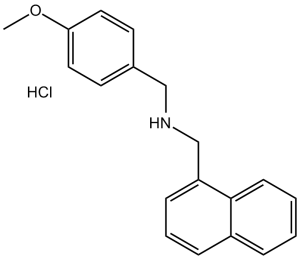
| Chemical Name | 1-(4-methoxyphenyl)-N-(naphthalen-1-ylmethyl)methanamine;hydrochloride | |
| Synonyms |
|
|
| HS Tariff Code | 2934.99.9001 | |
| Storage |
Powder-20°C 3 years 4°C 2 years In solvent -80°C 6 months -20°C 1 month Note: Please store this product in a sealed and protected environment, avoid exposure to moisture. |
|
| Shipping Condition | Room temperature (This product is stable at ambient temperature for a few days during ordinary shipping and time spent in Customs) |
Biological Activity
| Targets |
ML133 HCl (hydrochloride salt of ML133) is a selective inhibitor of the inward rectifier potassium channel K(ir)2 family, with potent activity against K(ir)2.1, K(ir)2.2, and K(ir)2.3 subtypes. Its IC50 values are: K(ir)2.1 (1.8 μM), K(ir)2.2 (2.5 μM), K(ir)2.3 (3.2 μM) (measured via whole-cell patch clamp in transfected HEK293 cells). It exhibits no significant activity against other potassium channels, including K(ir)1.1 (IC50 > 10 μM), K(v)1.5 (IC50 > 20 μM), K(ATP) (IC50 > 20 μM), and calcium channels (e.g., Ca(v)1.2, IC50 > 20 μM), confirming K(ir)2 family selectivity [1] |
| ln Vitro |
ML133 hydrochloride exhibits little selectivity for other Kir2.x family of channels, but it blocks Kir2.1 with an IC50 of 1.8 μM at pH 7.4 and 290 nM at pH 8.5 [1]. The hydrochloride ML133 demonstrates minimal efficacy against Kir1.1 (IC50 > 300 μM), Kir4.1 (76 μM), and Kir7.1 (33 μM) [1]. K(ir)2 channel current inhibition : 1. In HEK293 cells stably expressing K(ir)2.1: ML133 HCl dose-dependently inhibits inward rectifier current (I(Kir)2.1) under whole-cell patch clamp. At 1 μM, it inhibits current by 45%; at 10 μM, inhibition reaches 85%. The current-voltage relationship shows reduced inward current at hyperpolarizing potentials (-120 to -60 mV) without altering voltage dependence. 2. In primary rat ventricular myocytes: ML133 HCl inhibits the endogenous inward rectifier potassium current I(K1) (a major component of K(ir)2.1) with an IC50 of 2.2 μM. 5 μM ML133 HCl reduces I(K1) by 70% at -100 mV, leading to a small depolarization of resting membrane potential (from -85 mV to -78 mV) without affecting action potential duration. 3. Selectivity validation: In HEK293 cells expressing non-K(ir)2 channels (e.g., K(ir)1.1, K(v)1.5), 20 μM ML133 HCl inhibits current by <10%, confirming no off-target channel effects [1] - Cell viability : ML133 HCl shows low cytotoxicity to mammalian cells: (1) HEK293 cells (transfected with K(ir)2.1) treated with 0-20 μM ML133 HCl for 72 hours maintain >90% viability (MTT assay); (2) Primary rat ventricular myocytes treated with 10 μM ML133 HCl for 24 hours show no significant increase in apoptotic cells (Annexin V/PI staining, apoptotic rate <5% vs. control) [1] |
| Enzyme Assay |
Whole-cell patch clamp assay for K(ir)2 channel activity: 1. Cell preparation: HEK293 cells are stably transfected with human K(ir)2.1, K(ir)2.2, or K(ir)2.3 cDNA, then cultured in DMEM medium supplemented with 10% FBS and antibiotics. Cells are plated on glass coverslips 24 hours before experiments and maintained at 37°C (5% CO₂). 2. Patch clamp setup: Experiments are performed at room temperature (22-25°C) using a patch clamp amplifier in whole-cell voltage-clamp mode. Borosilicate glass electrodes (resistance: 2-4 MΩ) are filled with internal solution (in mM: 140 KCl, 10 HEPES, 1 EGTA, 2 MgATP, pH 7.2 with KOH). The external bath solution contains (in mM: 140 NaCl, 5 KCl, 2 CaCl₂, 1 MgCl₂, 10 HEPES, pH 7.4 with NaOH). 3. Current recording: After achieving whole-cell configuration, cells are clamped at a holding potential of -60 mV. Inward currents are elicited by a voltage protocol: 200-ms step potentials from -120 mV to +40 mV in 20-mV increments, followed by a return to -60 mV. Current amplitudes are measured at the end of each step. 4. Drug treatment: Serial concentrations of ML133 HCl (0.1-30 μM, dissolved in external solution with 0.1% DMSO) are perfused into the bath. Steady-state current inhibition is recorded after 5 minutes of perfusion per concentration. 5. Data analysis: Current inhibition rates are calculated as [(control current - drug-treated current)/control current] × 100%. IC50 values are determined by fitting dose-response curves with a four-parameter logistic model using GraphPad Prism [1] - Fluorescent membrane potential assay for K(ir)2 activity : 1. Cell seeding: K(ir)2.1-transfected HEK293 cells are seeded in 96-well black-walled plates (1×10⁴ cells/well) and cultured overnight. 2. Dye loading: Cells are incubated with 5 μM membrane potential-sensitive dye (e.g., DiBAC4(3)) in HBSS buffer for 30 minutes at 37°C (5% CO₂). 3. Drug treatment: Serial concentrations of ML133 HCl (0.3-30 μM) are added, and fluorescence intensity (excitation: 485 nm, emission: 535 nm) is measured every 2 minutes for 20 minutes using a microplate reader. 4. Data analysis: Fluorescence changes (ΔF/F0) are normalized to the maximum response induced by 50 mM KCl (positive control). EC50 for membrane depolarization (a surrogate for K(ir)2 inhibition) is consistent with the patch clamp-derived IC50 [1] |
| Cell Assay |
Primary rat ventricular myocyte isolation and I(K1) recording : 1. Myocyte isolation: Ventricular myocytes are isolated from adult male Sprague-Dawley rats (250-300 g) via retrograde aortic perfusion with collagenase-containing solution. Cells are filtered through a 200-μm mesh, washed with KB buffer, and stored at room temperature for 1-4 hours before use. 2. Patch clamp recording: Whole-cell voltage-clamp experiments are performed as described for HEK293 cells, with minor modifications to internal/external solutions (e.g., internal solution includes 5 mM EGTA to reduce calcium currents). I(K1) is recorded using a voltage protocol from -120 mV to -40 mV (20-mV steps) from a holding potential of -70 mV. 3. ML133 HCl treatment: 1-10 μM ML133 HCl is perfused, and I(K1) amplitude at -100 mV is measured to calculate inhibition rates [1] - MTT cell viability assay : 1. Cell seeding: HEK293 cells (K(ir)2.1-transfected or parental) and primary rat ventricular myocytes are seeded in 96-well plates (5×10³ cells/well for HEK293, 2×10³ cells/well for myocytes) and cultured overnight. 2. Drug treatment: Serial concentrations of ML133 HCl (0.1-20 μM) are added, with 3 replicates per concentration. Plates are incubated at 37°C (5% CO₂) for 72 hours (HEK293) or 24 hours (myocytes). 3. MTT reaction: 20 μL MTT solution (5 mg/mL in PBS) is added to each well, and plates are incubated for 4 hours. The supernatant is removed, and 150 μL DMSO is added to dissolve formazan crystals. 4. Absorbance measurement: Absorbance at 570 nm is measured using a microplate reader. Cell viability is calculated as (A570 of sample / A570 of vehicle control) × 100% [1] |
| Animal Protocol | N/A N/A |
| Toxicity/Toxicokinetics |
In vitro toxicity : As described in the In Vitro section, ML133 HCl (up to 20 μM) shows no significant cytotoxicity to HEK293 cells (viability >90%) or primary rat ventricular myocytes (apoptotic rate <5%). It does not induce changes in cell morphology (e.g., rounding, detachment) at concentrations up to 10 μM [1] |
| References |
[1]. Selective inhibition of the K(ir)2 family of inward rectifier potassium channels by a small molecule probe: the discovery, SAR, and pharmacological characterization of ML133. ACS Chem Biol. 2011 Aug 19;6(8):845-56. |
| Additional Infomation |
ML133 HCl is a first-in-class selective small-molecule probe for the K(ir)2 family of inward rectifier potassium channels, developed to address the lack of selective tools for studying K(ir)2 function. Unlike non-selective K(ir) inhibitors (e.g., Ba²⁺), it does not affect other ion channels, making it suitable for dissecting the role of K(ir)2 channels in physiology and disease [1] - Physiological relevance: K(ir)2 channels (especially K(ir)2.1) are critical for maintaining resting membrane potential in excitable cells (e.g., cardiomyocytes, neurons) and regulating cardiac rhythm. ML133 HCl is used to investigate the contribution of K(ir)2 channels to cardiac arrhythmias, epilepsy, and smooth muscle contraction [1] - Development status: ML133 HCl is a research tool compound, not intended for clinical development. It has no reported FDA approval or investigational new drug (IND) status, and its use is limited to in vitro and ex vivo studies [1] |
Solubility Data
| Solubility (In Vitro) |
|
|||
| Solubility (In Vivo) |
Solubility in Formulation 1: ≥ 2.08 mg/mL (6.63 mM) (saturation unknown) in 10% DMSO + 40% PEG300 + 5% Tween80 + 45% Saline (add these co-solvents sequentially from left to right, and one by one), clear solution. For example, if 1 mL of working solution is to be prepared, you can add 100 μL of 20.8 mg/mL clear DMSO stock solution to 400 μL PEG300 and mix evenly; then add 50 μL Tween-80 to the above solution and mix evenly; then add 450 μL normal saline to adjust the volume to 1 mL. Preparation of saline: Dissolve 0.9 g of sodium chloride in 100 mL ddH₂ O to obtain a clear solution. Solubility in Formulation 2: ≥ 2.08 mg/mL (6.63 mM) (saturation unknown) in 10% DMSO + 90% (20% SBE-β-CD in Saline) (add these co-solvents sequentially from left to right, and one by one), clear solution. For example, if 1 mL of working solution is to be prepared, you can add 100 μL of 20.8 mg/mL clear DMSO stock solution to 900 μL of 20% SBE-β-CD physiological saline solution and mix evenly. Preparation of 20% SBE-β-CD in Saline (4°C,1 week): Dissolve 2 g SBE-β-CD in 10 mL saline to obtain a clear solution. Solubility in Formulation 3: ≥ 2.08 mg/mL (6.63 mM) (saturation unknown) in 10% DMSO + 90% Corn Oil (add these co-solvents sequentially from left to right, and one by one), clear solution. For example, if 1 mL of working solution is to be prepared, you can add 100 μL of 20.8 mg/mL clear DMSO stock solution to 900 μL of corn oil and mix evenly. (Please use freshly prepared in vivo formulations for optimal results.) |
| Preparing Stock Solutions | 1 mg | 5 mg | 10 mg | |
| 1 mM | 3.1865 mL | 15.9327 mL | 31.8654 mL | |
| 5 mM | 0.6373 mL | 3.1865 mL | 6.3731 mL | |
| 10 mM | 0.3187 mL | 1.5933 mL | 3.1865 mL |