Licochalcone A is a naturally occuring chalconoid/phenol and estrogenic chalcone compound extracted from licorice root (Glycyrrhiza glabra or Glycyrrhiza inflata) with antimalarial, anticancer, antibacterial and antiviral activities. Licochalcone A markedly inhibits the in vitro growth of L. major amastigotes in human MDMs and U937 cells. Licochalcone A shows antibacterial effects against all gram-positive bacteria tested and especially against all Bacillus spp. In CT-26 colon cancer cells, Licochalcone A reduces the cell viability and DNA synthesis.
Physicochemical Properties
| Molecular Formula | C21H22O4 | |
| Molecular Weight | 338.3970 | |
| Exact Mass | 338.151 | |
| Elemental Analysis | C, 74.54; H, 6.55; O, 18.91 | |
| CAS # | 58749-22-7 | |
| Related CAS # |
|
|
| PubChem CID | 5318998 | |
| Appearance | Yellow to orange solid powder | |
| Density | 1.2±0.1 g/cm3 | |
| Boiling Point | 532.6±50.0 °C at 760 mmHg | |
| Melting Point | 100° | |
| Flash Point | 186.9±23.6 °C | |
| Vapour Pressure | 0.0±1.5 mmHg at 25°C | |
| Index of Refraction | 1.611 | |
| LogP | 4.95 | |
| Hydrogen Bond Donor Count | 2 | |
| Hydrogen Bond Acceptor Count | 4 | |
| Rotatable Bond Count | 6 | |
| Heavy Atom Count | 25 | |
| Complexity | 488 | |
| Defined Atom Stereocenter Count | 0 | |
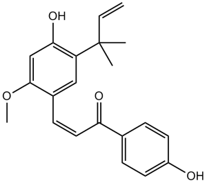
| SMILES | O([H])C1C([H])=C(C(/C(/[H])=C(\[H])/C(C2C([H])=C([H])C(=C([H])C=2[H])O[H])=O)=C([H])C=1C(C([H])=C([H])[H])(C([H])([H])[H])C([H])([H])[H])OC([H])([H])[H] |
|
| InChi Key | KAZSKMJFUPEHHW-DHZHZOJOSA-N | |
| InChi Code | InChI=1S/C21H22O4/c1-5-21(2,3)17-12-15(20(25-4)13-19(17)24)8-11-18(23)14-6-9-16(22)10-7-14/h5-13,22,24H,1H2,2-4H3/b11-8+ | |
| Chemical Name | (E)-3-(4-hydroxy-2-methoxy-5-(2-methylbut-3-en-2-yl)phenyl)-1-(4-hydroxyphenyl)prop-2-en-1-one | |
| Synonyms |
|
|
| HS Tariff Code | 2934.99.9001 | |
| Storage |
Powder-20°C 3 years 4°C 2 years In solvent -80°C 6 months -20°C 1 month |
|
| Shipping Condition | Room temperature (This product is stable at ambient temperature for a few days during ordinary shipping and time spent in Customs) |
Biological Activity
| Targets | Natural product; UGTs/UDP-glucuronosyltransferases | ||
| ln Vitro | Licochalcone A (LCA) demonstrates substantial inhibitory effects against UGT1A1, 1A3, 1A4, 1A6, 1A7, 1A9, and 2B7 (both IC50 and Ki values lower than 5 μM) [2]. | ||
| ln Vivo |
|
||
| Enzyme Assay |
Determination of lipid peroxidation. [3] The content of MDA, as an index of the extent of lipid peroxidation, was assayed in the form of thiobarbituric acid-reactive substances as previously described [31]. The reaction mixture (4 ml) consisted of 0.2 ml of 8.1% sodium dodecyl sulfate, 1.5 ml of 20% acetic acid (pH 3.5), 1.5 ml of 0.8% thiobarbituric acid, 0.2 ml of the homogenate, and distilled water. The mixture was incubated for 1 hr at 95°, cooled for 5 min with tap water, vigorously mixed with 5 ml of a n-butanol-pyridine (15-1, v/v) mixture, and centrifuged for 10 min at 1200 ×g. The absorbance of the organic layer (upper n-butanol phase) was determined at 532 nm. 1,1,3,3-Tetramethoxypropane was utilized to establish the standard curve, and the final MDA concentration was expressed as nmol MDA per mg protein.[3] Determination of GSH level. [3] To determine GSH content in accordance with the method described by Higach, 0.1 ml of the tissue homogenate was added to an equal volume of 10% trichloroacetic acid solution, then centrifuged for 20 min at 1200 ×g. 0.1 ml of the supernatant was added to 0.5 ml of a 0.2 N H2SO4 solution containing 1 mM NaNO2 and incubated for 5 min at room temperature, followed by the addition of 0.2 ml of a 0.5% sulfamic acid ammonium solution, 1 ml of a 0.4 N HCl solution containing 0.1% HgCl2 and 3% sulfanilamide, and 1 ml of a 0.4 N HCl solution containing 0.1% N-(1-naphthyl)ethylenediamine. Five minutes later, the absorbance was determined at 540 nm. The GSH content was expressed as nmol GSH per mg protein using GSH standard calibration curve. |
||
| Cell Assay |
Licochalcone A (LCA), a flavonoid isolated from the famous Chinese medicinal herb Glycyrrhiza uralensis Fisch, presents obvious anti-cancer effects. In this study, the anti-cancer effects and potential mechanisms of LCA in non-small cell lung cancer (NSCLC) cells were studied. LCA decreased cell viability, increased lactate dehydrogenase release, and induced apoptosis in a concentration-dependent manner in NSCLC cells while not in human embryonic lung fibroblast cells. The expression of phosphatidylethanolamine-modified microtubule-associated protein light-chain 3 (LC3-II) and formation of GFP-LC3 punta, two autophagic markers, were increased after treatment with LCA. LCA-induced LC3-II expression was increased when combined with chloroquine (CQ), while knock-down of autophagy related protein (ATG) 7 or ATG5 reversed LCA-induced LC3-II expression and GFP-LC3 punta formation, suggesting that LCA induced autophagy in NSCLC cells. Inhibition of autophagy could not reverse the LCA-induced cell viability decrease and apoptosis. In addition, LCA increased the expression of endoplasmic reticulum stress related proteins, such as binding immunoglobulin protein and C/EBP homologous protein (CHOP). Knock-down of CHOP reversed LCA-induced cell viability decrease, apoptosis, and autophagy. Taken together, LCA-induced autophagic effect is an accompanied phenomenon in NSCLC cells, and CHOP is critical for LCA-induced cell viability decrease, apoptosis, and autophagy.[1] Human erythrocytes drawn from healthy individuals were exposed for 24 hours to 1-10 µg/ml licochalcone A. Flow cytometry was subsequently employed to estimate phosphatidylserine exposure at the cell surface from annexin V binding, cell volume from forward scatter, [Ca2+]i from Fluo3-fluorescence, and ceramide utilizing specific antibodies. In addition, hemolysis was quantified from hemoglobin release.[2] |
||
| Animal Protocol |
|
||
| References |
[1]. Induction of C/EBP homologous protein-mediated apoptosis and autophagy by licochalcone A in non-small cell lung cancer cells. Sci Rep. 2016 May 17;6:26241. [2]. Licochalcone A Induced Suicidal Death of Human Erythrocytes. Cell Physiol Biochem. 2015;37(5):2060-70. [3]. Licochalcone A inhibits the growth of colon carcinoma and attenuates cisplatin-induced toxicity without a loss of chemotherapeutic efficacy in mice. Basic Clin Pharmacol Toxicol . 2008 Jul;103(1):48-54. |
||
| Additional Infomation |
Licochalcone A is a member of chalcones. Licochalcone a has been reported in Glycyrrhiza uralensis, Euphorbia helioscopia, and other organisms with data available. Licochalcone A is a derivative of the phenol chalconoid, found in and extracted from the roots of Glycyrrhiza species G. glabra and inflata, with potential anti-inflammatory, antibacterial, and anticancer activities. Upon administration, licochalcone A inhibits the phosphatidylinositol-3-kinase/Akt/mammalian target of rapamycin (PI3K/Akt/mTOR) signaling pathway and inhibits the activity of c-Jun N-terminal kinase 1 (JNK-1), a member of the mitogen-activated protein kinase (MAPK) family that plays a role in the MAPK-mediated signaling pathway. Inhibition of the PI3K/Akt/mTOR- and MAPK-signaling pathways induces cell cycle arrest and apoptosis, decreases migration and invasion of cancer cells, and inhibits tumor cell proliferation. Licochalcone A also prevents the production of reactive oxygen species (ROS), and reduces oxidative stress through the nuclear factor-erythroid 2-related factor 2 (Nrf2) pathway. |
Solubility Data
| Solubility (In Vitro) |
|
|||
| Solubility (In Vivo) |
Solubility in Formulation 1: ≥ 2.5 mg/mL (7.39 mM) (saturation unknown) in 10% DMSO + 90% Corn Oil (add these co-solvents sequentially from left to right, and one by one), clear solution. For example, if 1 mL of working solution is to be prepared, you can add 100 μL of 25.0 mg/mL clear DMSO stock solution to 900 μL of corn oil and mix evenly. Solubility in Formulation 2: 2.08 mg/mL (6.15 mM) in 10% DMSO + 40% PEG300 + 5% Tween80 + 45% Saline (add these co-solvents sequentially from left to right, and one by one), suspension solution; with ultrasonication. For example, if 1 mL of working solution is to be prepared, you can add 100 μL of 20.8 mg/mL clear DMSO stock solution to 400 μL PEG300 and mix evenly; then add 50 μL Tween-80 to the above solution and mix evenly; then add 450 μL normal saline to adjust the volume to 1 mL. Preparation of saline: Dissolve 0.9 g of sodium chloride in 100 mL ddH₂ O to obtain a clear solution. Solubility in Formulation 3: 2.08 mg/mL (6.15 mM) in 10% DMSO + 90% (20% SBE-β-CD in Saline) (add these co-solvents sequentially from left to right, and one by one), suspension solution; with ultrasonication. For example, if 1 mL of working solution is to be prepared, you can add 100 μL of 20.8 mg/mL clear DMSO stock solution to 900 μL of 20% SBE-β-CD physiological saline solution and mix evenly. Preparation of 20% SBE-β-CD in Saline (4°C,1 week): Dissolve 2 g SBE-β-CD in 10 mL saline to obtain a clear solution. (Please use freshly prepared in vivo formulations for optimal results.) |
| Preparing Stock Solutions | 1 mg | 5 mg | 10 mg | |
| 1 mM | 2.9551 mL | 14.7754 mL | 29.5508 mL | |
| 5 mM | 0.5910 mL | 2.9551 mL | 5.9102 mL | |
| 10 mM | 0.2955 mL | 1.4775 mL | 2.9551 mL |