Physicochemical Properties
| Molecular Formula | C17H15N3S |
| Molecular Weight | 293.386101961136 |
| Exact Mass | 293.099 |
| Elemental Analysis | C, 61.60; H, 4.68; N, 10.26; O, 15.63; S, 7.83 |
| CAS # | 894002-50-7 |
| PubChem CID | 7207425 |
| Appearance | White to gray solid powder |
| LogP | 4.139 |
| Hydrogen Bond Donor Count | 0 |
| Hydrogen Bond Acceptor Count | 4 |
| Rotatable Bond Count | 4 |
| Heavy Atom Count | 21 |
| Complexity | 312 |
| Defined Atom Stereocenter Count | 0 |
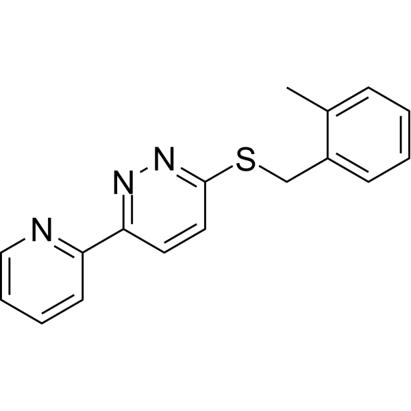
| SMILES | N1C(SCC2C(C)=CC=CC=2)=CC=C(C2C=CC=CN=2)N=1 |
| InChi Key | DUUQLWDHNYFUPP-UHFFFAOYSA-N |
| InChi Code | InChI=1S/C17H15N3S/c1-13-6-2-3-7-14(13)12-21-17-10-9-16(19-20-17)15-8-4-5-11-18-15/h2-11H,12H2,1H3 |
| Chemical Name | 3-[(2-methylphenyl)methylsulfanyl]-6-pyridin-2-ylpyridazine |
| Synonyms | LDN-0212320 Maleate |
| HS Tariff Code | 2934.99.9001 |
| Storage |
Powder-20°C 3 years 4°C 2 years In solvent -80°C 6 months -20°C 1 month |
| Shipping Condition | Room temperature (This product is stable at ambient temperature for a few days during ordinary shipping and time spent in Customs) |
Biological Activity
| Targets | GLT-1 (glutamate transporter); EAAT2 (excitatory amino acid transporter 2) |
| ln Vitro | Excitatory amino acid transporter 2 (EAAT2) is the major glutamate transporter and functions to remove glutamate from synapses. A thiopyridazine derivative has been found to increase EAAT2 protein levels in astrocytes. A structure-activity relationship study revealed that several components of the molecule were required for activity, such as the thioether and pyridazine. Modification of the benzylthioether resulted in several derivatives (7–13, 7–15 and 7–17) that enhanced EAAT2 levels by > 6 fold at concentrations < 5 μM after 24 h. In addition, one of the derivatives (7–22; LDN-212320) enhanced EAAT2 levels 3.5 – 3.9 fold after 24 h with an EC50 of 0.5 μM[2]. |
| ln Vivo | Formalin-induced nociceptive behaviors can be markedly reduced by intraperitoneal injection of LDN-212320 (10 or 20 mg/kg) [1]. The intraperitoneal injection of LDN-212320 (10 or 20 mg/kg) significantly restored the hippocampal-dependent behaviors that had been impaired by formalin. Moreover, GLT-1 expression is elevated in the ACC and hippocampal regions by LDN-212320 (10 or 20 mg/kg, i.p.) [1]. Formalin-induced ERK phosphorylation, a nociception marker, is markedly reduced in the ACC and hippocampal regions by LDN-212320 (20 mg/kg, i.p.) [1]. |
| Enzyme Assay | All of the derivatives of 1 were initially evaluated in PA-EAAT2 cells (a primary astrocyte line stably expressing EAAT2 mRNAs) following compound (10 μM) incubation for 4 and 24 h before harvesting and measuring EAAT2 levels by Western blot analysis. The fold increases in EAAT2 protein levels relative to DMSO controls are reported. [2] |
| Cell Assay |
Western blot analysis[1] Western blot analysis was performed as described previously (Xu et al., 2006) with minor modifications. Briefly, mice were euthanized through rapid decapitation; their anterior cingulate cortices (bregma 1.18 mm) and hippocampi (bregma −1.7 mm) were dissected from 1-mm coronal sections using mouse brain stereotaxic coordinates (Franklin and Paxinos, 2008) and stored at −80 °C until analysis. Tissue was homogenized in modified RIPA buffer containing Dulbecco’s phosphate-buffered saline (pH 7.4), 1% Igepal CA-630 and 0.1% sodium dodecyl sulphate (SDS). Protease inhibitor and phosphatase inhibitor were added to RIPA buffer and used as per manufacturer’s protocol. The samples were centrifuged (16,000 g, 20 min at 4 °C) and supernatants were collected. Protein concentration was determined by bicinchoninic acid assay using albumin as standard. Equal amounts of protein (60 μg) were loaded onto 10% gels for SDS polyacrylamide gel electrophoresis. Separated proteins were transferred onto nitrocellulose membranes at 60 V overnight at 4°C. Membranes were blocked with 5% non-fat dry milk in Tris-buffered saline and 0.1% Tween-20 (TBST) for 1 hr. Then, subsequently incubated overnight at 4°C with primary antibodies for GLT-1 (1:1,000, rabbit polyclonal), ERK1/2 and pERK1/2 (1:1000, rabbit polyclonal, Cell signaling technology, MA, USA) or β-tubulin (E7, 1:5,000, and mouse monoclonal). After incubation, membranes were washed in TBST, followed by incubation with appropriate horseradish peroxidase-conjugated secondary antibodies, diluted in blocking buffer at a concentration of 1:5,000. Bound antibodies were detected with ECL Prime reagent, and protein quantification was performed using densitometric analysis.[1] Immunohistochemistry[1] Immunohistochemistry study was performed as described previously (Zhang et al., 2018), with minor modifications. Briefly, mice were euthanized through rapid decapitation; their brains were removed and postfixed in 4% paraformaldehyde fixative overnight at room temperature. Brains were cryoprotected by immersion in 30% sucrose in 0.1 M PB at 4°C until the brains sank at the bottom. The brains were embedded with Tissue-Tek OCT and sectioned into 15 μm thick sections with Leica CM1850 cryostat. Three to five random sections per each group were blocked with 5% goat serum in 0.3% Triton X-100 in 1x BPS for 1 hour at room temperature then incubated overnight at 4°C with pERK1/2 (1:500, rabbit polyclonal) or GFAP (1:500, rabbit polyclonal). For double immunofluorescence, sections were incubated with a mixture of rabbit p-ERK1/2, and mouse monoclonal anti-neuronal specific nuclear protein (NeuN) (neuronal marker, 1:200) or mouse monoclonal anti-glial fibrillary acidic protein (GFAP) (astroglial marker, 1:200) overnight at 4°C, followed by a mixture of AF488 or AF647 fluorescence-conjugated secondary antibodies (1:200; ab 150077 or ab169348) for 1 hr at room temperature in dark place. The stained sections were then examined with Olympus AX70 Olympus microscope Epi-fluorescence attached with DP70 Digital Camera. For Quantification of immunofluorescence, integrated density was quantified using ImageJ software. The hippocampus and ACC coordinates were based on the stereotaxic plates of the atlas of Franklin and Paxinos (Franklin and Paxinos, 2008); anterior-posterior (AP) coordinates referred to bregma, lateral (ML) coordinates to the midsagittal suture line and ventral (DV) coordinates to the surface of the skull: CA1 (AP: −1.70 mm; ML: 1.17 mm and DV: 1.34 mm), DG (AP: −1.70 mm; ML: 0.70 mm and DV: 2.04 mm)and ACC (AP: 0.14 mm; ML: 0.25 mm and DV: 1.00 mm). |
| Animal Protocol |
Animal/Disease Models: Mouse[1]. Doses: 10 or 20 mg/kg. Route of Administration: IP 24 hrs (hrs (hours)) before injection of formalin. Experimental Results: Licking and biting behaviors were Dramatically attenuated in both Phase 1 and Phase 2 in a dose-dependent manner compared to formalin-injected mice. Significant (P < 0.01 or P < 0.001) reduction in licking and biting behavior. Preference for the displaced object was Dramatically increased compared to formalin-injected mice (F3,13 = 28.03, P < 0.01). Interaction time with displaced objects was Dramatically increased (P < 0.001) compared with formalin-injected mice. Formalin-induced nociceptive pain behaviour[1] The formalin-induced nociception model was used as described previously (Fisher and Coderre, 1996; Kim et al., 1999) with minor modifications. Briefly, mice were placed in a 15 × 12 × 10 cm Plexiglas chambers at least 30 min daily for three consecutive days to adapt to the experimental environment. Two mirrors were placed behind the chamber at a 45° angle to allow an unimpeded view of the paws. Mice were treated with the (vehicle, i.p) or (LDN-212320 5, 10 or 20 mg/kg, i.p) 24 hr before the injection of formalin. Control animals received (vehicle, 10 μl, i.pl + LDN-212320 20 mg/kg, i.p). For the antagonist study, DHK (10 mg/kg) was administered intraperitoneally 30 min before the behavioural tests (Yang et al., 2011). Mice were then injected subcutaneously with 10 μl of 2.5% formalin or 10 μl of vehicle into the dorsal surface of the hind paw using a 30-gauge needle Hamilton micro-syringe (Reno, NV, USA). Immediately after the injection, mice were put back into the chamber and the observational period started. For nociception responses, licking and biting of the injected paw were monitored by measuring the total duration of the responses in seconds during the two phases, early phase (0–5 min) and late phase (10–45 min) of the typical bi-phasic pain behaviour. The doses of LDN-212320, ceftriaxone or DHK were used as described previously (Rasmussen et al., 2011; Tallarida et al., 2013). LDN-212320 was dissolved in normal saline (0.9% NaCl) having 1% dimethyl sulphoxide (DMSO) and 0.5% Tween 80. |
| References |
[1]. Effects of glial glutamate transporter activator in formalin-induced pain behaviour in mice. Eur J Pain. 2019 Apr;23(4):765-783. [2]. Structure-activity relationship study of pyridazine derivatives as glutamate transporter EAAT2 activators. Bioorg Med Chem Lett. 2011 Oct 1;21(19):5774-7. |
Solubility Data
| Solubility (In Vitro) | DMSO : ~50 mg/mL (~170.42 mM) |
| Solubility (In Vivo) |
Solubility in Formulation 1: ≥ 2.5 mg/mL (8.52 mM) (saturation unknown) in 10% DMSO + 40% PEG300 + 5% Tween80 + 45% Saline (add these co-solvents sequentially from left to right, and one by one), clear solution. For example, if 1 mL of working solution is to be prepared, you can add 100 μL of 25.0 mg/mL clear DMSO stock solution to 400 μL PEG300 and mix evenly; then add 50 μL Tween-80 to the above solution and mix evenly; then add 450 μL normal saline to adjust the volume to 1 mL. Preparation of saline: Dissolve 0.9 g of sodium chloride in 100 mL ddH₂ O to obtain a clear solution. Solubility in Formulation 2: ≥ 2.5 mg/mL (8.52 mM) (saturation unknown) in 10% DMSO + 90% (20% SBE-β-CD in Saline) (add these co-solvents sequentially from left to right, and one by one), clear solution. For example, if 1 mL of working solution is to be prepared, you can add 100 μL of 25.0 mg/mL clear DMSO stock solution to 900 μL of 20% SBE-β-CD physiological saline solution and mix evenly. Preparation of 20% SBE-β-CD in Saline (4°C,1 week): Dissolve 2 g SBE-β-CD in 10 mL saline to obtain a clear solution. (Please use freshly prepared in vivo formulations for optimal results.) |
| Preparing Stock Solutions | 1 mg | 5 mg | 10 mg | |
| 1 mM | 3.4084 mL | 17.0422 mL | 34.0843 mL | |
| 5 mM | 0.6817 mL | 3.4084 mL | 6.8169 mL | |
| 10 mM | 0.3408 mL | 1.7042 mL | 3.4084 mL |