Kainic acid is a novel and potent excitatory amino acid and an agonist at excitatory amino acid receptor subtypes in the CNS. Kainate agonist; excitant and neurotoxin
Physicochemical Properties
| Molecular Formula | C10H15NO4.H2O |
| Molecular Weight | 231.24568 |
| Exact Mass | 213.1 |
| CAS # | 487-79-6 |
| Related CAS # | Kainic acid hydrate;58002-62-3 |
| PubChem CID | 10255 |
| Appearance | White to off-white solid powder |
| Density | 1.2±0.1 g/cm3 |
| Boiling Point | 439.9±45.0 °C at 760 mmHg |
| Melting Point | 253-254ºC |
| Flash Point | 219.8±28.7 °C |
| Vapour Pressure | 0.0±2.3 mmHg at 25°C |
| Index of Refraction | 1.509 |
| LogP | 0.5 |
| Hydrogen Bond Donor Count | 3 |
| Hydrogen Bond Acceptor Count | 5 |
| Rotatable Bond Count | 4 |
| Heavy Atom Count | 15 |
| Complexity | 300 |
| Defined Atom Stereocenter Count | 3 |
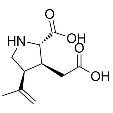
| SMILES | CC(=C)[C@H]1CN[C@@H]([C@H]1CC(=O)O)C(=O)O |
| InChi Key | VLSMHEGGTFMBBZ-OOZYFLPDSA-N |
| InChi Code | InChI=1S/C10H15NO4/c1-5(2)7-4-11-9(10(14)15)6(7)3-8(12)13/h6-7,9,11H,1,3-4H2,2H3,(H,12,13)(H,14,15)/t6-,7+,9-/m0/s1 |
| Chemical Name | (2S,3S,4S)-3-(carboxymethyl)-4-prop-1-en-2-ylpyrrolidine-2-carboxylic acid |
| HS Tariff Code | 2934.99.9001 |
| Storage |
Powder-20°C 3 years 4°C 2 years In solvent -80°C 6 months -20°C 1 month |
| Shipping Condition | Room temperature (This product is stable at ambient temperature for a few days during ordinary shipping and time spent in Customs) |
Biological Activity
| Targets |
Kainic acid is a cyclic analog of L-glutamate and an agonist of ionotropic kainate receptors (KARs). Specific subunits include KA1 (GluK4), KA2 (GluK5), GluR5 (GluK1), and GluR6 (GluK2). KARs are highly expressed in the hippocampus (especially CA3 pyramidal cells), amygdala, entorhinal cortex, basal ganglia, and cerebellum. [3] |
| ln Vitro |
Kainic acid induces robust depolarizations and eventually cell death in neurons, a central phenomenon in temporal lobe epilepsy. [3] Bath application of KA can induce high-amplitude gamma oscillations (30-80 Hz) in the CA3 region of hippocampal brain slices from non-epileptic animals. [3] In brain slices from chronic epileptic mice (after unilateral hippocampal KA injection), gamma activity occurs in the CA3 region, and the discharge frequency of dendrite-inhibiting OLM interneurons shifts from the theta to the gamma frequency band. [3] In vitro, synchronized network-driven activities in the dentate gyrus of epileptic animals can be inhibited by pharmacological blockade of kainate receptors. [3] |
| ln Vivo |
Program plan implementation status for alginic acid (5 mg/kg; intraperitoneal injection; at least once for at least three hours, formula regimen continuous status) [1]. Systemic, intrahippocampal, or intra-amygdaloid administration of kainic acid induces acute status epilepticus (SE) characterized by behavioral seizures (e.g., facial clonus, wet-dog shakes, forelimb clonus, rearing and falling). [3] Following the initial SE, a latent period (5-40 days depending on species and administration route) occurs before the onset of spontaneous recurrent seizures, mimicking human temporal lobe epilepsy. [3] KA administration leads to neuropathological changes similar to hippocampal sclerosis in human TLE, including selective neuronal loss in CA1/CA3/hilus, granule cell dispersion, and aberrant mossy fiber sprouting in the dentate gyrus. [3] The hippocampus is often the seizure onset zone, even when KA is administered at distant sites (e.g., amygdala), suggesting a central role in seizure generation and propagation. [3] EEG features include interictal spikes, gamma oscillations (30-80 Hz), and ictal discharges originating from the hippocampus or amygdala. [3] Age affects susceptibility: very young (up to P15) and old rats (P60 and older) are more sensitive to KA-induced seizures and have shorter latency to SE compared to young adults (P20-P60). Seizures in immature brains cause less neuronal damage but may lead to long-term alterations in GABAergic signaling. [3] |
| Animal Protocol |
Animal/Disease Models: 8 weeks, 200-250 g male adult Wistar rats[1]: 5 mg/kg Route of Administration: intraperitoneal (ip) injection; at least 3 hrs (hrs (hours)) every hour until status epilepticus occurs. Experimental Results: Induced epileptic seizures in rats. Systemic Administration (intraperitoneal, i.p.): In rats, a single dose of 6-15 mg/kg can induce status epilepticus (SE). Alternatively, multiple lower doses (e.g., 5 mg/kg/h) can be administered until SE occurs to reduce mortality. SE typically occurs about 1 hour after injection. Diazepam (20 mg/kg) and ketamine (50 mg/kg) can be used to terminate SE. Mortality rates range from 5% to 30%. [3] Intrahippocampal Administration: In rats, doses ranging from 0.4 to 2.0 µg (in a small volume, e.g., 0.2 µL) are injected directly into the hippocampus. This induces convulsive SE within 5-60 minutes. Similar protocols are used in mice and guinea pigs. [3] Intra-amygdaloid Administration: In rats, doses of 0.4–2 µg are injected into the amygdala, inducing acute seizures with symptoms similar to intrahippocampal injection, sometimes with additional signs like salivation and exophthalmos. In monkeys, doses of 0.5–10 µg/µl of saline are used, producing focal seizures with oral automatisms. [3] Post-SE Monitoring: Following SE induction, animals enter a latent period. The development of chronic epilepsy is assessed via long-term video-EEG monitoring to detect spontaneous recurrent seizures. Neuropathological examination is performed at various time points after SE to assess neuronal damage. [3] |
| ADME/Pharmacokinetics |
The provided text does not contain detailed pharmacokinetic data (e.g., absorption, distribution, metabolism, excretion, half-life, oral bioavailability) for kainic acid. [3] It is noted that systemic administration offers no control over the bioavailability of KA in the brain. [3] KA can enhance the permeability of the blood-brain barrier, an effect observed within 1 hour of administration and before SE onset. This increased permeability may lead to enhanced glutamate release in the hippocampus, favoring seizure occurrence. [3] |
| Toxicity/Toxicokinetics |
Mortality rates following systemic administration of kainic acid in rats range from 5% to 30%. [3] KA is an excitotoxin. Its administration induces neuronal death, particularly in the CA3 and CA1 regions of the hippocampus, amygdala, and other limbic structures. The extent of damage correlates with the severity and propagation of seizure activity rather than solely direct toxin action. [3] Age-specific toxicity is observed: young (P15) and old rats are more sensitive, showing shorter latency to SE and more severe seizures compared to young adults (P20-P60). Interestingly, in very young animals (less than 3 weeks), KA induces severe seizures but causes minimal brain damage, attributed to immature neural connectivity. [3] Distant neuropathological changes (outside the injection site) are attributed to the propagation of epileptiform activity during SE, not to the direct neurotoxic effect of KA itself. Pretreatment with diazepam can prevent hippocampal damage from intra-amygdaloid KA without affecting local amygdala damage. [3] |
| References |
[1]. Chronic levetiracetam decreases hippocampal and testicular aromatase expression in normal but not kainic acid-induced experimental model of acute seizures in rats. Neuroreport. 2017 Sep 27;28(14):903-909. [2]. Kainic acid-mediated excitotoxicity as a model for neurodegeneration. Mol Neurobiol. 2005;31(1-3):3-16. [3]. The kainic acid model of temporal lobe epilepsy. Neurosci Biobehav Rev. 2013 Dec;37(10 Pt 2):2887-99. [4]. Scherer-Singler U, McGeer EG. Distribution and persistence of kainic acid in brain. Life Sci. 1979 Mar 12;24(11):1015-22. [5]. The effect of dipeptidyl peptidase IV on disease-associated microglia phenotypic transformation in epilepsy. J Neuroinflammation. 2021 May 11;18(1):112. [6]. Melatonin attenuates kainic acid-induced hippocampal neurodegeneration and oxidative stress through microglial inhibition. J Pineal Res. 2003 Mar;34(2):95-102. |
| Additional Infomation |
Kainic acid is a dicarboxylic acid, a pyrrolidinecarboxylic acid, a L-proline derivative and a non-proteinogenic L-alpha-amino acid. It has a role as an antinematodal drug and an excitatory amino acid agonist. It is a conjugate acid of a kainate(1-). Kainic acid has been reported in Digenea simplex, Apis cerana, and other organisms with data available. (2S-(2 alpha,3 beta,4 beta))-2-Carboxy-4-(1-methylethenyl)-3-pyrrolidineacetic acid. Ascaricide obtained from the red alga Digenea simplex. It is a potent excitatory amino acid agonist at some types of excitatory amino acid receptors and has been used to discriminate among receptor types. Like many excitatory amino acid agonists it can cause neurotoxicity and has been used experimentally for that purpose. Kainic acid was originally isolated from the red algae Digenea simplex and was initially intended as an ascaricide. [3] It became a crucial tool in neuroscience for studying glutamate receptors, excitotoxicity, and for developing animal models of temporal lobe epilepsy (TLE). [3] The KA model replicates key features of human TLE: a latent period after an initial injury (SE), spontaneous recurrent seizures, and hippocampal sclerosis. [3] It is compared to other TLE models like pilocarpine and electrical kindling. The pilocarpine model is noted for high reliability in inducing epilepsy, while kindling allows controlled network targeting but rarely produces spontaneous seizures or hippocampal sclerosis. [3] The choice between intracerebral and systemic KA administration depends on the research question: intracerebral for focal network studies, systemic for studying widespread vulnerability and disease. [3] The model has contributed to understanding epileptogenesis, ictogenesis, and testing potential therapies. [3] |
Solubility Data
| Solubility (In Vitro) |
DMSO : ~50 mg/mL (~234.49 mM) H2O : ~25 mg/mL (~117.24 mM) |
| Solubility (In Vivo) |
Solubility in Formulation 1: ≥ 5 mg/mL (23.45 mM) (saturation unknown) in 10% DMSO + 40% PEG300 + 5% Tween80 + 45% Saline (add these co-solvents sequentially from left to right, and one by one), clear solution. For example, if 1 mL of working solution is to be prepared, you can add 100 μL of 50.0 mg/mL clear DMSO stock solution to 400 μL PEG300 and mix evenly; then add 50 μL Tween-80 to the above solution and mix evenly; then add 450 μL normal saline to adjust the volume to 1 mL. Preparation of saline: Dissolve 0.9 g of sodium chloride in 100 mL ddH₂ O to obtain a clear solution. Solubility in Formulation 2: ≥ 5 mg/mL (23.45 mM) (saturation unknown) in 10% DMSO + 90% (20% SBE-β-CD in Saline) (add these co-solvents sequentially from left to right, and one by one), clear solution. For example, if 1 mL of working solution is to be prepared, you can add 100 μL of 50.0 mg/mL clear DMSO stock solution to 900 μL of 20% SBE-β-CD physiological saline solution and mix evenly. Preparation of 20% SBE-β-CD in Saline (4°C,1 week): Dissolve 2 g SBE-β-CD in 10 mL saline to obtain a clear solution. Solubility in Formulation 3: ≥ 5 mg/mL (23.45 mM) (saturation unknown) in 10% DMSO + 90% Corn Oil (add these co-solvents sequentially from left to right, and one by one), clear solution. For example, if 1 mL of working solution is to be prepared, you can add 100 μL of 50.0 mg/mL clear DMSO stock solution to 900 μL of corn oil and mix evenly. (Please use freshly prepared in vivo formulations for optimal results.) |
| Preparing Stock Solutions | 1 mg | 5 mg | 10 mg | |
| 1 mM | 4.3243 mL | 21.6216 mL | 43.2432 mL | |
| 5 mM | 0.8649 mL | 4.3243 mL | 8.6486 mL | |
| 10 mM | 0.4324 mL | 2.1622 mL | 4.3243 mL |