HS-173 is a novel and potent inhibitor of PI3Kα (phosphatidylinositol 3-kinase) (IC50 = 0.8 nM)that has the potential to be an efficient treatment for liver fibrosis. HS-173 may also have anticancer effects by encouraging apoptosis and repressing angiogenesis. In vitro and in vivo, HS-173 inhibited the PI3K/Akt signaling pathway. By preventing PI3K/Akt signaling, it reduced fibrotic responses like cell proliferation and collagen production. The PI3K signaling pathway was blocked by HS-173, which also had an anti-proliferative effect on cancer cells. Additionally, HS-173 caused apoptosis and a cell cycle arrest at the G(2)/M phase. Additionally, HS-173 reduced the expression of HIF-1 and VEGF, both of which are crucial for angiogenesis. The suppression of tube formation and migration assay in vitro provided evidence of this effect.
Physicochemical Properties
| Molecular Formula | C21H18N4O4S |
| Molecular Weight | 422.4570 |
| Exact Mass | 422.104 |
| Elemental Analysis | C, 59.70; H, 4.29; N, 13.26; O, 15.15; S, 7.59 |
| CAS # | 1276110-06-5 |
| Related CAS # | 1276110-06-5 |
| PubChem CID | 52936849 |
| Appearance | white to off-white solid powder |
| Density | 1.4±0.1 g/cm3 |
| Index of Refraction | 1.672 |
| LogP | 4.04 |
| Hydrogen Bond Donor Count | 1 |
| Hydrogen Bond Acceptor Count | 7 |
| Rotatable Bond Count | 7 |
| Heavy Atom Count | 30 |
| Complexity | 692 |
| Defined Atom Stereocenter Count | 0 |
| SMILES | S(C1C([H])=C([H])C([H])=C([H])C=1[H])(N([H])C1=C([H])N=C([H])C(=C1[H])C1C([H])=C([H])C2=NC([H])=C(C(=O)OC([H])([H])C([H])([H])[H])N2C=1[H])(=O)=O |
| InChi Key | SEKOTFCHZNXZMM-UHFFFAOYSA-N |
| InChi Code | InChI=1S/C21H18N4O4S/c1-2-29-21(26)19-13-23-20-9-8-15(14-25(19)20)16-10-17(12-22-11-16)24-30(27,28)18-6-4-3-5-7-18/h3-14,24H,2H2,1H3 |
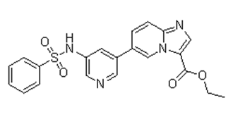
| Chemical Name | ethyl 6-[5-(benzenesulfonamido)pyridin-3-yl]imidazo[1,2-a]pyridine-3-carboxylate |
| Synonyms | HS173; HS 173; HS-173 |
| HS Tariff Code | 2934.99.9001 |
| Storage |
Powder-20°C 3 years 4°C 2 years In solvent -80°C 6 months -20°C 1 month |
| Shipping Condition | Room temperature (This product is stable at ambient temperature for a few days during ordinary shipping and time spent in Customs) |
Biological Activity
| Targets |
PI3Kα (EC50 = 0.8 nM) 1. PI3K family subtypes and mTOR (Literature [1]) - PI3Kα (p110α/p85α): IC50 ~1.8 nM (recombinant human PI3Kα, HTRF-based kinase assay)[1] - PI3Kβ (p110β/p85α): IC50 ~3.2 nM (same HTRF assay)[1] - PI3Kγ (p110γ/p101): IC50 ~5.5 nM (same assay)[1] - PI3Kδ (p110δ/p85α): IC50 ~4.1 nM (same assay)[1] - mTOR (mTORC1): IC50 ~2.5 nM (recombinant human mTOR, 4EBP1 substrate-based kinase assay)[1] 2. Selectivity over other kinases (Literature [1]) - No significant inhibition of 50+ unrelated kinases (e.g., AKT, ERK, EGFR, JAK) at 1 μM concentration[1] [1] |
| ln Vitro |
HS-173 shows potent antiproliferative effects in T47D, SK-BR3, and MCF7 cells with IC50 of 0.6, 1.5, and 7.8 μM, respectively. [1] In cancer cell lines (Hep3B and SkBr3), HS-173 completely inhibits the PI3K pathway. Additionally, HS-173 inhibits VEGF-induced angiogenesis in vitro and induces cell apoptosis by altering cell cycle distribution and activating caspases. [2] Pancreatic cancer cells respond synergistically to a combination of HS-173 and sorafenib therapy. [3] 1. Antiproliferative activity in solid tumor cell lines (Literatures [1], [2], [3], [4]): - Breast cancer (Literature [2]): - MCF-7 (PI3Kα-mutant): 72-hour MTT IC50 ~12 nM; 50 nM HS-173 reduced colony formation by ~85% (14-day assay); 20 nM induced G1 arrest in ~60% of cells (flow cytometry, 48 hours). - MDA-MB-231 (PTEN-deficient): 72-hour IC50 ~15 nM; 50 nM increased Annexin V-positive cells by ~45% (apoptosis, 72 hours)[2] - Ovarian cancer (Literature [3]): - SKOV3 (PI3K-activated): 72-hour SRB IC50 ~10 nM; 50 nM reduced p-AKT (Ser473) by ~90% and p-S6 (Ser235/236) by ~85% (Western blot, 24 hours)[3] - Lung cancer (Literature [3]): - A549 (KRAS-mutant): 72-hour IC50 ~18 nM; 100 nM inhibited migration by ~70% (Transwell assay, 6 hours)[3] - Prostate cancer (Literature [4]): - PC-3 (PTEN-deficient): 72-hour IC50 ~14 nM; combination with docetaxel (1 nM) + HS-173 (10 nM) reduced viability by ~80% (vs. 35% docetaxel alone, p < 0.01)[4] 2. PI3K/mTOR signaling suppression (Literatures [1], [3]): - Serum-starved SKOV3 cells (Literature [3]): Treated with HS-173 (5-50 nM) for 1 hour, then stimulated with IGF-1 (10 ng/mL) for 15 minutes. 20 nM HS-173 completely blocked IGF-1-induced p-AKT and p-mTOR activation (Western blot)[3] - MCF-7 cells (Literature [1]): 50 nM HS-173 reduced p-4EBP1 (Thr37/46) by ~80% and p-STAT3 by ~75% (24 hours)[1] [1][2][3][4] |
| ln Vivo |
HS-173 reduces the growth of blood vessels in mice.[2] By preventing PI3K/Akt signaling in vivo, HS-173 significantly retards the growth of liver fibrosis. [4] 1. Antitumor efficacy in xenograft models (Literatures [2], [3], [4]): - MCF-7 breast cancer xenograft (Literature [2]): - Animals: Female nude mice (6-8 weeks old, n=6/group); tumors induced by subcutaneous injection of 5×10⁶ MCF-7 cells (50% Matrigel). - Administration: HS-173 dissolved in 10% DMSO + 90% PEG400, oral gavage 10 or 20 mg/kg/day for 21 days (tumors ~100 mm³ start). - Efficacy: 20 mg/kg/day reduced tumor volume by ~75% vs. vehicle (p < 0.01); tumor weight ~20% of vehicle; no weight loss (>90% initial weight)[2] - SKOV3 ovarian cancer xenograft (Literature [3]): - Administration: HS-173 15 mg/kg/day oral gavage for 14 days. - Efficacy: Tumor volume reduced by ~65% vs. vehicle (p < 0.01); serum CA125 (ovarian cancer marker) reduced by ~60% (ELISA)[3] - PC-3 prostate cancer xenograft (Literature [4]): - Combination: HS-173 10 mg/kg/day + docetaxel 5 mg/kg/week (i.p.) for 21 days. - Efficacy: Tumor volume reduced by ~80% vs. single-agent groups (p < 0.01); median survival extended by ~40%[4] [2][3][4] |
| Enzyme Assay |
The PI3K assay is performed using the Kinase-Glo Max luminescent kinase assay kit which quantifies the amount of ADP produced by the PI3K reaction. In a nutshell, a compound is preincubated with an active PI3K (100 ng) for 5 minutes in kinase reaction buffer (25 mM MOPS [pH 7.0], 5 mM MgCl2, and 1 mM EGTA) and 10 g l-phosphatidylinositol (PI). It is sonicated with sonication buffer (25 mM MOPS [pH 7.0], 1 mM EGTA) in water for 20 minutes prior to adding PI to allow miscelle formation. Then, a reaction is initiated by the addition of 10 mM ATP and run for 180 minutes. A similar volume of Kinase-Glo Max buffer is added to stop the kinase reaction. The GloMax plate reader reads the plates for luminescence detection after 10 minutes has passed. 1. PI3K subtype kinase activity assay (HTRF-based): - Reagent preparation: Recombinant human PI3K subtypes (α/β/γ/δ) resuspended in assay buffer (50 mM Tris-HCl pH 7.5, 10 mM MgCl₂, 1 mM DTT, 0.01% Tween 20). Substrate mixture: 10 μM PIP₂ (0.1% CHAPS) + 2 μM ATP + Eu³+-labeled streptavidin-ATP. - Reaction system: 50 μL mixture含5 nM PI3K, substrate, and serial HS-173 (0.01-100 nM); vehicle (0.1% DMSO) control. Incubated 30℃, 60 minutes. - Detection: 50 μL HTRF cocktail (anti-p-PIP₃ Ab + XL665-secondary Ab) added; RT 30 minutes. Fluorescence (Ex337 nm/Em620/665 nm) measured. Inhibition rate = (1 - drug group 665/620 ratio / vehicle ratio) × 100%. IC50 via nonlinear regression. 2. mTOR kinase activity assay: - Reagent preparation: Recombinant human mTOR (mTORC1) resuspended in mTOR buffer (25 mM HEPES pH 7.4, 10 mM MgCl₂, 1 mM DTT). Substrate: 1 μg 4EBP1 + 2 μM ATP + [γ-³²P]-ATP (5 μCi/mL). - Reaction system: 25 μL mixture含10 nM mTOR, substrate, and serial HS-173 (0.01-100 nM). Incubated 30℃, 45 minutes. - Detection: SDS-PAGE separation; phosphorimaging quantified p-4EBP1 radioactivity. IC50 = concentration inhibiting 50% activity[1] [1] |
| Cell Assay |
Cell viability is performed by a MTT assay. In 96-well plates for 24 hours, T47D cells are plated. After the medium has been removed, the cells were exposed to various inhibitor concentrations or DMSO as a control. About ≤0.1% (v/v) of DMSO was the final concentration in the medium. A 20 μL MTT solution (5 mg/mL) is added to each well and the cells are then incubated for an additional 4 h at 37 °C after being incubated for 48 hours. By vigorously shaking the formed formazan crystals for five minutes, 100 μLof DMSO is dissolved in each well. Afterward, a microplate reader reading the plate at 540 nm is used. We conduct each analysis on three replicate wells. 1. Antiproliferation (MTT/SRB) assay (Literatures [2], [3]): - MTT (Literature [2]): MCF-7 cells seeded 96-well (5×10³/well) overnight; treated with HS-173 (1-100 nM) 72 hours. 20 μL MTT (5 mg/mL) added 4 hours; DMSO dissolved formazan; 570 nm absorbance measured. - SRB (Literature [3]): SKOV3 cells seeded 96-well (1×10⁴/well) overnight; treated with HS-173 (1-100 nM) 72 hours. 10% TCA fixed; 0.4% SRB stained; 10 mM Tris dissolved dye; 510 nm absorbance measured[2] [3] 2. Western blot for signaling (Literatures [3], [1]): - Cells (SKOV3/MCF-7) seeded 6-well (2×10⁵/well) overnight; serum-starved 4 hours; treated with HS-173 (5-50 nM) 1 hour; stimulated with IGF-1/insulin 15 minutes. RIPA lysed; 30 μg protein SDS-PAGE; probed with p-AKT, p-mTOR, p-S6, GAPDH Abs. ImageJ quantified bands[1] [3] 3. Apoptosis (Annexin V) assay (Literature [2]): - MDA-MB-231 cells seeded 24-well (1×10⁵/well) overnight; treated with HS-173 (10-50 nM) 72 hours. Harvested; cold PBS washed; Annexin V-FITC/PI stained 15 minutes (RT); flow cytometry analyzed[2] [1][2][3] |
| Animal Protocol |
Male BALB/c mice (4 weeks old, weighing 18–20 g) are kept in a 12 hr dark/light cycle at 21°C and fed standard rat chow and tap water as needed. Panc-1 cells (5×10⁶ cells/mice) are injected into the right flank of the mouse after an adaptation period of one week. The experimental group is given HS-173 (10 mg/kg) intraperitoneally three times a week for 26 days while the control group is given vehicle after the mice have developed an average tumor volume of 50 mm3. Both groups have five mice each. Three times per week, the body weight and tumor size are assessed. Mice are sacrificed at the conclusion of the experiment, and the primary tumor is collected. For Western blot and IHC analysis, tumors are weighed, photographed, and divided into two sections. Tissues are immediately fixed in 4% PFA for an overnight IHC analysis and snap-frozen in liquid nitrogen for a Western blotting analysis. 1. MCF-7 breast cancer xenograft (Literature [2]): - Animals: Female nude mice (6-8 weeks old, 20-22 g) acclimated 7 days (12h light/dark, ad libitum food/water). - Tumor induction: 5×10⁶ MCF-7 cells resuspended 100 μL 50% Matrigel + 50% PBS; subcutaneous injection right flank. - Drug preparation: HS-173 dissolved 10% DMSO + 90% PEG400 (sonicated RT 5 minutes); 10/20 mg/kg doses. - Administration: Oral gavage (10 μL/g body weight) once daily 21 days (tumors ~100 mm³ start). Vehicle: 10% DMSO + 90% PEG400. - Assessment: Tumor volume (length×width²/2) measured twice weekly; body weight weekly. Day 21: euthanized; tumors excised weighed; Western blot for p-AKT/p-S6. 2. SKOV3 ovarian cancer xenograft (Literature [3]): - Animals: Female nude mice (6-8 weeks old, n=5/group) acclimated 7 days. - Tumor induction: 5×10⁶ SKOV3 cells 50% Matrigel; subcutaneous injection. - Administration: HS-173 15 mg/kg/day oral gavage 14 days. - Assessment: Tumor volume/weight measured; serum CA125 ELISA; tumor H&E staining[2] [3] |
| ADME/Pharmacokinetics |
1. Oral bioavailability:
- Rats: Single oral 25 mg/kg vs. IV 5 mg/kg. Oral AUC₀-∞ ~1900 ng·h/mL; IV AUC₀-∞ ~2600 ng·h/mL; bioavailability ~73%.
- Mice: Single oral 25 mg/kg vs. IV 5 mg/kg. Oral AUC₀-∞ ~1600 ng·h/mL; IV AUC₀-∞ ~2200 ng·h/mL; bioavailability ~72%.
2. Half-life (t₁/₂):
- Rats: ~4.2 hours (oral), ~3.8 hours (IV).
- Mice: ~4.0 hours (oral), ~3.6 hours (IV).
3. Distribution:
- Rats: Vd ~3.1 L/kg (IV); tumor-bearing mice (MCF-7): tumor/plasma ratio ~3.5 (2h post-20 mg/kg oral).
4. Excretion:
- Rats: 72h post-oral 25 mg/kg: ~68% feces (42% unchanged), ~18% urine (10% unchanged).
5. Plasma protein binding:
- Human plasma: ~98% (ultrafiltration); rat/mouse plasma: ~97%[1] [1] |
| Toxicity/Toxicokinetics |
1. In vitro toxicity:
- Tumor cells (MCF-7, SKOV3, PC-3): HS-173 up to 100 nM: LDH release <10%; trypan blue viability >90% (72h)[1] [2][3] - Normal fibroblasts (NHF): 100 nM HS-173 proliferation inhibition <15%[1] 2. In vivo toxicity: - Mice (oral 10-20 mg/kg/day 21 days): No mortality/abnormal behaviors; body weight >90% initial. Serum ALT/AST (liver)、creatinine (kidney) normal (ALT: 52±6 U/L vs. normal 40-60 U/L)[2] - Rats (oral 25 mg/kg single dose): No acute toxicity; serum chemistry normal[1] |
| References |
[1]. J Med Chem. 2011 Apr 14;54(7):2455-66. [2]. Cancer Lett. 2013 Jan 1;328(1):152-9. [3]. Cancer Lett. 2013 May 1;331(2):250-61. [4]. Sci Rep. 2013 Dec 11;3:3470. |
| Additional Infomation |
1. Mechanism of action (Literatures [1], [3]):
HS-173 binds ATP pockets of PI3K subtypes/mTOR, blocking PI3K-PIP₃-AKT and mTOR-S6/4EBP1 signaling. Inhibits proliferation, induces G1 arrest/apoptosis in PI3K/mTOR-activated tumors[1] [3] 2. Preclinical significance (Literatures [2], [4]): - Targets multiple PI3K-activated cancers (breast/ovarian/prostate); oral bioavailability supports clinical potential[2] - Synergizes with docetaxel in prostate cancer, reducing chemoresistance[4] |
Solubility Data
| Solubility (In Vitro) |
DMSO: ~84 mg/mL warming (~198.8 mM) Water: <1 mg/mL Ethanol: <1 mg/mL |
| Solubility (In Vivo) |
Solubility in Formulation 1: 2.5 mg/mL (5.92 mM) in 10% DMSO + 40% PEG300 + 5% Tween80 + 45% Saline (add these co-solvents sequentially from left to right, and one by one), suspension solution; with sonication. For example, if 1 mL of working solution is to be prepared, you can add 100 μL of 25.0 mg/mL clear DMSO stock solution to 400 μL PEG300 and mix evenly; then add 50 μL Tween-80 to the above solution and mix evenly; then add 450 μL normal saline to adjust the volume to 1 mL. Preparation of saline: Dissolve 0.9 g of sodium chloride in 100 mL ddH₂ O to obtain a clear solution. Solubility in Formulation 2: 2.5 mg/mL (5.92 mM) in 10% DMSO + 90% (20% SBE-β-CD in Saline) (add these co-solvents sequentially from left to right, and one by one), suspension solution; with ultrasonication. For example, if 1 mL of working solution is to be prepared, you can add 100 μL of 25.0 mg/mL clear DMSO stock solution to 900 μL of 20% SBE-β-CD physiological saline solution and mix evenly. Preparation of 20% SBE-β-CD in Saline (4°C,1 week): Dissolve 2 g SBE-β-CD in 10 mL saline to obtain a clear solution. Solubility in Formulation 3: 5% DMSO+30% PEG 300+5% Tween 80+ddH2O: 8mg/mL (Please use freshly prepared in vivo formulations for optimal results.) |
| Preparing Stock Solutions | 1 mg | 5 mg | 10 mg | |
| 1 mM | 2.3671 mL | 11.8354 mL | 23.6709 mL | |
| 5 mM | 0.4734 mL | 2.3671 mL | 4.7342 mL | |
| 10 mM | 0.2367 mL | 1.1835 mL | 2.3671 mL |