GV-58 is a pupin analog that acts as a novel, potent and selective agonist of N- and P/Q-type Ca2+ channels with EC50 of 7.21/8.81 uM for N-type/P-Q-type Ca2+ channel. It has the potential to be used for treating neuromuscular weakness.
Physicochemical Properties
| Molecular Formula | C₁₈H₂₆N₆OS |
| Molecular Weight | 374.50 |
| Exact Mass | 374.188 |
| CAS # | 1402821-41-3 |
| PubChem CID | 71463101 |
| Appearance | Off-white to light brown solid powder |
| Density | 1.3±0.1 g/cm3 |
| Boiling Point | 599.6±60.0 °C at 760 mmHg |
| Flash Point | 316.4±32.9 °C |
| Vapour Pressure | 0.0±1.8 mmHg at 25°C |
| Index of Refraction | 1.674 |
| LogP | 2.01 |
| Hydrogen Bond Donor Count | 3 |
| Hydrogen Bond Acceptor Count | 7 |
| Rotatable Bond Count | 9 |
| Heavy Atom Count | 26 |
| Complexity | 431 |
| Defined Atom Stereocenter Count | 1 |
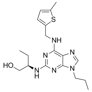
| SMILES | CCCN1C=NC2=C(N=C(N=C21)N[C@H](CC)CO)NCC3=CC=C(S3)C |
| InChi Key | DPTXJOUVBMUSGY-CYBMUJFWSA-N |
| InChi Code | InChI=1S/C18H26N6OS/c1-4-8-24-11-20-15-16(19-9-14-7-6-12(3)26-14)22-18(23-17(15)24)21-13(5-2)10-25/h6-7,11,13,25H,4-5,8-10H2,1-3H3,(H2,19,21,22,23)/t13-/m1/s1 |
| Chemical Name | (2R)-2-[[6-[(5-methylthiophen-2-yl)methylamino]-9-propylpurin-2-yl]amino]butan-1-ol |
| Synonyms | GV-58GV 58GV58 |
| HS Tariff Code | 2934.99.9001 |
| Storage |
Powder-20°C 3 years 4°C 2 years In solvent -80°C 6 months -20°C 1 month |
| Shipping Condition | Room temperature (This product is stable at ambient temperature for a few days during ordinary shipping and time spent in Customs) |
Biological Activity
| Targets |
N-type (Cav2.2) calcium channels (EC50 = 7.21 ± 0.86 µM) P/Q-type (Cav2.1) calcium channels (EC50 = 8.81 ± 1.07 µM) Cyclin-dependent kinase 1 (Cdk1) (IC50 >20 µM) Cyclin-dependent kinase 2 (Cdk2) (IC50 = 3.29 ± 0.43 µM) Cyclin-dependent kinase 5 (Cdk5) (IC50 = 3.03 ± 0.32 µM) No agonist activity on L-type (Cav1.3) calcium channels up to 100 µM. [1] |
| ln Vitro |
The ability of LEMS to passively transfer neuromuscular junctions is restored by GV-58 (50 μM; 30 minutes) [1]. In whole-cell patch-clamp recordings using tsA-201 cells expressing N-type (Cav2.2) calcium channels, GV-58 increased the tail current integral by approximately 32-fold compared to control, with an EC50 of 7.21 ± 0.86 µM. [1] In tsA-201 cells expressing P/Q-type (Cav2.1) calcium channels, GV-58 increased the tail current integral by approximately 33-fold compared to control, with an EC50 of 8.81 ± 1.07 µM. [1] In a commercial kinase screen, GV-58 showed reduced inhibitory activity against Cdks compared to (R)-roscovitine, with IC50 values of >20 µM for Cdk1, 3.29 ± 0.43 µM for Cdk2, and 3.03 ± 0.32 µM for Cdk5. No significant inhibition of MAPK1 or MLCK was observed at concentrations up to 20 µM. [1] |
| ln Vivo |
In a passive transfer mouse model of Lambert-Eaton myasthenic syndrome (LEMS), application of 50 µM GV-58 to ex vivo neuromuscular junction (NMJ) preparations significantly increased endplate potential (EPP) amplitude from 13.00 ± 0.56 mV to 19.44 ± 0.98 mV, and quantal content (measured by peak) from 38.0 ± 12.8 to 56.0 ± 15.2. Quantal content measured by EPP area increased from 38.3 ± 12.7 to 65.6 ± 15.0. [1] GV-58 (50 µM) significantly increased the frequency of miniature endplate potentials (mEPPs) from 3.27 ± 0.15 s⁻¹ to 10.45 ± 0.64 s⁻¹, but did not significantly change mEPP amplitude. [1] GV-58 (50 µM) partially restored short-term plasticity characteristics during a 50 Hz stimulus train in LEMS model NMJs, changing the response from facilitation to a pattern more resembling depression. [1] The potent Cdk inhibitor olomoucine (50 µM) did not significantly increase transmitter release, indicating that the effects of GV-58 are due to Ca²⁺ channel agonism, not Cdk inhibition. [1] |
| Enzyme Assay |
A kinase screen was performed using a commercial service. The inhibitory activity of GV-58 and reference compounds was tested at three concentrations (0.2, 2, and 20 µM) against five kinases: Cdk1/cyclinB, Cdk2/cyclinA, Cdk5/p35, MAPK1, and MLCK. All assays were conducted in the presence of 10 µM ATP. IC50 values were determined from the resulting dose-response curves. [1] |
| Cell Assay |
Cell Viability Assay[1] Cell Types: Upper arm muscle isolated from LEMS mice Tested Concentrations: 50 μM Incubation Duration: 30 minutes Experimental Results: mEPP frequency increased from 3.27 s−1 in vehicle control to 10.45 s−1. A slight boost was shown in the final EPP on the train, which subsequently dropped to 94%. Whole-cell perforated patch-clamp recordings were performed to assess calcium channel activity. Pipettes were filled with a solution containing Cs₂SO₄, CsCl, MgCl₂, and HEPES (pH 7.4). Cells were bathed in a saline containing choline chloride, TEA-Cl, CaCl₂, MgCl₂, and HEPES (pH 7.4). Pipette tips were dipped in antibiotic-free solution, then backfilled with solution containing amphotericin-B to achieve perforated patch access. Currents were recorded using an amplifier, filtered at 5 kHz, and digitized at 10 kHz. Tail current integrals were measured before and after compound application, with each trace normalized to its peak. Compounds were bath-applied from DMSO stock solutions. Control experiments with DMSO alone showed no significant effects. [1] |
| Animal Protocol |
A LEMS passive-transfer mouse model was established. Adult female CFW mice received an intraperitoneal injection of cyclophosphamide (300 mg/kg) on day 1 to suppress immune responses. For the next 24-30 consecutive days, mice received daily intraperitoneal injections of either 1.5 ml of serum from LEMS patients or 1.5 ml of control human serum. LEMS patient sera were screened for voltage-gated calcium channel antibodies and for their ability to reduce quantal content in mice. Serum from patient aBC2 was selected for consistent efficacy. [1] For ex vivo electrophysiology, the epitrochleoanconeus (ETA) muscle with its nerve was dissected after the passive transfer protocol. The preparation was placed in a physiological saline bath. The nerve was stimulated via a suction electrode, and muscle contractions were blocked by μ-conotoxin GIIIB (1 µM). Intracellular recordings were made using microelectrodes filled with potassium acetate. Spontaneous mEPPs and nerve-evoked EPPs (single and 50 Hz trains) were recorded. [1] To test drug effects, baseline recordings were taken in vehicle (0.05–0.1% DMSO), followed by a 30-minute incubation in 50 µM GV-58 and subsequent recordings from the same NMJs. [1] |
| References |
[1]. Evaluation of a novel calcium channel agonist for therapeutic potential in Lambert-Eaton myasthenic syndrome. J Neurosci. 2013 Jun 19;33(25):10559-67. [2]. Complete reversal of Lambert-Eaton myasthenic syndrome synaptic impairment by the combined use of a K+ channel blocker and a Ca2+ channel agonist. J Physiol. 2014 Aug 15;592(16):3687-96. [3]. Lambert-Eaton myasthenic syndrome: mouse passive-transfer model illuminates disease pathology and facilitates testing therapeutic leads. Ann N Y Acad Sci. 2018 Jan;1412(1):73-81. |
| Additional Infomation |
GV-58 is a novel analog of (R)-roscovitine, developed through strategic medicinal chemistry modifications to the purine scaffold, specifically modifying the benzylamine and isopropyl side chains, with the goal of reducing cyclin-dependent kinase (Cdk) antagonist activity while enhancing calcium channel agonist activity. [1] Its primary mechanism of action is as an agonist of presynaptic N- and P/Q-type voltage-gated calcium channels, slowing channel deactivation kinetics to increase calcium influx during action potentials, thereby enhancing neurotransmitter release at synapses. [1] GV-58 demonstrates promising therapeutic potential for strengthening weakened neuromuscular synapses in Lambert-Eaton myasthenic syndrome (LEMS) and represents a new direct calcium channel targeting strategy, as opposed to the standard indirect therapy with potassium channel blockers like 3,4-diaminopyridine. [1] It may also have potential as a treatment for other neuromuscular diseases characterized by presynaptic weakness, such as certain congenital myasthenic syndromes. [1] Beyond therapeutics, GV-58 serves as a valuable experimental tool for studying the basic properties of P/Q- and N-type calcium channels and the calcium control of neurotransmitter release. [1] |
Solubility Data
| Solubility (In Vitro) | DMSO : ~75 mg/mL (~200.27 mM) |
| Solubility (In Vivo) |
Solubility in Formulation 1: ≥ 2.5 mg/mL (6.68 mM) (saturation unknown) in 10% DMSO + 40% PEG300 + 5% Tween80 + 45% Saline (add these co-solvents sequentially from left to right, and one by one), clear solution. For example, if 1 mL of working solution is to be prepared, you can add 100 μL of 25.0 mg/mL clear DMSO stock solution to 400 μL PEG300 and mix evenly; then add 50 μL Tween-80 to the above solution and mix evenly; then add 450 μL normal saline to adjust the volume to 1 mL. Preparation of saline: Dissolve 0.9 g of sodium chloride in 100 mL ddH₂ O to obtain a clear solution. Solubility in Formulation 2: ≥ 2.5 mg/mL (6.68 mM) (saturation unknown) in 10% DMSO + 90% Corn Oil (add these co-solvents sequentially from left to right, and one by one), clear solution. For example, if 1 mL of working solution is to be prepared, you can add 100 μL of 25.0 mg/mL clear DMSO stock solution to 900 μL of corn oil and mix evenly. (Please use freshly prepared in vivo formulations for optimal results.) |
| Preparing Stock Solutions | 1 mg | 5 mg | 10 mg | |
| 1 mM | 2.6702 mL | 13.3511 mL | 26.7023 mL | |
| 5 mM | 0.5340 mL | 2.6702 mL | 5.3405 mL | |
| 10 mM | 0.2670 mL | 1.3351 mL | 2.6702 mL |