GSK2110183 analog HCl; (Afuresertib-F HCl), an analog of Afuresertib, is a potent, orally bioavailable and ATP-competitive Akt inhibitor with Ki of 0.08 nM, 2 nM, and 2.6 nM for Akt1, Akt2, and Akt3, respectively. GSK2110183 HCl is an analog of afuresertib. The serine/threonine protein kinase Akt (protein kinase B) inhibitor GSK2110183 may have anti-cancer effects. The PI3K/Akt signaling pathway, tumor cell proliferation, and tumor cell apoptosis may all be inhibited by the Akt inhibitor GSK2110183, which binds to and inhibits the activity of Akt. The PI3K/Akt signaling pathway is frequently involved in the development of tumors, and aberrant PI3K/Akt signaling may play a role in the development of tumor resistance to a range of antineoplastic agents.
Physicochemical Properties
| Molecular Formula | C18H17CL3F2N4OS |
| Molecular Weight | 481.774586439133 |
| Exact Mass | 480.015 |
| Elemental Analysis | C, 44.88; H, 3.56; Cl, 22.07; F, 7.89; N, 11.63; O, 3.32; S, 6.65 |
| CAS # | 2070009-64-0 |
| Related CAS # | GSK2110183 analog 1;1047634-63-8; GSK2110183 analog 1 hydrochloride;2070009-64-0; 1047644-62-1 ; 1047645-82-8 (HCl); |
| PubChem CID | 92044396 |
| Appearance | White to off-white solid powder |
| Hydrogen Bond Donor Count | 3 |
| Hydrogen Bond Acceptor Count | 6 |
| Rotatable Bond Count | 6 |
| Heavy Atom Count | 29 |
| Complexity | 552 |
| Defined Atom Stereocenter Count | 1 |
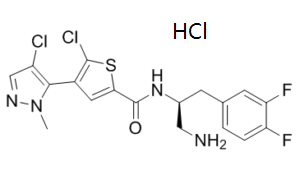
| SMILES | CN1C(=C(C=N1)Cl)C2=C(SC(=C2)C(=O)N[C@@H](CC3=CC(=C(C=C3)F)F)CN)Cl.Cl |
| InChi Key | BCVQLTLODHPIJM-PPHPATTJSA-N |
| InChi Code | InChI=1S/C18H16Cl2F2N4OS.ClH/c1-26-16(12(19)8-24-26)11-6-15(28-17(11)20)18(27)25-10(7-23)4-9-2-3-13(21)14(22)5-9;/h2-3,5-6,8,10H,4,7,23H2,1H3,(H,25,27);1H/t10-;/m0./s1 |
| Chemical Name | N-[(2S)-1-Amino-3-(3,4-difluorophenyl)propan-2-yl]-5-chloro-4-(4-chloro-2-methylpyrazol-3-yl)thiophene-2-carboxamide hydrochloride |
| Synonyms | Afuresertib-F hydrochloride; Afuresertib-F; GSK2110183-analog; GSK-2110183-analog; GSK 2110183-analog; Afuresertib-F HCl |
| HS Tariff Code | 2934.99.9001 |
| Storage |
Powder-20°C 3 years 4°C 2 years In solvent -80°C 6 months -20°C 1 month Note: Please store this product in a sealed and protected environment, avoid exposure to moisture. |
| Shipping Condition | Room temperature (This product is stable at ambient temperature for a few days during ordinary shipping and time spent in Customs) |
Biological Activity
| Targets | Akt | ||
| ln Vitro |
|
||
| ln Vivo |
|
||
| Enzyme Assay |
Kinase Assays[1] The potency of compounds against AKT enzymes was measured as described before. Since GSK2110183 and GSK2141795 are highly potent inhibitors of the 3 isoforms of AKT, the true potency (Ki *) of the inhibitors was initially determined at low enzyme concentrations (0.1 nM AKT1, 0.7 nM AKT2, and 0.2 nM AKT3) using a filter binding assay and then confirmed with progress curve analysis. In the filter binding assay, a pre-mix of enzyme plus inhibitor was incubated for 1 h and then added to a GSKα peptide (Ac-KKGGRARTSSFAEPG-amide) and [γ33P] ATP. Reactions were terminated after 2 h and the radio labeled AKT peptide product was captured in a phospho-cellulose filter plate. Progress curve analysis utilized continuous real-time fluorescence detection of product formation using the Sox-AKT-tide substrate (Ac-ARKRERAYSF-d-Pro-Sox-Gly-NH2).[1] GSK2110183 and GSK2141795 were tested against a diverse panel of kinase assays. Initially, the compounds were tested at 0.5 and 10 µM in all available kinase assays and were followed up with full IC50 curves against a subset of enzymes that showed strong inhibition against 0.5 µM, for which in-house assay were not available. |
||
| Cell Assay |
poptosis assay[2] Apoptosis was evaluated by performing AxV–FITC/PI double staining‐based FACS analysis, as described previously 25. Briefly, ACC‐MESO‐4 and MSTO‐211H cells were seeded in six‐well plates (cell density, 1 × 10⁵ cells/well) and were incubated for 24 h at 37°C. Next, the cells were incubated with indicated concentrations of afuresertib, followed by incubation with AxV–FITC and PI (10 μg/mL) for 15 min at room temperature. Fluorescence intensities were determined by performing FACS with FACSCantoII. Cell cycle analysis[2] Cell cycle was evaluated by performing PI‐staining‐based FACS analysis, as described previously 26. ACC‐MESO‐4 and MSTO‐211H cells were seeded in a six‐well culture plate (cell density, 1 × 10⁵ cells/well) and were incubated for 24 h. Next, the cells were incubated with the indicated concentrations of afuresertib for 24 h. For FACS analysis, the cells were detached using trypsin after 24 h of serum treatment and were fixed overnight in ice‐cold 70% ethanol. After fixation, the cells were treated with RNase A (100 μg/mL) and stained with PI (10 μg/mL). The percentages of cells in the sub‐G1, G1, S, and G2‐M phases of the cell cycle were measured using FlowJo software. A 3-day proliferation assay using CellTiter-Glo is performed to measure the growth inhibition by the compounds at 0-30 μM. The rate of cell growth is measured in comparison to untreated (DMSO) controls. In the Assay Client application, EC50 values are calculated from inhibition curves using a 4- or 6-parameter fitting algorithm.[1] |
||
| Animal Protocol |
|
||
| References | :Cancer Chemother Pharmacol.2015 Jan;75(1):183-9;Blood.2014Oct 2;124(14):2162-3. |
Solubility Data
| Solubility (In Vitro) |
|
|||
| Solubility (In Vivo) |
Solubility in Formulation 1: ≥ 2.5 mg/mL (5.19 mM) (saturation unknown) in 10% DMSO + 40% PEG300 + 5% Tween80 + 45% Saline (add these co-solvents sequentially from left to right, and one by one), clear solution. For example, if 1 mL of working solution is to be prepared, you can add 100 μL of 25.0 mg/mL clear DMSO stock solution to 400 μL PEG300 and mix evenly; then add 50 μL Tween-80 to the above solution and mix evenly; then add 450 μL normal saline to adjust the volume to 1 mL. Preparation of saline: Dissolve 0.9 g of sodium chloride in 100 mL ddH₂ O to obtain a clear solution. Solubility in Formulation 2: ≥ 2.5 mg/mL (5.19 mM) (saturation unknown) in 10% DMSO + 90% (20% SBE-β-CD in Saline) (add these co-solvents sequentially from left to right, and one by one), clear solution. For example, if 1 mL of working solution is to be prepared, you can add 100 μL of 25.0 mg/mL clear DMSO stock solution to 900 μL of 20% SBE-β-CD physiological saline solution and mix evenly. Preparation of 20% SBE-β-CD in Saline (4°C,1 week): Dissolve 2 g SBE-β-CD in 10 mL saline to obtain a clear solution. Solubility in Formulation 3: ≥ 2.5 mg/mL (5.19 mM) (saturation unknown) in 10% DMSO + 90% Corn Oil (add these co-solvents sequentially from left to right, and one by one), clear solution. For example, if 1 mL of working solution is to be prepared, you can add 100 μL of 25.0 mg/mL clear DMSO stock solution to 900 μL of corn oil and mix evenly. Solubility in Formulation 4: ≥ 2.5 mg/mL (5.19 mM) (saturation unknown) in 10% DMSO + 90% Saline (add these co-solvents sequentially from left to right, and one by one), clear solution. Preparation of saline: Dissolve 0.9 g of sodium chloride in 100 mL ddH₂ O to obtain a clear solution. (Please use freshly prepared in vivo formulations for optimal results.) |
| Preparing Stock Solutions | 1 mg | 5 mg | 10 mg | |
| 1 mM | 2.0757 mL | 10.3784 mL | 20.7568 mL | |
| 5 mM | 0.4151 mL | 2.0757 mL | 4.1514 mL | |
| 10 mM | 0.2076 mL | 1.0378 mL | 2.0757 mL |