Physicochemical Properties
| Molecular Formula | C16H10FN3 |
| Molecular Weight | 262.271641254425 |
| Exact Mass | 262.088 |
| CAS # | 1522051-90-6 |
| PubChem CID | 70957463 |
| Appearance | Typically exists as solid at room temperature |
| LogP | 3.917 |
| Hydrogen Bond Donor Count | 1 |
| Hydrogen Bond Acceptor Count | 3 |
| Rotatable Bond Count | 1 |
| Heavy Atom Count | 20 |
| Complexity | 351 |
| Defined Atom Stereocenter Count | 0 |
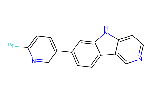
| SMILES | [18F]C1=CC=C(C=N1)C1C=CC2C3C=NC=CC=3NC=2C=1 |
| InChi Key | GETAAWDSFUCLBS-SJPDSGJFSA-N |
| InChi Code | InChI=1S/C16H10FN3/c17-16-4-2-11(8-19-16)10-1-3-12-13-9-18-6-5-14(13)20-15(12)7-10/h1-9,20H/i17-1 |
| Chemical Name | 7-(6-(18F)fluoranylpyridin-3-yl)-5H-pyrido[4,3-b]indole |
| HS Tariff Code | 2934.99.9001 |
| Storage |
Powder-20°C 3 years 4°C 2 years In solvent -80°C 6 months -20°C 1 month |
| Shipping Condition | Room temperature (This product is stable at ambient temperature for a few days during ordinary shipping and time spent in Customs) |
Biological Activity
| ADME/Pharmacokinetics |
Absorption, Distribution and Excretion Flortaucipir F-18 is administered as an intravenous bolus injection, and peak brain uptake in mice of 4.16% ID/g is achieved by 2 minutes. Fast transfer from the peripheral circulation to the brain was corroborated by human studies that demonstrated peak SUV in gray matter >2 across subjects approximately 5 minutes after administration. Pharmacokinetic studies in humans suggest that equilibrium is achieved by 55 minutes (Logan DVR) and by 80 - 100 minutes (SUVR), and current guidelines recommend initiating imaging approximately 80 minutes after initial administration. The major route of elimination for Flortaucipir F-18 is via the kidneys. Flortaucipir F-18 injected into mice accumulates primarily in the kidneys (14.99 ± 0.39 %ID/g at five minutes and 5.52 ± 0.91 %ID/g at 30 minutes post-injection) and liver (4.44 ± 0.16/5.99 ± 0.42 %ID/g at five/30 minutes, respectively). It is able to cross the blood-brain barrier, with relatively high penetration early (4.43 ± 0.91 %ID/g at five minutes) and low residual penetration later (0.62 ± 0.06 %ID/g at 30 minutes). Detectable amounts of Flortaucipir F-18 are found in the systemic circulation, as well as in muscle and bone. Metabolism / Metabolites Initial studies in mice described the parent compound and four uncharacterized metabolites, one of which, termed metabolite 1, is presumed to be [18F]fluoride. Plasma radioactivity corresponded only to the parent compound and metabolite 1. All metabolites were detected in the liver while all metabolites except metabolite 2 were found in the kidneys. Biological Half-Life Flortaucipir F-18 plasma half-life was calculated at 17.0 ± 4.2 minutes; correction for metabolite half-life yielded a biexponential distribution with half-lives of 18.1 ± 5.8 and 2.4 ± 0.5 minutes. |
| Additional Infomation |
Flortaucipir F-18, also known as 18F-T807 and 18F-AV-1451, is a small indole molecule synthesized with a radioactive fluorine isotope. It is used as a marker in positron emission tomography (PET) imaging of patients suspected of having Alzheimer's disease. After crossing the blood-brain barrier, flortaucipir F-18 binds to aggregated tau protein, a hallmark of Alzheimer's disease whose incidence correlates well with disease progression. Although flortaucipir F-18 displays low levels of background binding throughout the brain, it does display off-target binding to monoamine oxidase MAO-A and MAO-B, as well as to regions containing high levels of melanin, neuromelanin, and iron.. It was approved by the FDA on May 28, 2020, for sale by Avid Radiopharmaceuticals under the name TAUVID™ and is the first FDA-approved molecule for imaging aggregated tau protein in the brain. Flortaucipir F-18 is a radioconjugate composed of the paired helical filament (PHF) tau binding agent flortaucipir, a benzimidazole pyrimidine derivative, conjugated to the radioisotope fluorine F 18, with potential tau positron emission tomography (PET) imaging activity. Upon administration, flortaucipir F-18 targets and binds to PHF-tau in the brain, which allows PET imaging of PHF-tau and analysis of phosphorylated PHF-tau aggregates (neurofibrillary tangles). Increased levels of PHF-tau and neurofibrillary tangles are correlated with the cognitive decline seen in neurodegenerative diseases, such as dementia, Alzheimer's disease (AD) and chemotherapy-induced cognitive impairment (CICI). Drug Indication Flortaucipir F-18 is a radioactive agent indicated for positron emission tomography (PET) imaging of aggregated tau neurofibrillary tangles (NFTs) in adult patients under evaluation for Alzheimer's disease. Flortaucipir F-18 is not indicated for use in patients under evaluation for chronic traumatic encephalopathy. FDA Label Diagnosis of Alzheimer's disease Mechanism of Action Alzheimer's disease is a progressive neurodegenerative disease characterized by the build-up of hyperphosphorylated tau protein aggregates. Hyperphosphorylated tau forms dimers termed paired helical filaments (PHFs), which aggregate further to form neurofibrillary tangles (NFTs) associated with neurodegeneration and severity of Alzheimer's symptoms. Flortaucipir F-18 is a small molecule that contains radioactive 18F, which decays by positron emission to 18O with a half-life of 109.8 minutes. As a small relatively lipophilic molecule, flortaucipir F-18, following intravenous injection, quickly passes through systemic circulation, crosses the blood-brain barrier, and binds to NFTs. Once bound, the ensuing radioactive decay emits pairs of 511 keV gamma photons useful in diagnostic imaging. The pattern and intensity of emission during imaging is used in the diagnosis of Alzheimer's disease. Pharmacodynamics Flortaucipir F-18 is a radioactive molecule that binds to and accumulates in tau NFT deposits allowing for imaging detection; however, non-specific binding and other factors allow for the possibility of misdiagnosis. As Flortaucipir F-18 is a radioactive substance, standard precautionary measures for protecting both patients and health care workers are advised. The safety and efficacy of flortaucipir F-18 in patients being evaluated for chronic traumatic encephalopathy are unknown and hence is not recommended. |
Solubility Data
| Solubility (In Vitro) | May dissolve in DMSO (in most cases), if not, try other solvents such as H2O, Ethanol, or DMF with a minute amount of products to avoid loss of samples |
| Solubility (In Vivo) |
Note: Listed below are some common formulations that may be used to formulate products with low water solubility (e.g. < 1 mg/mL), you may test these formulations using a minute amount of products to avoid loss of samples. Injection Formulations (e.g. IP/IV/IM/SC) Injection Formulation 1: DMSO : Tween 80: Saline = 10 : 5 : 85 (i.e. 100 μL DMSO stock solution → 50 μL Tween 80 → 850 μL Saline) *Preparation of saline: Dissolve 0.9 g of sodium chloride in 100 mL ddH ₂ O to obtain a clear solution. Injection Formulation 2: DMSO : PEG300 :Tween 80 : Saline = 10 : 40 : 5 : 45 (i.e. 100 μL DMSO → 400 μLPEG300 → 50 μL Tween 80 → 450 μL Saline) Injection Formulation 3: DMSO : Corn oil = 10 : 90 (i.e. 100 μL DMSO → 900 μL Corn oil) Example: Take the Injection Formulation 3 (DMSO : Corn oil = 10 : 90) as an example, if 1 mL of 2.5 mg/mL working solution is to be prepared, you can take 100 μL 25 mg/mL DMSO stock solution and add to 900 μL corn oil, mix well to obtain a clear or suspension solution (2.5 mg/mL, ready for use in animals). Injection Formulation 4: DMSO : 20% SBE-β-CD in saline = 10 : 90 [i.e. 100 μL DMSO → 900 μL (20% SBE-β-CD in saline)] *Preparation of 20% SBE-β-CD in Saline (4°C,1 week): Dissolve 2 g SBE-β-CD in 10 mL saline to obtain a clear solution. Injection Formulation 5: 2-Hydroxypropyl-β-cyclodextrin : Saline = 50 : 50 (i.e. 500 μL 2-Hydroxypropyl-β-cyclodextrin → 500 μL Saline) Injection Formulation 6: DMSO : PEG300 : castor oil : Saline = 5 : 10 : 20 : 65 (i.e. 50 μL DMSO → 100 μLPEG300 → 200 μL castor oil → 650 μL Saline) Injection Formulation 7: Ethanol : Cremophor : Saline = 10: 10 : 80 (i.e. 100 μL Ethanol → 100 μL Cremophor → 800 μL Saline) Injection Formulation 8: Dissolve in Cremophor/Ethanol (50 : 50), then diluted by Saline Injection Formulation 9: EtOH : Corn oil = 10 : 90 (i.e. 100 μL EtOH → 900 μL Corn oil) Injection Formulation 10: EtOH : PEG300:Tween 80 : Saline = 10 : 40 : 5 : 45 (i.e. 100 μL EtOH → 400 μLPEG300 → 50 μL Tween 80 → 450 μL Saline) Oral Formulations Oral Formulation 1: Suspend in 0.5% CMC Na (carboxymethylcellulose sodium) Oral Formulation 2: Suspend in 0.5% Carboxymethyl cellulose Example: Take the Oral Formulation 1 (Suspend in 0.5% CMC Na) as an example, if 100 mL of 2.5 mg/mL working solution is to be prepared, you can first prepare 0.5% CMC Na solution by measuring 0.5 g CMC Na and dissolve it in 100 mL ddH2O to obtain a clear solution; then add 250 mg of the product to 100 mL 0.5% CMC Na solution, to make the suspension solution (2.5 mg/mL, ready for use in animals). Oral Formulation 3: Dissolved in PEG400 Oral Formulation 4: Suspend in 0.2% Carboxymethyl cellulose Oral Formulation 5: Dissolve in 0.25% Tween 80 and 0.5% Carboxymethyl cellulose Oral Formulation 6: Mixing with food powders Note: Please be aware that the above formulations are for reference only. InvivoChem strongly recommends customers to read literature methods/protocols carefully before determining which formulation you should use for in vivo studies, as different compounds have different solubility properties and have to be formulated differently. (Please use freshly prepared in vivo formulations for optimal results.) |
| Preparing Stock Solutions | 1 mg | 5 mg | 10 mg | |
| 1 mM | 3.8129 mL | 19.0643 mL | 38.1286 mL | |
| 5 mM | 0.7626 mL | 3.8129 mL | 7.6257 mL | |
| 10 mM | 0.3813 mL | 1.9064 mL | 3.8129 mL |