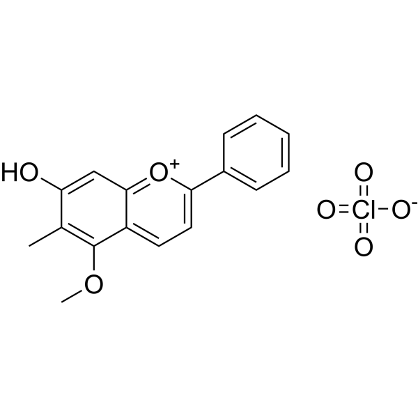
Physicochemical Properties
| Molecular Formula | C17H15O3+.CH4 |
| Molecular Weight | 283.34166 |
| Exact Mass | 366.05 |
| CAS # | 125536-25-6 |
| Related CAS # | Dracorhodin;643-56-1 |
| PubChem CID | 74787691 |
| Appearance | Yellow to orange solid powder |
| LogP | 4.617 |
| Hydrogen Bond Donor Count | 1 |
| Hydrogen Bond Acceptor Count | 6 |
| Rotatable Bond Count | 2 |
| Heavy Atom Count | 25 |
| Complexity | 409 |
| Defined Atom Stereocenter Count | 0 |
| InChi Key | KRTYZFUODYMZPG-UHFFFAOYSA-N |
| InChi Code | InChI=1S/C17H14O3.ClHO4/c1-11-14(18)10-16-13(17(11)19-2)8-9-15(20-16)12-6-4-3-5-7-12;2-1(3,4)5/h3-10H,1-2H3;(H,2,3,4,5) |
| Chemical Name | 5-methoxy-6-methyl-2-phenylchromenylium-7-ol;perchlorate |
| HS Tariff Code | 2934.99.9001 |
| Storage |
Powder-20°C 3 years 4°C 2 years In solvent -80°C 6 months -20°C 1 month Note: Please store this product in a sealed and protected environment, avoid exposure to moisture. |
| Shipping Condition | Room temperature (This product is stable at ambient temperature for a few days during ordinary shipping and time spent in Customs) |
Biological Activity
| Targets |
Extracellular signal-regulated kinase (ERK) signaling pathway activator (indirectly, by increasing phosphorylation). The mechanism may involve activation of the Epidermal Growth Factor Receptor (EGFR), but this is speculative based on the discussion of similar flavonoids.[1] |
| ln Vitro |
Dracorhodin perchlorate (DP) promoted the proliferation of NIH/3T3 fibroblasts in a concentration-dependent manner. Treatment for 24 hours at concentrations ranging from 0.625 to 10 μg/mL significantly increased cell viability, with a peak effect observed at 2.5 μg/mL. Concentrations above 20 μg/mL inhibited fibroblast proliferation.[1] Treatment of NIH/3T3 fibroblasts with 2.5 μg/mL DP significantly increased the level of phosphorylated ERK (p-ERK) in a time-dependent manner (0 to 120 minutes), without changing the total ERK protein level.[1] RNA interference-mediated knockdown of ERK1/2 expression using siRNA completely blocked DP-induced fibroblast proliferation, confirming the essential role of the ERK pathway.[1] Similarly, pretreatment with the ERK1/2 inhibitor PD98059 significantly reduced DP-induced ERK phosphorylation and attenuated DP-induced fibroblast proliferation.[1] |
| ln Vivo |
In a rat full-thickness wound model, topical application of Dracorhodin perchlorate (DP) ointment (at concentrations of 2.5, 5, and 10 μg/mL) twice daily significantly accelerated wound healing in a dose-dependent manner compared to the control (Vaseline) and vehicle (DMSO) groups. The healing rate was significantly higher at day 7, and wounds in the high-dose group (10 μg/mL) showed the greatest healing extent by day 14.[1] Immunohistochemical staining of wound tissues at day 7 using SERPINH1 (a fibroblast marker) antibody showed a significant, dose-dependent increase in fibroblast density in DP-treated groups compared to the control.[1] Western blot analysis of wound tissues at day 7 confirmed a significant, dose-dependent upregulation of phosphorylated ERK (p-ERK) levels in DP-treated groups compared to the control group.[1] |
| Cell Assay |
For cell viability and proliferation assays, NIH/3T3 fibroblasts were seeded in 96-well plates. After serum starvation for 3 hours, cells were treated with various concentrations of Dracorhodin perchlorate (DP) (0 to 40 μg/mL) or 50 ng/mL EGF (positive control) for different time periods (6, 12, 24, 36 hours). Cell viability was assessed using the CCK-8 assay by measuring absorbance at 450 nm.[1] For Western blot analysis to detect ERK phosphorylation, NIH/3T3 fibroblasts were treated with 2.5 μg/mL DP for various durations (0, 15, 30, 60, 120 minutes). Cells were lysed, and proteins were separated by SDS-PAGE, transferred to PVDF membranes, and probed with antibodies against p-ERK, total ERK, and GAPDH. Band densities were quantified using Image J software.[1] For RNA interference experiments, NIH/3T3 fibroblasts were transfected with ERK-specific siRNA (100 nM final concentration) or control siRNA using a transfection reagent. After 48 hours, transfected cells were treated with or without 2.5 μg/mL DP. ERK expression was analyzed by Western blot after 120 minutes, and cell viability was assessed by CCK-8 after 24 hours.[1] To inhibit ERK pharmacologically, fibroblasts were pretreated with the ERK inhibitor PD98059 for 24 hours before adding 2.5 μg/mL DP for 120 minutes. Levels of p-ERK and cell viability were then assessed.[1] |
| Animal Protocol |
A rat full-thickness wound healing model was established using one hundred Wistar rats (6-8 weeks old). After anesthesia, two circular full-thickness wounds (10 mm diameter) were created on the shaved dorsum of each rat.[1] Dracorhodin perchlorate (DP) ointment was prepared by first dissolving DP in dimethyl sulfoxide (DMSO) and then mixing this solution with Vaseline (petroleum jelly). Final ointment concentrations were 2.5, 5, and 10 μg/mL DP. Control groups received Vaseline only or Vaseline mixed with DMSO (vehicle control).[1] Rats were randomly divided into groups (n=20/group): control (Vaseline), vehicle (DMSO in Vaseline), low-dose DP (2.5 μg/mL), middle-dose DP (5 μg/mL), and high-dose DP (10 μg/mL). Ointments were applied topically to the wounds twice daily.[1] Wound areas were photographed on days 0, 3, 7, and 14, and the wound healing rate was calculated using image analysis software. Skin tissue samples were collected on day 7 for immunohistochemical analysis of SERPINH1 (fibroblast marker) and for Western blot analysis of p-ERK.[1] |
| References |
[1]. Dracorhodin perchlorate regulates fibroblast proliferation to promote rat's wound healing. J Pharmacol Sci. 2018 Feb;136(2):66-72. [2]. Dracorhodin perchlorate suppresses proliferation and induces apoptosis in human prostate cancer cell line PC-3. J Huazhong Univ Sci Technolog Med Sci. 2011 Apr;31(2):215. [3]. Dracorhodin perchlorate induces the apoptosis of glioma cells. Oncol Rep. 2016 Apr;35(4):2364-72. |
| Additional Infomation |
Dracorhodin perchlorate (DP) is a stable salt form of Dracorhodin, an active compound derived from the traditional medicine "Dragon's Blood" resin.[1] The study demonstrates that DP promotes wound healing by enhancing fibroblast proliferation through activation of the ERK signaling pathway both in vitro and in vivo.[1] It is suggested (based on literature discussion) that DP, as a flavonoid, might exert its effects by initially activating cell surface receptors such as EGFR, which then triggers downstream pathways including ERK.[1] |
Solubility Data
| Solubility (In Vitro) | DMSO : ~100 mg/mL (~272.67 mM) |
| Solubility (In Vivo) |
Solubility in Formulation 1: 2.5 mg/mL (6.82 mM) in 10% DMSO + 90% (20% SBE-β-CD in Saline) (add these co-solvents sequentially from left to right, and one by one), suspension solution; with sonication. For example, if 1 mL of working solution is to be prepared, you can add 100 μL of 25.0 mg/mL clear DMSO stock solution to 900 μL of 20% SBE-β-CD physiological saline solution and mix evenly. Preparation of 20% SBE-β-CD in Saline (4°C,1 week): Dissolve 2 g SBE-β-CD in 10 mL saline to obtain a clear solution. (Please use freshly prepared in vivo formulations for optimal results.) |
| Preparing Stock Solutions | 1 mg | 5 mg | 10 mg | |
| 1 mM | 3.5293 mL | 17.6466 mL | 35.2933 mL | |
| 5 mM | 0.7059 mL | 3.5293 mL | 7.0587 mL | |
| 10 mM | 0.3529 mL | 1.7647 mL | 3.5293 mL |