Physicochemical Properties
| Molecular Formula | C22H20N2O8-4.2[CA+2] |
| Molecular Weight | 524.59 |
| Exact Mass | 520.047 |
| CAS # | 94088-85-4 |
| Related CAS # | Doxycycline;564-25-0;Doxycycline hydrochloride;10592-13-9;Doxycycline hyclate;24390-14-5 |
| PubChem CID | 73415789 |
| Appearance | Typically exists as solid at room temperature |
| Boiling Point | 685.2ºC at 760mmHg |
| Flash Point | 368.2ºC |
| LogP | 0.824 |
| Hydrogen Bond Donor Count | 6 |
| Hydrogen Bond Acceptor Count | 9 |
| Rotatable Bond Count | 2 |
| Heavy Atom Count | 34 |
| Complexity | 956 |
| Defined Atom Stereocenter Count | 6 |
| SMILES | CC1C2C(C3C(C(=O)C(=C(C3(C(=O)C2=C(C4=C1C=CC=C4[O-])[O-])O)[O-])C(=O)N)N(C)C)[O-].[Ca+2].[Ca+2] |
| InChi Key | CPLUWNIKPLOTRG-QFWOMMJSSA-N |
| InChi Code | InChI=1S/C22H24N2O8.2Ca/c1-7-8-5-4-6-9(25)11(8)16(26)12-10(7)17(27)14-15(24(2)3)18(28)13(21(23)31)20(30)22(14,32)19(12)29;;/h4-7,10,14-15,17,25-27,30,32H,1-3H3,(H2,23,31);;/q;2*+2/t7-,10+,14+,15-,17-,22-;;/m0../s1 |
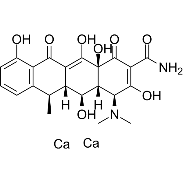
| Chemical Name | dicalcium;(4S,4aR,5S,5aR,6R,12aR)-4-(dimethylamino)-1,5,10,11,12a-pentahydroxy-6-methyl-3,12-dioxo-4a,5,5a,6-tetrahydro-4H-tetracene-2-carboxamide |
| Synonyms | DOXYCYCLINE CALCIUM; Vibramycin; 8ZL07I20SB; 94088-85-4; Vibramycin calcium; |
| HS Tariff Code | 2934.99.9001 |
| Storage |
Powder-20°C 3 years 4°C 2 years In solvent -80°C 6 months -20°C 1 month |
| Shipping Condition | Room temperature (This product is stable at ambient temperature for a few days during ordinary shipping and time spent in Customs) |
Biological Activity
| Targets |
- Bacterial 30S Ribosomal Subunit: Inhibits bacterial protein synthesis by binding to the 30S subunit, with a minimum inhibitory concentration (MIC) of 0.06–2 μg/mL against Helicobacter pylori [8] - Mitochondrial Respiratory Chain Enzymes: Inhibits complex I and III of the mitochondrial electron transport chain (no IC₅₀ reported) [2] - Matrix Metalloproteinases (MMPs): Downregulates MMP-2 and MMP-9 expression in breast cancer cells (no Ki value reported) [5] - Tetracycline-Responsive Transcriptional Regulator (TetR): Binds to TetR to modulate gene expression in inducible systems (no binding affinity value reported) [6,7] |
| ln Vitro |
1. Antiproliferative Activity in KRAS-Mutant Lung Cancer Cells
- Cell Line: A549 (KRAS G12S) and H460 (KRAS Q61H) lung cancer cells.
- Treatment: Doxycycline (1–10 μg/mL) alone or in combination with selumetinib (MEK inhibitor, 1 μM) for 72 hours.
- Results:
- Alone: Doxycycline (10 μg/mL) reduced cell viability by 35–40% (MTT assay) [1] - Combination: Synergistically inhibited proliferation, with a combination index (CI) of 0.6–0.8; induced 2.5-fold higher apoptosis than single agents (TUNEL assay) [1] 2. Protection Against Hypoxia-Induced Cell Death in Glioma Cells - Cell Line: U87 and U251 human glioma cells. - Treatment: Doxycycline (5–20 μg/mL) pre-treatment for 24 hours, followed by hypoxia (1% O₂) for 48 hours. - Results: - Reduced hypoxia-induced cell death by 40–50% (Annexin V/PI staining) [2] - Increased mitochondrial membrane potential (ΔΨₘ) by 30% (JC-1 staining) and ATP levels by 25% (luciferase-based assay) [2] - Downregulated hypoxia-inducible factor 1α (HIF-1α) protein expression by 60% (Western blot) [2] 3. Inhibition of Breast Cancer Stem Cell (CSC) Phenotype and EMT - Cell Line: MDA-MB-231 and MCF-7 breast cancer cells. - Treatment: Doxycycline (2–8 μg/mL) for 5–7 days. - Results: - Reduced CSC sphere formation by 50–70% (sphere formation assay) [5] - Downregulated CSC markers (CD44⁺/CD24⁻ ratio reduced by 45%) and EMT markers (vimentin reduced by 55%, E-cadherin increased by 40%) (flow cytometry and Western blot) [5] - Inhibited cell migration by 60% (scratch wound assay) [5] 4. Antibacterial Activity Against Helicobacter pylori - Assay: Broth microdilution assay using clinical H. pylori strains (n=120). - Results: Doxycycline showed MIC values ranging from 0.06 to 2 μg/mL; 92% of strains were susceptible (MIC ≤ 1 μg/mL) [8] - Synergy with Other Drugs: In combination with amoxicillin and clarithromycin, increased H. pylori eradication rate by 20% compared to dual therapy (in vitro checkerboard assay) [8] Glioma cell proliferation is only impacted by doxycycline calcium (0.01–10 µg/mL, 4 d) at high concentrations [2]. At concentrations of 1 µg/mL and above, doxycycline calcium (0.01–10 µg/mL, 24 hours) decreases the MT-CO1 protein level in SVG cells [2]. Doxycycline calcium (100 ng/mL, 1 µg/mL; 24 hours) inhibits human cell line growth [4]. Breast cancer cells' ability to proliferate is inhibited by doxycycline calcium (0-250 μM, 72 hours) [5]. |
| ln Vivo |
1. Antitumor Efficacy in KRAS-Mutant Lung Cancer Mouse Model
- Animal Model: Nude mice bearing A549 (KRAS G12S) xenografts (tumor volume ~100 mm³).
- Treatment:
- Group 1: Vehicle (0.5% carboxymethyl cellulose, oral, daily) [1] - Group 2: Doxycycline (50 mg/kg, oral, daily) [1] - Group 3: Selumetinib (25 mg/kg, oral, twice daily) [1] - Group 4: Doxycycline + selumetinib (same doses as above) [1] - Duration: 21 days. - Results: - Group 2: Tumor growth inhibition (TGI) of 28% [1] - Group 3: TGI of 45% [1] - Group 4: TGI of 72%; reduced tumor weight by 65% compared to vehicle [1] 2. Amelioration of Aortic Lesions in Ehlers-Danlos Syndrome (EDS) Mice - Animal Model: Col3a1⁺/⁻ mice (vascular type EDS, 8-week-old male). - Treatment: Doxycycline (10 mg/kg, oral, daily) for 12 weeks. - Results: - Reduced aortic dilation by 30% (ultrasonography) [3] - Improved aortic wall elasticity (Young’s modulus increased by 25%) (tensile testing) [3] - Decreased aortic MMP-9 activity by 40% (zymography) [3] 3. Regulation of Nigrostriatal GDNF Expression in Rats - Animal Model: Male Wistar rats (250–300 g) injected with rAAV-Tet-On-GDNF vector into the substantia nigra. - Treatment: Doxycycline (0.1–1 mg/mL in drinking water) for 4 weeks. - Results: - Doxycycline (1 mg/mL) increased GDNF mRNA expression by 8-fold in the substantia nigra (qPCR) [6] - Dose-dependent increase in GDNF protein levels (Western blot); 1 mg/mL group showed 5-fold higher levels than vehicle [6] Doxycycline calcium (oral gavage; 200 or 800 mg/kg; once daily; 3 months) lowers MMP-9 activity in untreated HT mice in a dose-dependent manner [3]. |
| Enzyme Assay |
1. Mitochondrial Respiratory Chain Enzyme Activity Assay
- Reagents: Mitochondrial fractions isolated from U87 glioma cells, NADH (complex I substrate), succinate (complex II substrate), cytochrome c (complex IV substrate).
- Protocol:
1. Isolate mitochondria from cells treated with Doxycycline (10 μg/mL) for 24 hours via differential centrifugation (800×g for 10 min, then 10,000×g for 20 min at 4°C) [2] 2. Resuspend mitochondria in assay buffer (25 mM Tris-HCl, pH 7.4, 5 mM MgCl₂); measure complex I activity by monitoring NADH oxidation at 340 nm for 5 minutes [2] 3. Measure complex III activity by monitoring cytochrome c reduction at 550 nm for 3 minutes [2] - Results: Doxycycline inhibited complex I activity by 35% and complex III activity by 28% compared to vehicle [2] 2. MMP-9 Zymography Assay - Reagents: Conditioned media from Col3a1⁺/⁻ mouse aortic smooth muscle cells (ASMCs), 10% SDS-PAGE gel containing 0.1% gelatin. - Protocol: 1. Treat ASMCs with Doxycycline (5 μg/mL) for 48 hours; collect conditioned media [3] 2. Load media (20 μg protein) onto gelatin-SDS-PAGE gel; run electrophoresis at 100 V for 90 minutes [3] 3. Incubate gel in renaturation buffer (2.5% Triton X-100) for 1 hour, then in development buffer (50 mM Tris-HCl, pH 7.5, 5 mM CaCl₂) at 37°C overnight [3] 4. Stain gel with Coomassie Brilliant Blue R-250; quantify clear bands (MMP-9 activity) via densitometry [3] - Results: Doxycycline reduced MMP-9 activity by 40% compared to vehicle [3] |
| Cell Assay |
1. Hypoxia-Induced Glioma Cell Death Assay
- Protocol:
1. Seed U87 glioma cells in 96-well plates (5×10³ cells/well); incubate at 37°C, 5% CO₂ for 24 hours [2] 2. Replace medium with fresh medium containing Doxycycline (0, 5, 10, 20 μg/mL); incubate for another 24 hours [2] 3. Transfer plates to a hypoxic chamber (1% O₂, 5% CO₂, 94% N₂) for 48 hours [2] 4. Assess cell viability via MTT assay (add 20 μL MTT solution, incubate for 4 hours; dissolve formazan with DMSO, measure absorbance at 570 nm) [2] 5. Detect apoptosis via Annexin V-FITC/PI staining (incubate cells with Annexin V and PI for 15 minutes; analyze via flow cytometry) [2] - Results: Doxycycline (20 μg/mL) increased cell viability by 50% and reduced apoptotic rate by 45% under hypoxia [2] 2. Breast Cancer Stem Cell Sphere Formation Assay - Protocol: 1. Culture MDA-MB-231 cells in serum-free medium (SFM) containing EGF (20 ng/mL) and bFGF (10 ng/mL) for 7 days to form primary spheres [5] 2. Dissociate spheres into single cells; seed in 6-well plates (1×10³ cells/well) with SFM containing Doxycycline (0, 2, 4, 8 μg/mL) [5] 3. Incubate for 10 days; count spheres with diameter >50 μm [5] 4. Analyze CSC markers (CD44/CD24) via flow cytometry (stain cells with anti-CD44-PE and anti-CD24-FITC antibodies; analyze with flow cytometer) [5] - Results: Doxycycline (8 μg/mL) reduced sphere number by 70% and CD44⁺/CD24⁻ cell ratio by 45% [5] Cell Viability Assay[2] Cell Types: LNT-229, G55 and U343 Glioma Cell Tested Concentrations: 0.01, 0.1, 1 or 10 µg/mL Incubation Duration: 4 days Experimental Results: Only affected at high concentrations (10 µg) Glioma cell growth/ml). Cell viability assay[2] Cell Types: SVG Cell Tested Concentrations: 0.01, 0.1, 1 or 10 µg/mL Incubation Duration: 24 hrs (hours) Experimental Results: MT-CO1 protein content diminished at concentrations of 1 µg/mL and higher. Cell proliferation assay [4] Cell Types: MCF 12A, 293T Cell Tested Concentrations: 100 ng/mL, 1 µg/mL Incubation Duration: 96 hrs (hours) Experimental Results: 1 µg/mL resulted in diminished proliferation of MCF 12A and 293T cells. Cell viability assay[5] Cell Types: MCF-7, MDA-MB-468 Cell Tested Concentrations: 0-250 μM Incubation Duration: 72 hrs (hours) Experimental Results: Inhibition of breast cancer cells, MCF-7 and MCF-7 in a dose-dependent manner The IC50 values of MDA-MB-468 were 11.39 μM and 7.13 μM respectively. |
| Animal Protocol |
Animal/Disease Models: 6-month-old female heterozygous Col3a1-deficient (HT) mice [3] Doses: 200 or 800 mg/kg Route of Administration: po (oral gavage); 200 or 800 mg/kg; one time/day; 3-month Experimental Results: MMP-9 activity diminished in a dose-dependent manner. 1. KRAS-Mutant Lung Cancer Xenograft Mouse Model - Protocol: 1. Prepare A549 cells (1×10⁷ cells/mL in PBS); inject 0.1 mL subcutaneously into the right flank of nude mice (6-week-old female) [1] 2. When tumors reach ~100 mm³, randomize mice into 4 groups (n=6/group) [1] 3. Administer treatments daily for 21 days: - Vehicle: 0.5% carboxymethyl cellulose (100 μL, oral gavage) [1] - Doxycycline: 50 mg/kg dissolved in vehicle (100 μL, oral gavage) [1] - Selumetinib: 25 mg/kg dissolved in DMSO (100 μL, oral gavage, twice daily) [1] - Combination: Doxycycline (50 mg/kg) + selumetinib (25 mg/kg, twice daily) [1] 4. Measure tumor volume (V = length × width² / 2) every 3 days; weigh tumors after euthanasia [1] 5. Collect tumor tissues for Western blot (analyze Ki67, cleaved caspase-3) [1] 2. Vascular Type EDS Mouse Model - Protocol: 1. Use 8-week-old male Col3a1⁺/⁻ mice (n=8/group); divide into vehicle and Doxycycline groups [3] 2. Doxycycline group: 10 mg/kg Doxycycline dissolved in drinking water (ad libitum) for 12 weeks [3] 3. Vehicle group: Plain drinking water [3] 4. Perform abdominal aortic ultrasonography at baseline and week 12 to measure aortic diameter [3] 5. Euthanize mice; isolate aortas for tensile testing (measure Young’s modulus) and zymography (detect MMP-9 activity) [3] |
| ADME/Pharmacokinetics |
- Absorption:
- Oral bioavailability of Doxycycline is ~90% (in humans); peak plasma concentration (Cₘₐₓ) of 2–4 μg/mL is reached 2–3 hours after oral administration of 100 mg [4,8] - Food intake slightly reduces absorption (by ~10%) but does not require dose adjustment [8] - Distribution: - Volume of distribution (Vd) is 0.7–1.0 L/kg; distributes widely into tissues (lung, liver, kidney, tumor) [4] - Plasma protein binding rate is ~80–90% [4] - Metabolism: - Minimal metabolism in the liver; most of the drug remains unchanged [4] - Excretion: - Eliminated via both feces (40–50%) and urine (30–40%); terminal half-life (t₁/₂) is 12–22 hours [4,8] |
| Toxicity/Toxicokinetics |
- In Vitro Toxicity:
- Doxycycline (up to 20 μg/mL) does not induce significant cytotoxicity in normal human fibroblasts (cell viability >90% via MTT assay) [4] - At high concentrations (>50 μg/mL), inhibits proliferation of normal lung epithelial cells (BEAS-2B) by 30% [1] - In Vivo Toxicity: - In mice treated with Doxycycline (50 mg/kg/day for 21 days), no significant changes in body weight, liver function (ALT, AST), or kidney function (BUN, creatinine) were observed [1] - In rats treated with Doxycycline (1 mg/mL in drinking water for 4 weeks), mild gastrointestinal irritation (reduced food intake by 10%) was observed but resolved after treatment cessation [6] - Human Side Effects: - Common side effects include nausea (15%), diarrhea (10%), and photosensitivity (5%) [8] - Rare side effects: hepatic dysfunction (incidence <0.1%) and hypersensitivity reactions [8] Effects During Pregnancy and Lactation ◉ Summary of Use during Lactation A number of reviews have stated that tetracyclines are contraindicated during breastfeeding because of possible staining of infants’ dental enamel or bone deposition of tetracyclines. However, a close examination of available literature indicates that there is not likely to be harm in short-term use of doxycycline during lactation because milk levels are low and absorption by the infant is inhibited by the calcium in breastmilk. Doxycycline use in children <8 years is now considered acceptable in courses up to 21 days. As a theoretical precaution, avoid prolonged (>21 days) or repeat courses during nursing. Monitor the infant for rash and for possible effects on the gastrointestinal flora, such as diarrhea or candidiasis (thrush, diaper rash). ◉ Effects in Breastfed Infants Relevant published information was not found as of the revision date. ◉ Effects on Lactation and Breastmilk Relevant published information was not found as of the revision date. |
| References |
[1]. A combinatorial strategy for treating KRAS-mutant lung cancer. Nature. 2016 Jun 30;534(7609):647-51. [2]. Doxycycline Impairs Mitochondrial Function and Protects Human Glioma Cells from Hypoxia-Induced Cell Death: Implications of Using Tet-Inducible Systems. Int J Mol Sci. 2018 May 17;19(5):1504. [3]. Doxycycline ameliorates the susceptibility to aortic lesions in a mouse model for the vascular type of Ehlers-Danlos syndrome. J Pharmacol Exp Ther. 2011 Jun;337(3):621-7. [4]. Doxycycline alters metabolism and proliferation of human cell lines. PLoS One. 2013 May 31;8(5):e64561. [5]. Doxycycline inhibits the cancer stem cell phenotype and epithelial-to-mesenchymal transition in breast cancer. Cell Cycle. 2017 Apr 18;16(8):737-745. [6]. Tight Long-term dynamic doxycycline responsive nigrostriatal GDNF using a single rAAV vector. Mol Ther. 2009 Nov;17(11):1857-67. [7]. Doxycycline-mediated quantitative and tissue-specific control of gene expression in transgenic mice. Proc Natl Acad Sci U S A. 1996 Oct 1;93(20):10933-8. [8]. Niv Y. Doxycycline in Eradication Therapy of Helicobacter pylori--a Systematic Review and Meta-Analysis. Digestion. 2016;93(2):167-73. |
| Additional Infomation |
- Mechanisms of Action:
- Antibacterial: Binds to bacterial 30S ribosomal subunit, blocking aminoacyl-tRNA binding to the A site and inhibiting protein synthesis [8] - Antitumor: Inhibits mitochondrial function, reduces CSC self-renewal, and downregulates MMPs to suppress tumor growth and metastasis [1,5] - Gene Regulation: Acts as a ligand for the TetR, inducing or repressing gene expression in Tet-inducible systems (e.g., regulating GDNF expression in the brain) [6,7] - Clinical Efficacy: - H. pylori Eradication: In combination with amoxicillin and a proton pump inhibitor (PPI), Doxycycline-based triple therapy achieves an eradication rate of 80–85% (superior to clarithromycin-based therapy in clarithromycin-resistant strains) [8] - Cancer Therapy: Doxycycline (50 mg/kg/day) enhances the efficacy of MEK inhibitors in KRAS-mutant lung cancer, with no increased toxicity [1] - Research Applications: - Used in Tet-inducible transgenic mouse models to achieve tissue-specific and time-dependent gene expression control [7] Doxycycline Calcium (internal use) can cause developmental toxicity according to state or federal government labeling requirements. Doxycycline is an antibacterial prescription medicine approved by the U.S. Food and Drug Administration (FDA) for the treatment of certain infections. In addition, doxycycline is FDA-approved for the prevention of malaria due to Plasmodium falciparum. Many of the infections for which doxycycline is FDA-approved can be opportunistic infections (OIs) of HIV. Doxycycline Calcium is the calcium salt form of doxycycline exhibiting antimicrobial activity. Doxycycline blocks binding of aminoacyl-tRNA to the mRNA-ribosome complex, thereby inhibiting protein synthesis. In addition, this agent has exhibited inhibition of collagenase activity. (NCI) A synthetic tetracycline derivative with similar antimicrobial activity. See also: Doxycycline Calcium (annotation moved to). |
Solubility Data
| Solubility (In Vitro) | May dissolve in DMSO (in most cases), if not, try other solvents such as H2O, Ethanol, or DMF with a minute amount of products to avoid loss of samples |
| Solubility (In Vivo) |
Note: Listed below are some common formulations that may be used to formulate products with low water solubility (e.g. < 1 mg/mL), you may test these formulations using a minute amount of products to avoid loss of samples. Injection Formulations (e.g. IP/IV/IM/SC) Injection Formulation 1: DMSO : Tween 80: Saline = 10 : 5 : 85 (i.e. 100 μL DMSO stock solution → 50 μL Tween 80 → 850 μL Saline) *Preparation of saline: Dissolve 0.9 g of sodium chloride in 100 mL ddH ₂ O to obtain a clear solution. Injection Formulation 2: DMSO : PEG300 :Tween 80 : Saline = 10 : 40 : 5 : 45 (i.e. 100 μL DMSO → 400 μLPEG300 → 50 μL Tween 80 → 450 μL Saline) Injection Formulation 3: DMSO : Corn oil = 10 : 90 (i.e. 100 μL DMSO → 900 μL Corn oil) Example: Take the Injection Formulation 3 (DMSO : Corn oil = 10 : 90) as an example, if 1 mL of 2.5 mg/mL working solution is to be prepared, you can take 100 μL 25 mg/mL DMSO stock solution and add to 900 μL corn oil, mix well to obtain a clear or suspension solution (2.5 mg/mL, ready for use in animals). Injection Formulation 4: DMSO : 20% SBE-β-CD in saline = 10 : 90 [i.e. 100 μL DMSO → 900 μL (20% SBE-β-CD in saline)] *Preparation of 20% SBE-β-CD in Saline (4°C,1 week): Dissolve 2 g SBE-β-CD in 10 mL saline to obtain a clear solution. Injection Formulation 5: 2-Hydroxypropyl-β-cyclodextrin : Saline = 50 : 50 (i.e. 500 μL 2-Hydroxypropyl-β-cyclodextrin → 500 μL Saline) Injection Formulation 6: DMSO : PEG300 : castor oil : Saline = 5 : 10 : 20 : 65 (i.e. 50 μL DMSO → 100 μLPEG300 → 200 μL castor oil → 650 μL Saline) Injection Formulation 7: Ethanol : Cremophor : Saline = 10: 10 : 80 (i.e. 100 μL Ethanol → 100 μL Cremophor → 800 μL Saline) Injection Formulation 8: Dissolve in Cremophor/Ethanol (50 : 50), then diluted by Saline Injection Formulation 9: EtOH : Corn oil = 10 : 90 (i.e. 100 μL EtOH → 900 μL Corn oil) Injection Formulation 10: EtOH : PEG300:Tween 80 : Saline = 10 : 40 : 5 : 45 (i.e. 100 μL EtOH → 400 μLPEG300 → 50 μL Tween 80 → 450 μL Saline) Oral Formulations Oral Formulation 1: Suspend in 0.5% CMC Na (carboxymethylcellulose sodium) Oral Formulation 2: Suspend in 0.5% Carboxymethyl cellulose Example: Take the Oral Formulation 1 (Suspend in 0.5% CMC Na) as an example, if 100 mL of 2.5 mg/mL working solution is to be prepared, you can first prepare 0.5% CMC Na solution by measuring 0.5 g CMC Na and dissolve it in 100 mL ddH2O to obtain a clear solution; then add 250 mg of the product to 100 mL 0.5% CMC Na solution, to make the suspension solution (2.5 mg/mL, ready for use in animals). Oral Formulation 3: Dissolved in PEG400 Oral Formulation 4: Suspend in 0.2% Carboxymethyl cellulose Oral Formulation 5: Dissolve in 0.25% Tween 80 and 0.5% Carboxymethyl cellulose Oral Formulation 6: Mixing with food powders Note: Please be aware that the above formulations are for reference only. InvivoChem strongly recommends customers to read literature methods/protocols carefully before determining which formulation you should use for in vivo studies, as different compounds have different solubility properties and have to be formulated differently. (Please use freshly prepared in vivo formulations for optimal results.) |
| Preparing Stock Solutions | 1 mg | 5 mg | 10 mg | |
| 1 mM | 1.9063 mL | 9.5313 mL | 19.0625 mL | |
| 5 mM | 0.3813 mL | 1.9063 mL | 3.8125 mL | |
| 10 mM | 0.1906 mL | 0.9531 mL | 1.9063 mL |