Demethyleneberberine is a potent and naturally occuring mitochondria-targeted antioxidant. Demethyleneberberine alleviates mice colitis and inhibits the inflammatory responses by inhibiting NF-κB pathway and regulating the balance of Th cells. Demethyleneberberine could serve as a AMPK activator for treating non-alcoholic fatty liver disease (NAFLD).
Physicochemical Properties
| Molecular Formula | C19H18NO4 |
| Molecular Weight | 324.3505 |
| Exact Mass | 323.116 |
| CAS # | 25459-91-0 |
| Related CAS # | Demethyleneberberine chloride;16705-03-6 |
| PubChem CID | 363209 |
| Appearance | Light yellow to yellow solid powder |
| LogP | 1.649 |
| Hydrogen Bond Donor Count | 2 |
| Hydrogen Bond Acceptor Count | 4 |
| Rotatable Bond Count | 2 |
| Heavy Atom Count | 24 |
| Complexity | 447 |
| Defined Atom Stereocenter Count | 0 |
| InChi Key | HVTCKKMWZDDWOY-UHFFFAOYSA-O |
| InChi Code | InChI=1S/C19H17NO4/c1-23-18-4-3-11-7-15-13-9-17(22)16(21)8-12(13)5-6-20(15)10-14(11)19(18)24-2/h3-4,7-10,22H,5-6H2,1-2H3/p+1 |
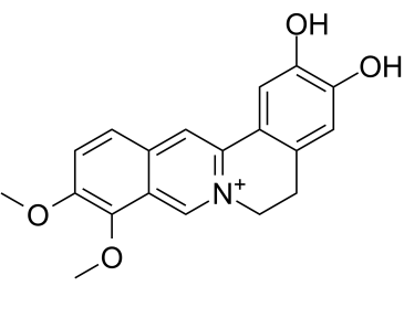
| Chemical Name | 9,10-dimethoxy-5,6-dihydroisoquinolino[2,1-b]isoquinolin-7-ium-2,3-diol |
| HS Tariff Code | 2934.99.9001 |
| Storage |
Powder-20°C 3 years 4°C 2 years In solvent -80°C 6 months -20°C 1 month Note: This product requires protection from light (avoid light exposure) during transportation and storage. |
| Shipping Condition | Room temperature (This product is stable at ambient temperature for a few days during ordinary shipping and time spent in Customs) |
Biological Activity
| Targets |
Demethyleneberberine targets AMP-activated protein kinase (AMPK) [3] Demethyleneberberine targets nuclear factor kappa B (NF-κB) signaling pathway [2] Demethyleneberberine acts as a mitochondria-targeted antioxidant [1] |
| ln Vitro |
#### From Literature [1] - In ethanol-treated HepG2 cells, Demethyleneberberine (10, 20, 40 μM) dose-dependently reduced intracellular reactive oxygen species (ROS) production and mitochondrial superoxide levels [1] - It improved mitochondrial membrane potential (ΔΨm) and increased ATP production, alleviating ethanol-induced mitochondrial dysfunction [1] - Downregulated the expression of lipogenic genes (SREBP-1c, FASN) and upregulated fatty acid oxidation-related gene (PPARα) in HepG2 cells [1] #### From Literature [2] - In LPS-induced RAW264.7 macrophages, Demethyleneberberine (5, 10, 20 μM) inhibited the production of pro-inflammatory cytokines (TNF-α, IL-6, IL-1β) and nitric oxide (NO) [2] - It suppressed NF-κB activation by inhibiting IκBα phosphorylation and p65 nuclear translocation [2] - In TNF-α-treated Caco-2 intestinal epithelial cells, the compound enhanced tight junction protein (ZO-1, occludin) expression, improving intestinal barrier integrity [2] #### From Literature [3] - In palmitic acid (PA)-treated HepG2 cells, Demethyleneberberine (10, 20, 40 μM) reduced lipid accumulation (Oil Red O staining) and intracellular triglyceride (TG) content [3] - It activated AMPK phosphorylation (p-AMPK) and upregulated downstream energy metabolism-related genes (PGC-1α, CPT1A) [3] - Decreased PA-induced ROS generation and malondialdehyde (MDA) levels, while increasing superoxide dismutase (SOD) and glutathione (GSH) activities [3] |
| ln Vivo |
#### From Literature [1] (Alcoholic Liver Disease Model) - In C57BL/6 mice fed an ethanol-containing Lieber-DeCarli diet for 4 weeks, oral administration of Demethyleneberberine (25, 50 mg/kg/day) dose-dependently reduced hepatic steatosis (H&E staining) and TG content [1] - Improved mitochondrial function in liver tissue, as evidenced by increased mitochondrial DNA copy number and complex I/III/IV activities [1] - Reduced hepatic oxidative stress (decreased MDA, increased SOD/GSH-Px activities) and inhibited hepatocyte apoptosis (TUNEL staining) [1] #### From Literature [2] (Inflammatory Bowel Disease Model) - In DSS-induced colitis mice, oral administration of Demethyleneberberine (25, 50 mg/kg/day) for 7 days alleviated weight loss, colon shortening, and histological damage (H&E staining) [2] - Reduced colonic pro-inflammatory cytokine (TNF-α, IL-6, IL-1β) levels and NF-κB p65 nuclear translocation [2] - Restored T-helper cell homeostasis by increasing Treg cell percentage and decreasing Th1/Th17 cell percentages in mesenteric lymph nodes [2] #### From Literature [3] (Non-Alcoholic Fatty Liver Disease Model) - In ob/ob mice or high-fat diet (HFD)-fed C57BL/6 mice, oral administration of Demethyleneberberine (25, 50 mg/kg/day) for 8 weeks reduced hepatic steatosis and liver weight [3] - Activated hepatic AMPK phosphorylation and upregulated fatty acid oxidation-related proteins (CPT1A, PGC-1α) [3] - Decreased hepatic oxidative stress and inflammation (reduced TNF-α, IL-6 mRNA expression) [3] |
| Enzyme Assay |
#### From Literature [1] (Mitochondrial Function Assays) - Mitochondrial membrane potential (ΔΨm) assay: HepG2 cells were loaded with a ΔΨm-sensitive fluorescent dye, treated with Demethyleneberberine (10, 20, 40 μM) and ethanol, and fluorescence intensity was measured by flow cytometry [1] - ATP assay: Ethanol-treated HepG2 cells were lysed, and ATP content was quantified using a luciferase-based assay kit; results were normalized to protein concentration [1] - Oxidative stress assays: Cells/tissues were homogenized, and SOD, GSH-Px activities, and MDA levels were measured using colorimetric assay kits [1] #### From Literature [3] (AMPK Activity Assay) - HepG2 cells were treated with Demethyleneberberine (10, 20, 40 μM) and palmitic acid, then lysed [3] - AMPK activity was determined by measuring the phosphorylation of its substrate (ACC) via Western blot, with p-ACC/ACC ratio reflecting AMPK activation [3] |
| Cell Assay |
- HepG2 cells were cultured in DMEM medium with 10% fetal bovine serum, treated with Demethyleneberberine (10, 20, 40 μM) for 2 hours, then exposed to ethanol (100 mM) for 24 hours [1] - ROS detection: Cells were loaded with DCFH-DA fluorescent probe, incubated for 30 minutes, and fluorescence intensity was measured by flow cytometry [1] - Lipid accumulation assay: Cells were stained with Oil Red O, eluted with isopropanol, and absorbance was measured at 510 nm [1] - Western blot: Cells were lysed, proteins separated by SDS-PAGE, and blotted with antibodies against SREBP-1c, FASN, PPARα, and β-actin [1] #### From Literature [2] - RAW264.7 cells were treated with Demethyleneberberine (5, 10, 20 μM) for 1 hour, then stimulated with LPS (1 μg/mL) for 24 hours [2] - Cytokine detection: Culture supernatants were collected, and TNF-α, IL-6, IL-1β levels were measured by ELISA [2] - NF-κB translocation assay: Cells were fixed, permeabilized, stained with p65 antibody and DAPI, and observed under confocal microscopy [2] - Caco-2 cells were treated with Demethyleneberberine (5, 10, 20 μM) and TNF-α (10 ng/mL) for 24 hours; tight junction proteins were detected by Western blot [2] #### From Literature [3] - HepG2 cells were treated with Demethyleneberberine (10, 20, 40 μM) and palmitic acid (200 μM) for 24 hours [3] - Triglyceride assay: Cells were lysed, and TG content was measured by colorimetric assay [3] - Western blot: Blotted with antibodies against p-AMPK, AMPK, PGC-1α, CPT1A, and β-actin [3] |
| Animal Protocol |
#### From Literature [1] (Alcoholic Liver Disease) - Male C57BL/6 mice (6–8 weeks old) were randomly divided into control, ethanol model, and Demethyleneberberine (25, 50 mg/kg/day) groups (n=8 per group) [1] - Mice were fed a Lieber-DeCarli ethanol diet (5% ethanol) for 4 weeks; the compound was dissolved in 0.5% carboxymethylcellulose sodium and administered by oral gavage once daily [1] - At the end of treatment, mice were sacrificed; liver tissues were collected for H&E staining, TG assay, mitochondrial function analysis, and Western blot [1] - Serum was collected to detect ALT, AST levels [1] #### From Literature [2] (Inflammatory Bowel Disease) - Male C57BL/6 mice (6–8 weeks old) were randomly divided into control, DSS model, and Demethyleneberberine (25, 50 mg/kg/day) groups (n=8 per group) [2] - Colitis was induced by adding 3% DSS to drinking water for 7 days; the compound was administered by oral gavage once daily during DSS exposure [2] - Body weight and colon length were measured; colon tissues were collected for H&E staining, cytokine assay, and Western blot [2] - Mesenteric lymph nodes were isolated for flow cytometry analysis of T-cell subsets [2] #### From Literature [3] (Non-Alcoholic Fatty Liver Disease) - Male ob/ob mice or HFD-fed C57BL/6 mice (6–8 weeks old) were randomly divided into control, model, and Demethyleneberberine (25, 50 mg/kg/day) groups (n=8 per group) [3] - The compound was dissolved in 0.5% carboxymethylcellulose sodium and administered by oral gavage once daily for 8 weeks [3] - Mice were sacrificed; liver tissues were collected for H&E staining, TG assay, and Western blot [3] - Serum was collected to detect glucose, insulin, and lipid profiles [3] |
| Toxicity/Toxicokinetics |
- In all animal models, oral administration of Demethyleneberberine (25–50 mg/kg/day) for 4–8 weeks did not cause significant changes in body weight, liver function (ALT, AST), or kidney function (BUN, Cr) [1][2][3] - No obvious clinical toxicity signs (lethargy, loss of appetite, organ damage) were observed in treated mice [1][2][3] |
| References |
[1]. Demethyleneberberine, a natural mitochondria-targeted antioxidant, inhibits mitochondrial dysfunction, oxidative stress, and steatosis in alcoholic liver disease mouse model. J Pharmacol Exp Ther. 2015 Jan;352(1):139-47. [2]. Demethyleneberberine alleviates inflammatory bowel disease in mice through regulating NF-κB signaling and T-helper cell homeostasis. Inflamm Res. 2017 Feb;66(2):187-196. [3]. Demethyleneberberine attenuates non-alcoholic fatty liver disease with activation of AMPK and inhibition of oxidative stress. Biochem Biophys Res Commun. 2016 Apr 15;472(4):603-9. |
| Additional Infomation |
Demethyleneberberine is a member of isoquinolines. Demethyleneberberine has been reported in Thalictrum javanicum with data available. Demethyleneberberine is a natural isoquinoline alkaloid isolated from plants of the Berberidaceae family [1][2][3] Its core mechanisms include: mitochondria-targeted antioxidant activity, AMPK activation, and NF-κB signaling inhibition [1][2][3] It exhibits therapeutic potential for metabolic and inflammatory diseases, including alcoholic liver disease, non-alcoholic fatty liver disease, and inflammatory bowel disease [1][2][3] The compound exerts tissue-protective effects by regulating lipid metabolism, reducing oxidative stress, inhibiting inflammation, and maintaining cellular barrier function [1][2][3] |
Solubility Data
| Solubility (In Vitro) | DMSO : ~10 mg/mL (~30.83 mM) |
| Solubility (In Vivo) |
Solubility in Formulation 1: ≥ 2.5 mg/mL (7.71 mM) (saturation unknown) in 10% DMSO + 90% (20% SBE-β-CD in Saline) (add these co-solvents sequentially from left to right, and one by one), clear solution. For example, if 1 mL of working solution is to be prepared, you can add 100 μL of 25.0 mg/mL clear DMSO stock solution to 900 μL of 20% SBE-β-CD physiological saline solution and mix evenly. Preparation of 20% SBE-β-CD in Saline (4°C,1 week): Dissolve 2 g SBE-β-CD in 10 mL saline to obtain a clear solution. (Please use freshly prepared in vivo formulations for optimal results.) |
| Preparing Stock Solutions | 1 mg | 5 mg | 10 mg | |
| 1 mM | 3.0831 mL | 15.4154 mL | 30.8309 mL | |
| 5 mM | 0.6166 mL | 3.0831 mL | 6.1662 mL | |
| 10 mM | 0.3083 mL | 1.5415 mL | 3.0831 mL |