Physicochemical Properties
| Molecular Formula | C10H13N5O3 |
| Molecular Weight | 251.2419 |
| Exact Mass | 251.102 |
| CAS # | 33231-14-0 |
| PubChem CID | 11658872 |
| Appearance | White to off-white solid powder |
| LogP | -0.5 |
| Hydrogen Bond Donor Count | 2 |
| Hydrogen Bond Acceptor Count | 7 |
| Rotatable Bond Count | 5 |
| Heavy Atom Count | 18 |
| Complexity | 303 |
| Defined Atom Stereocenter Count | 0 |
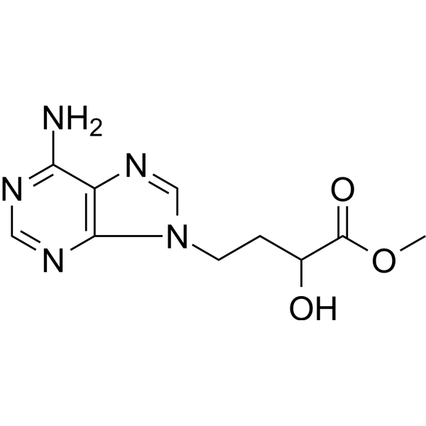
| SMILES | O([H])C([H])(C(=O)OC([H])([H])[H])C([H])([H])C([H])([H])N1C([H])=NC2=C(N([H])[H])N=C([H])N=C12 |
| InChi Key | HNKGMGPCSSJYOT-UHFFFAOYSA-N |
| InChi Code | InChI=1S/C10H13N5O3/c1-18-10(17)6(16)2-3-15-5-14-7-8(11)12-4-13-9(7)15/h4-6,16H,2-3H2,1H3,(H2,11,12,13) |
| Chemical Name | methyl 4-(6-aminopurin-9-yl)-2-hydroxybutanoate |
| HS Tariff Code | 2934.99.9001 |
| Storage |
Powder-20°C 3 years 4°C 2 years In solvent -80°C 6 months -20°C 1 month |
| Shipping Condition | Room temperature (This product is stable at ambient temperature for a few days during ordinary shipping and time spent in Customs) |
Biological Activity
| Targets | S-adenosyl-L-homocysteine hydrolase (SAHH) (IC50 = 0.15 μM for enzyme activity inhibition) [1] |
| ln Vitro |
The inhibitor of mixed lymphocyte reaction (MLR) responses is DZ2002 (0.1, 1, 10 µM; 96 hours) [1]. DZ2002 (0.1, 1, 10 µM; 24 hours) prevents human THP-1 cells and mouse peritoneal exudate cells from producing TNF-α and IL-12 [1]. On differentiated THP-1 cells, DZ2002 (0.1, 1, 10 µM; 64 hours) inhibits B7 (CD80/CD86) expression [1]. - DZ2002 exhibits potent SAHH inhibitory activity and immunosuppressive effects. It dose-dependently inhibited recombinant human SAHH activity with an IC50 of 0.15 μM. In mouse splenic lymphocytes, DZ2002 (0.1-10 μM) suppressed Concanavalin A (ConA)-induced T lymphocyte proliferation and Lipopolysaccharide (LPS)-induced B lymphocyte proliferation, with IC50 values of 0.8 μM and 1.2 μM, respectively. It also reduced the secretion of pro-inflammatory cytokines (IL-2, IFN-γ, TNF-α) from activated lymphocytes (ELISA assay) [1] - DZ2002 exerts anti-fibrotic and anti-inflammatory effects in vitro. In human dermal fibroblasts (HDFs) activated by TGF-β1, DZ2002 (0.5-5 μM) dose-dependently downregulated the expression of fibrosis-related markers (α-SMA, Col1A1, Col3A1) at both mRNA and protein levels (qPCR, Western blot). It also inhibited LPS-induced TNF-α and IL-6 production in human peripheral blood mononuclear cells (PBMCs) (ELISA) [2] |
| ln Vivo |
The DNFB-induced DTH response is blocked by DZ2002 (2, 10, 50 mg/kg; intraperitoneal injection; twice). (DNFB-induced DTH is a Th1 cell-mediated immunological response; macrophages have been demonstrated to play a significant part in this process and IL-12 is significantly produced) [1]. DZ2002 (0.08, 2 mg/kg; intraperitoneal injection; once daily for 7 days) substantially reduces the production of antibodies and delayed-type hypersensitivity reactions [1]. In SSc mice models, DZ2002 (50, 100 mg/kg; oral; once daily for 4 weeks) effectively inhibits the formation of collagen, promotes its breakdown, and controls the expression of many soluble factors. Fibrosis [2]. - In mouse models of , intraperitoneal injection of DZ2002 (10, 20 mg/kg/day) significantly prolonged the survival time of transplanted skin grafts. The 20 mg/kg dose extended graft survival from ~7 days (control) to ~18 days, accompanied by reduced infiltration of T lymphocytes and macrophages in graft tissues (immunohistochemistry) and decreased serum levels of IL-2 and IFN-γ (ELISA) [1] - In experimental systemic sclerosis (SSc) models (bleomycin-induced skin fibrosis and Tsk-1 mice), DZ2002 (15 mg/kg/day, intraperitoneal injection for 4 weeks) ameliorated skin fibrosis: reduced skin thickness, decreased collagen deposition (Masson's trichrome staining), and downregulated α-SMA and Col1A1 expression in skin tissues (Western blot). It also improved lung fibrosis, reduced pulmonary collagen content, and alleviated lung inflammation (HE staining, ELISA for TNF-α/IL-6). Additionally, it ameliorated vasculopathy by improving microvascular density and reducing vascular wall thickening in SSc mice [2] |
| Enzyme Assay | - SAHH activity inhibition assay: Recombinant human SAHH was dissolved in reaction buffer containing its substrate S-adenosyl-L-homocysteine. DZ2002 (0.01-10 μM) was added, and the mixture was incubated at 37°C for 60 minutes. The reaction was terminated by adding trichloroacetic acid, and the amount of adenosine (product of SAHH-catalyzed reaction) was quantified by high-performance liquid chromatography (HPLC) to calculate the inhibition rate of SAHH activity. IC50 value was derived from dose-response curves [1] |
| Cell Assay |
Cell Proliferation Assay[1] Cell Types: BALB/c and C57BL/6 splenocytes (mitomycin C pre-treated; mixed lymphocytes) Tested Concentrations: 0.1, 1, 10 µM Incubation Duration: 96 hrs (hours) Experimental Results: MLR inhibition 24.5, 42.3 and 46.0% at doses of 0.1, 1 and 10 µM respectively. Cell viability assay[1] Cell Types: TG-stimulated mouse peritoneal macrophages and human THP-1 cells Tested Concentrations: 0.1, 1, 10 µM Incubation Duration: 24 hrs (hours) Experimental Results: Significant blocking of approximately 1800 pg/mL of IL -12 p40 production in untreated cells dropped to approximately 850 pg/ml at 10 µM and Dramatically diminished the active p70 form in untreated cells from approximately 1200 pg/mL to approximately 50 pg/mL. TNF-α levels were diminished by 45%. Cell viability assay[1] Cell Types: THP-1 Cell Tested Concentrations: 0.1, 1, 10 µM Incubation Duration: 64 hrs (hours) Experimental Results: CD80, especially CD86 expression was Dramatically downregulated in a dose-dependent manner. - Lymphocyte proliferation assay: Mouse splenic lymphocytes were isolated and seeded in 96-well plates. DZ2002 (0.1-10 μM) was added 1 hour before stimulation with ConA (for T cells) or LPS (for B cells). After 72 hours of incubation, cell proliferation was measured by MTT assay, and culture supernatants were collected for ELISA detection of IL-2, IFN-γ, and TNF-α [1] - Fibroblast activation assay: Human dermal fibroblasts (HDFs) were seeded in 6-well plates and activated with TGF-β1 (10 ng/mL) for 24 hours. DZ2002 (0.5-5 μM) was added, and cells were cultured for another 48 hours. Total RNA was extracted for qPCR analysis of Col1A1, Col3A1, and α-SMA mRNA levels; total proteins were extracted for Western blot detection of corresponding proteins [2] - PBMC cytokine production assay: Human PBMCs were isolated and treated with DZ2002 (0.5-5 μM) for 1 hour, then stimulated with LPS (1 μg/mL) for 24 hours. ELISA was used to quantify TNF-α and IL-6 levels in supernatants [2] |
| Animal Protocol |
Animal/Disease Models: Male and female BALB/c and C57BL/6 mice (6 to 8 weeks old; DNFB-induced ear swelling model) [1]. Doses: 2, 10, 50 mg/kg Route of Administration: intraperitoneal (ip) injection; twice (1 hour before challenge and 24 hrs (hrs (hours)) after challenge) Experimental Results: Ear swelling was suppressed by 19.1%, 28.7% and 33.1%, respectively, in a dose-dependent manner . Animal/Disease Models: Male and female BALB/c and C57BL/6 mice (6 to 8 weeks old; DNFB-induced ear swelling model) [1]. Doses: 0.08, 2 mg/kg Route of Administration: intraperitoneal (ip) injection; single dose per day for 7 days. Experimental Results: Doses of 0.08 mg/kg and 2 mg/kg inhibited hemolysis by 24.5% and 18.4%, respectively, thereby reducing the production of anti-SRBC antibodies in the body. Animal/Disease Models: Wild-type C57BL/6 mice (8 to 12 weeks old; BLM-induced SSc mouse model) [2]. Doses: 50, 100 mg/kg Route of Administration: po (oral gavage); one time/day for 4 weeks. Experimental Results: BLM induced significant reductions in skin thickness and dermal thickness in mice. Dramatically diminished co - Allogeneic skin transplantation model: C57BL/6 mice (recipients) received skin grafts from BALB/c mice (donors). After transplantation, mice were randomly divided into control and DZ2002 treatment groups (10, 20 mg/kg/day). DZ2002 was dissolved in dimethyl sulfoxide (DMSO) and diluted with normal saline (final DMSO concentration ≤ 1%), administered via intraperitoneal injection once daily for 14 days. Graft survival time was recorded, and graft tissues were collected for immunohistochemistry; serum was collected for cytokine detection [1] - Systemic sclerosis models: 1) Bleomycin-induced skin fibrosis: C57BL/6 mice were subcutaneously injected with bleomycin (50 μg/day) for 4 weeks to induce SSc-like skin fibrosis. DZ2002 (15 mg/kg/day) was administered via intraperitoneal injection during weeks 2-4. 2) Tsk-1 mice (genetic SSc model): DZ2002 (15 mg/kg/day) was intraperitoneally injected for 4 weeks starting at 8 weeks of age. For both models, skin and lung tissues were collected for histopathological staining (HE, Masson's trichrome) and Western blot; serum was collected for ELISA [2] |
| References |
[1]. Wu QL, et al. Inhibition of S-adenosyl-L-homocysteine hydrolase induces immunosuppression. J Pharmacol Exp Ther. 2005 May;313(2):705-11. [2]. Zhang Z, et al. DZ2002 ameliorates fibrosis, inflammation, and vasculopathy in experimental systemic sclerosis models. Arthritis Res Ther. 2019 Dec 16;21(1):290. |
| Additional Infomation |
- DZ2002 is a synthetic small-molecule inhibitor of S-adenosyl-L-homocysteine hydrolase (SAHH) [1][2] - Its core mechanisms include: inhibiting SAHH to accumulate intracellular S-adenosyl-L-homocysteine, which suppresses methyltransferase activity and subsequent immune cell activation (immunosuppressive effect) [1]; downregulating TGF-β1-mediated fibroblast activation and pro-inflammatory cytokine production, thereby alleviating fibrosis and inflammation (anti-fibrotic and anti-inflammatory effects) [2] - DZ2002 shows potential therapeutic value in immune-related diseases (e.g., organ transplantation rejection) and autoimmune diseases associated with fibrosis (e.g., systemic sclerosis) [1][2] |
Solubility Data
| Solubility (In Vitro) | DMSO : ≥ 61 mg/mL (~242.80 mM) |
| Solubility (In Vivo) |
Solubility in Formulation 1: ≥ 2.5 mg/mL (9.95 mM) (saturation unknown) in 10% DMSO + 90% (20% SBE-β-CD in Saline) (add these co-solvents sequentially from left to right, and one by one), clear solution. For example, if 1 mL of working solution is to be prepared, you can add 100 μL of 25.0 mg/mL clear DMSO stock solution to 900 μL of 20% SBE-β-CD physiological saline solution and mix evenly. Preparation of 20% SBE-β-CD in Saline (4°C,1 week): Dissolve 2 g SBE-β-CD in 10 mL saline to obtain a clear solution. Solubility in Formulation 2: ≥ 2.5 mg/mL (9.95 mM) (saturation unknown) in 10% DMSO + 90% Corn Oil (add these co-solvents sequentially from left to right, and one by one), clear solution. For example, if 1 mL of working solution is to be prepared, you can add 100 μL of 25.0 mg/mL clear DMSO stock solution to 900 μL of corn oil and mix evenly. (Please use freshly prepared in vivo formulations for optimal results.) |
| Preparing Stock Solutions | 1 mg | 5 mg | 10 mg | |
| 1 mM | 3.9803 mL | 19.9013 mL | 39.8026 mL | |
| 5 mM | 0.7961 mL | 3.9803 mL | 7.9605 mL | |
| 10 mM | 0.3980 mL | 1.9901 mL | 3.9803 mL |