Physicochemical Properties
| Molecular Formula | C18H13N4O2F |
| Molecular Weight | 336.31982 |
| Exact Mass | 336.102 |
| CAS # | 484049-04-9 |
| PubChem CID | 660311 |
| Appearance | White to off-white solid powder |
| LogP | 2.474 |
| Hydrogen Bond Donor Count | 2 |
| Hydrogen Bond Acceptor Count | 4 |
| Rotatable Bond Count | 3 |
| Heavy Atom Count | 25 |
| Complexity | 517 |
| Defined Atom Stereocenter Count | 0 |
| InChi Key | MPQWYPLPWGUMJE-UHFFFAOYSA-N |
| InChi Code | InChI=1S/C18H13FN4O2/c19-12-8-6-11(7-9-12)15-14(16-17(24)21-18(25)20-16)10-23(22-15)13-4-2-1-3-5-13/h1-10,16H,(H2,20,21,24,25) |
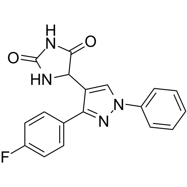
| Chemical Name | 5-[3-(4-fluorophenyl)-1-phenylpyrazol-4-yl]imidazolidine-2,4-dione |
| HS Tariff Code | 2934.99.9001 |
| Storage |
Powder-20°C 3 years 4°C 2 years In solvent -80°C 6 months -20°C 1 month |
| Shipping Condition | Room temperature (This product is stable at ambient temperature for a few days during ordinary shipping and time spent in Customs) |
Biological Activity
| Targets |
- DPH targets c-Abl kinase (binds to its myristoyl binding site as an activator), with an EC50 of ~1.2 μM for c-Abl activation and a dissociation constant (KD) of ~0.8 μM. [1] - DPH targets Abl2/Arg kinase (acts as an activator). [2] |
| ln Vitro |
In contrast to the previously discovered allosteric c-Abl with GNF-2, DPH binds to the myristoyl binding site and uses steric hindrance to stop the αI helix from forming its bent conformation. The myristyl acyl binding site is another area in which Silhouette interacts. The first tool chemical for c-Abl activation that penetrates cells is DPH [1]. - c-Abl Kinase Activation (Recombinant Enzyme & K562 Cells): 1. Recombinant c-Abl assay: DPH (0.1–10 μM) dose-dependently activated c-Abl kinase. At 1.2 μM, it achieved 50% maximum activation, confirmed by increased phosphorylation of substrate CrkL via [γ-³²P]ATP autoradiography. At 10 μM, DPH enhanced CrkL phosphorylation by ~2.5-fold vs. vehicle control. [1] 2. K562 cell assay: DPH (1–10 μM) treatment for 30 minutes increased CrkL phosphorylation (Tyr207) by ~20%–80% (western blot) without altering total c-Abl protein levels. [1] - Abl2/Arg-Mediated Neuroprotection (Primary Cortical Neurons): 1. Dendrite loss prevention: Primary cortical neurons were pretreated with DPH (0.5–5 μM) for 1 hour, then exposed to dexamethasone (1 μM) for 48 hours. DPH dose-dependently reversed dendrite loss: 5 μM DPH increased total dendrite length by ~60% and branch points by ~50% (MAP2 immunofluorescence) vs. dexamethasone-only group. [2] 2. Neuronal viability: DPH (0.5–5 μM) alone had no effect on neuron viability (MTT assay), but restored dexamethasone-induced viability reduction (from ~60% to ~90% of control). [2] |
| ln Vivo |
- Neuroprotection in Dexamethasone-Treated Mice: 1. 8-week-old C57BL/6 mice were divided into 3 groups: Control, Dexamethasone (DEX), DEX + DPH. [2] 2. DEX group: Intraperitoneal (i.p.) injection of DEX (10 mg/kg) once daily for 5 days. [2] 3. DEX + DPH group: I.p. injection of DPH (5 mg/kg) 30 minutes before each DEX injection (same DEX schedule). [2] 4. Behavioral tests: Open Field Test showed DPH increased central zone time by ~40% (reduced anxiety); Novel Object Recognition Test showed DPH increased discrimination index by ~35% (improved memory) vs. DEX group. [2] 5. Brain analysis: DPH increased hippocampal CA1 neuron dendrite length by ~50% and branch points by ~45% (MAP2 staining), and restored Abl2/Arg activity (increased phospho-Abl substrate levels by ~60%, western blot) vs. DEX group. [2] |
| Enzyme Assay |
- c-Abl Kinase Activity Activation Assay: Recombinant human c-Abl kinase (wild-type) was incubated with ATP (10 μM), CrkL peptide substrate (50 μM), and DPH (0.1–10 μM) in kinase buffer (pH 7.5) at 30°C for 30 minutes. The reaction was terminated with SDS sample buffer, and phosphorylated CrkL was quantified via [γ-³²P]ATP autoradiography to measure kinase activity. [1] - c-Abl Myristoyl Binding Site Interaction Assay: Isothermal Titration Calorimetry (ITC) was used to measure DPH binding to c-Abl’s myristoyl site. Recombinant c-Abl kinase domain (10 μM) was titrated with DPH (100 μM) in buffer at 25°C. A KD of ~0.8 μM confirmed direct, specific binding to the target site. [1] - Abl2/Arg Kinase Activity Activation Assay: Recombinant human Abl2/Arg kinase was incubated with ATP (10 μM), Abl peptide substrate (50 μM), and DPH (0.5–5 μM) in kinase buffer (pH 7.5) at 30°C for 30 minutes. Phosphorylated substrate was detected via Fluorescence Resonance Energy Transfer (FRET), showing 5 μM DPH increased Abl2/Arg activity by ~70% vs. vehicle control. [2] |
| Cell Assay |
- c-Abl Activation in K562 Cells: 1. K562 cells were seeded in 6-well plates (2×10⁶ cells/well) and cultured in RPMI 1640 medium (10% FBS) at 37°C (5% CO₂) for 24 hours. [1] 2. Cells were treated with DPH (1–10 μM) for 30 minutes (vehicle: DMSO). [1] 3. Cells were lysed, total protein was extracted, and western blot was performed with anti-phospho-CrkL (Tyr207) and anti-total c-Abl antibodies to assess c-Abl activation. [1] - Neuroprotection in Primary Cortical Neurons: 1. Primary cortical neurons (E18 mouse embryos) were seeded on poly-L-lysine-coated coverslips (5×10⁴ cells/coverslip) and cultured in neurobasal medium (B27 supplement) for 7 days. [2] 2. Neurons were pretreated with DPH (0.5–5 μM) for 1 hour, then treated with dexamethasone (1 μM) for 48 hours (controls: vehicle or dexamethasone alone). [2] 3. Neurons were fixed, immunostained with anti-MAP2 (dendrite marker) and DAPI (nuclei), imaged via confocal microscopy, and dendrite length/branch points were quantified with image analysis software. [2] |
| Animal Protocol |
- Dexamethasone-Induced Neurotoxicity Mouse Model: 1. Animal preparation: 8-week-old male C57BL/6 mice (20–22 g) were acclimated for 1 week (free access to food/water). [2] 2. Drug preparation: DPH was dissolved in DMSO (10% v/v) and diluted with saline; DEX was dissolved in saline. [2] 3. Administration: - Control: I.p. injection of saline (equal volume to drug groups) once daily for 5 days. [2] - DEX: I.p. injection of DEX (10 mg/kg) once daily for 5 days. [2] - DEX + DPH: I.p. injection of DPH (5 mg/kg) 30 minutes before DEX (10 mg/kg) once daily for 5 days. [2] 4. Sample collection: Mice were euthanized 24 hours after the last injection; hippocampi were dissected for immunofluorescence (dendrites) and western blot (Abl2/Arg activity). [2] |
| References |
[1]. Discovery and characterization of a cell-permeable, small-molecule c-Abl kinase activator that binds to the myristoyl binding site. Chem Biol. 2011 Feb 25;18(2):177-86. [2]. Corticosteroid-induced dendrite loss and behavioral deficiencies can be blocked by activation of Abl2/Arg kinase. Mol Cell Neurosci. 2017 Oct 26;85:226-234. |
| Additional Infomation |
- c-Abl Activation Mechanism: DPH binds to c-Abl’s myristoyl binding site, which normally maintains c-Abl in an autoinhibited conformation. This binding disrupts autoinhibition, inducing c-Abl activation and downstream substrate (e.g., CrkL) phosphorylation. [1] - Abl2/Arg-Mediated Neuroprotection Mechanism: DPH activates Abl2/Arg, which promotes actin cytoskeleton remodeling—critical for dendrite maintenance. This reverses dexamethasone-induced actin depolymerization, preventing dendrite loss and improving neuronal function/behavior. [2] - Cell Permeability: DPH is cell-permeable, as demonstrated by its ability to activate intracellular c-Abl (K562 cells) and Abl2/Arg (primary neurons) without transfection or membrane permeabilization. [1][2] |
Solubility Data
| Solubility (In Vitro) | DMSO : ~50 mg/mL (~148.67 mM) |
| Solubility (In Vivo) |
Solubility in Formulation 1: ≥ 2.5 mg/mL (7.43 mM) (saturation unknown) in 10% DMSO + 40% PEG300 + 5% Tween80 + 45% Saline (add these co-solvents sequentially from left to right, and one by one), clear solution. For example, if 1 mL of working solution is to be prepared, you can add 100 μL of 25.0 mg/mL clear DMSO stock solution to 400 μL PEG300 and mix evenly; then add 50 μL Tween-80 to the above solution and mix evenly; then add 450 μL normal saline to adjust the volume to 1 mL. Preparation of saline: Dissolve 0.9 g of sodium chloride in 100 mL ddH₂ O to obtain a clear solution. Solubility in Formulation 2: ≥ 2.5 mg/mL (7.43 mM) (saturation unknown) in 10% DMSO + 90% (20% SBE-β-CD in Saline) (add these co-solvents sequentially from left to right, and one by one), clear solution. For example, if 1 mL of working solution is to be prepared, you can add 100 μL of 25.0 mg/mL clear DMSO stock solution to 900 μL of 20% SBE-β-CD physiological saline solution and mix evenly. Preparation of 20% SBE-β-CD in Saline (4°C,1 week): Dissolve 2 g SBE-β-CD in 10 mL saline to obtain a clear solution. Solubility in Formulation 3: ≥ 2.5 mg/mL (7.43 mM) (saturation unknown) in 10% DMSO + 90% Corn Oil (add these co-solvents sequentially from left to right, and one by one), clear solution. For example, if 1 mL of working solution is to be prepared, you can add 100 μL of 25.0 mg/mL clear DMSO stock solution to 900 μL of corn oil and mix evenly. (Please use freshly prepared in vivo formulations for optimal results.) |
| Preparing Stock Solutions | 1 mg | 5 mg | 10 mg | |
| 1 mM | 2.9734 mL | 14.8668 mL | 29.7336 mL | |
| 5 mM | 0.5947 mL | 2.9734 mL | 5.9467 mL | |
| 10 mM | 0.2973 mL | 1.4867 mL | 2.9734 mL |