DMH1 (DMH-1; DMH 1) is a selective and 2nd-generation small molecule inhibitor of BMP (bone morphogenetic protein) receptor inhibitor with potential antitumor activity. It inhibits ALK2 with an IC50 of 107.9 nM and exhibits no effects against other kinases such as AMPK, ALK5, KDR (VEGFR-2) or PDGFR.
Physicochemical Properties
| Molecular Formula | C24H20N4O | |
| Molecular Weight | 380.44 | |
| Exact Mass | 380.163 | |
| Elemental Analysis | C, 75.77; H, 5.30; N, 14.73; O, 4.21 | |
| CAS # | 1206711-16-1 | |
| Related CAS # |
|
|
| PubChem CID | 50997747 | |
| Appearance | Off-white to yellow solid powder | |
| Density | 1.2±0.1 g/cm3 | |
| Index of Refraction | 1.672 | |
| LogP | 3.62 | |
| Hydrogen Bond Donor Count | 0 | |
| Hydrogen Bond Acceptor Count | 4 | |
| Rotatable Bond Count | 4 | |
| Heavy Atom Count | 29 | |
| Complexity | 535 | |
| Defined Atom Stereocenter Count | 0 | |
| InChi Key | JMIFGARJSWXZSH-UHFFFAOYSA-N | |
| InChi Code | InChI=1S/C24H20N4O/c1-16(2)29-19-9-7-17(8-10-19)18-13-26-24-22(14-27-28(24)15-18)20-11-12-25-23-6-4-3-5-21(20)23/h3-16H,1-2H3 | |
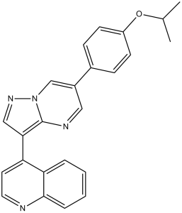
| Chemical Name | 4-(6-(4-isopropoxyphenyl)pyrazolo[1,5-a]pyrimidin-3-yl)quinoline | |
| Synonyms |
|
|
| HS Tariff Code | 2934.99.9001 | |
| Storage |
Powder-20°C 3 years 4°C 2 years In solvent -80°C 6 months -20°C 1 month |
|
| Shipping Condition | Room temperature (This product is stable at ambient temperature for a few days during ordinary shipping and time spent in Customs) |
Biological Activity
| Targets |
Bone morphogenetic protein (BMP) signaling cascade; ALK1 (IC50 = 27 nM); ALK2 (IC50 = 107.9 nM); ALK3 (IC50 < 5 nM); ALK6 (IC50 = 47.6 nM)[1] DMH1 is a selective inhibitor of bone morphogenetic protein (BMP) type I receptors ALK2 and ALK3 (ALK2 IC50 = 1.0 nM; ALK3 IC50 = 36 nM) [1] DMH1 shows weak or no inhibition of other ALK receptors (ALK1, ALK4-6: IC50 > 1 μM) and unrelated kinases (PKA, PKC: IC50 > 10 μM) [1] |
| ln Vitro |
The expression of OCT4, Nanog, and PAX6 proteins is regulated by DMH-1 (0.5 μM). In SM3 and CA6 cells, DMH-1 dramatically decreased the proportion of cells expressing the pluripotency marker proteins OCT4 and Nanog. On days 5 and 7, respectively, there was a substantial upregulation of PAX6 expression in CA6 and SM3 cells. DMH-1 controls the mRNA for the neural precursor marker and causes pluripotency. During the neural induction process of hiPSCs, PAX6 can independently control the expression of SOX1 by controlling the concentration of DMH-1 [2]. In HeLa cells, DMH-1 (5 μM and 10 μM) inhibits autophagy induced by CDDP and increases CDDP's capacity to reduce cell viability; in MCF-7 cells, it inhibits autophagy induced by Tamoxifen and increases Tamoxifen's capacity to reduce MCF-7 cell viability; in MCF-7 and HeLa cells, it inhibits autophagy induced by 5-FU, but it has no effect on the inhibitory effect of 5-FU on the viability of MCF-7 and HeLa cells. After 24 hours of treatment, DMH-1 increased the effect of CDDP on HeLa cells that causes apoptosis. HeLa and MCF-7 cell growth is inhibited by DMH-1 [3]. Smads 1, 5, and 9 had less canonical phosphorylation when exposed to DMH-1 (20 μM). In OVCAR8 cells, the combination of DMH-1 and cisplatin dramatically decreased Ki-67 positive staining. In OVCAR8 and NCI-RES cells, DMH-1 (20 μM) upregulates JAG1, decreases CYP1B1, and enhances HAPLN1 expression [4]. In human ovarian cancer cells (SKOV3, A2780), DMH1 (5 μM) inhibits cell proliferation by 65-70% after 72 hours (MTT assay). It induces G2/M cell cycle arrest (G2/M phase cells increased from 20% to 42% in SKOV3) and apoptosis (Annexin V-positive cells increased from 6% to 35% after 48 hours), and downregulates BMP target genes (ID1, ID3) by 60-65% at mRNA level [4] - In human non-small cell lung cancer (NSCLC) cells (A549, H1299), DMH1 (10 μM) reduces cell proliferation by 62-68% after 72 hours (CCK-8 assay) and inhibits cell migration (wound-healing assay: 65% reduction) and invasion (Transwell assay: 70% reduction) after 48 hours. It downregulates p-Smad1/5/8 (75% reduction) and pro-invasive gene MMP2 by 60% [5] - In human induced pluripotent stem cells (hiPSCs), DMH1 (2 μM) promotes neurogenesis during neural induction. It upregulates neural progenitor markers PAX6 (2.8-fold) and SOX1 (3.2-fold) at mRNA level after 7 days, with increased βIII-tubulin-positive neural cells (68% vs. 32% in control) [3] - In human cervical cancer HeLa cells treated with cisplatin (10 μM), DMH1 (5 μM) inhibits chemotherapeutic drug-induced autophagy. It reduces LC3-II/LC3-I ratio (60% reduction) and downregulates autophagy-related gene Beclin1 by 55% at protein level [2] - In normal human bronchial epithelial cells (HBECs) and foreskin fibroblasts, DMH1 shows low toxicity at concentrations up to 25 μM (cell viability > 85% vs. control) [4][5] |
| ln Vivo |
Treatment with DMH1 (5 mg/kg, ip) dramatically inhibited the growth of tumors in a human lung cancer xenograft model [5]. DMH1 attenuates xenograft lung tumor growth in mice[5] Researchers next examined the effect of DMH1 on lung tumor cell growth in vivo. The A549 cells were subcutaneously inoculated in the two sides of lower rear flanks of Severe combined immunodeficiency (SCID) mice. Intraperitoneal (i.p.) injections of vehicle (12.5% 2-hydroxypropyl-β-cyclodextrin, n = 5) or 5 mg/kg DMH1 (n = 5) were initiated on the same day of tumor cell implantation and were performed every other day for 4 weeks. Tumor volumes were measured regularly starting on the sixth day after implantation. The tumor growth was fit into an exponential growth curve ( Figure 4A ) (R2 = 0.87 and 0.84 for the DMH1 treated and control mice, respectively). The result indicated that the rate for doubling tumor size in DMH1-treated mice was about one day longer than the controls (5.6 versus 4.7 days in the DMH1 treated and control mice, respectively) ( Figure 4A ). As the initial tumor volumes were similar, no statistical differences between the two groups were observed until day 25. At the end of 4-week treatment, DMH1 treatment resulted in a statistically significant reduction in tumor volumes by about 50% compared to the vehicle control group (p-value <0.05) ( Figure 4B ). The mouse body weights were measured every other day throughout the experiment, and no notable weight changes were observed in both the control and DMH1 treated groups, suggesting an absence of DMH1 toxic effect at the administered dose (data not shown). To further examine the effect of DMH1 on tumor cell proliferation in vivo, tumor tissue samples from both the vehicle control and DMH1 treatment groups were subjected to Hematoxylin and eosin-stained (H&E) and human specific Ki67 staining. H&E sections were examined for regions that contained tumor and stromal cells, and the result indicated both the vehicle and DMH1 treated groups consisted of a morphologically similar differentiated adenocarcinoma (data not shown). However, immunohistochemical study showed a conspicuously significant decrease of human proliferation marker Ki67 in the DMH1 treated versus vehicle groups, suggesting that DMH1 treatment may attenuate human A549 cancer cell proliferation in vivo ( Figure 4C ). In nude mice bearing subcutaneous SKOV3 ovarian cancer xenografts, oral administration of DMH1 (50 mg/kg/day for 28 days) significantly inhibits tumor growth. Tumor volume was reduced by 63% compared to vehicle controls, and tumor weight decreased by 58%. Tumor tissues show downregulated p-Smad1/5/8 (70% reduction) and Ki-67 (55% reduction) [4] - In nude mice bearing subcutaneous A549 lung cancer xenografts, intraperitoneal injection of DMH1 (75 mg/kg/day for 21 days) inhibits tumor growth (volume reduced by 65%) and lung metastasis (nodule number reduced by 72% vs. vehicle). It suppresses BMP/Smad signaling in tumor tissues (ID1 mRNA downregulated by 60%) [5] |
| Enzyme Assay |
ALK2/ALK3 kinase activity assay: Purified recombinant human ALK2 or ALK3 was incubated with Smad1-derived substrate peptide and DMH1 (0.1 nM-100 nM) in assay buffer (50 mM Tris-HCl, pH 7.5, 10 mM MgCl₂, 1 mM DTT, 0.1 mM ATP) at 30°C for 60 minutes. Phosphorylated substrate was detected by radiolabeled ATP counting, and IC50 values were calculated from dose-response curves [1] - Kinase selectivity assay: DMH1 (10 μM) was screened against a panel of 40+ kinases (including ALK1, ALK4-6, PKA, PKC, ERK1/2) using respective substrate peptides and assay buffers. Kinase activity was quantified by colorimetric assay, with no significant off-target inhibition (>50% activity reduction) observed [1] |
| Cell Assay |
Cell scratch-wound Assay[4] A549 and H460 cells were seeded in 35 mm dishes to create a confluent monolayer. The dishes were allowed to incubate overnight in order to allow the cells to attach to the bottom of the dish. On the following day, wounds were created by a straight scratch from a pipette tip in the center of the culture. The cells were then treated with DMSO or DMH1 at 1 µM and 3 µM concentrations. Photographs were taken when wounds were created and after 24 hour's incubation using phase-contrast microscopy, and gap distances were quantitatively evaluated using software ImageJ (NIH). The gap distances after 24 h incubation were normalized with the gap distance at 0 hr as the migration rates. Cell Proliferation Assay[4] About 10,000 A549 cells per well were seeded in 96-well plates and incubated for overnight. The culture medium was then changed to fresh medium containing DMSO or DMH1 at various concentrations. The cells were then incubated for 48 hours and 96 hours before treatment termination by replacing the medium with 100 μL of 10% trichloroacetic acid in 1× PBS, followed by incubation at 4°C for at least 1 hour. Subsequently, the plates were washed with water and air dried. The plates were stained with 50 μL 0.4% sulphorhodamine assay in 1% acetic acid for 30 minutes at room temperature. Unbound dye was washed off with 1% acetic acid. After air drying and solubilization of the protein-bound dye in 10 mM Tris solution, absorbance was read in a microplate reader at 565 nm. In this study, researchers aimed to investigate the effects of DMH1 on chemotherapeutic drug-induced autophagy as well as the efficacy of chemotherapeutic drugs in different cancer cells. They found that DMH1 inhibited tamoxifen- and cispcis-diaminedichloroplatinum (II) (CDDP)-induced autophagy responses in MCF-7 and HeLa cells, and potentiated the anti-tumor activity of tamoxifen and CDDP for both cells. DMH1 inhibited 5-fluorouracil (5-FU)-induced autophagy responses in MCF-7 and HeLa cells, but did not affect the anti-tumor activity of 5-FU for these two cell lines. DMH1 itself did not induce cell death in MCF-7 and HeLa cells, but inhibited the proliferation of these cells. In conclusion, DMH1 inhibits chemotherapeutic drug-induced autophagy response and the enhancement of efficacy of chemotherapeutic drugs by DMH1 is dependent on the cell sensitivity to drugs.[2] In this study, researchers tested the efficacy of DMH1, a highly selective small molecule BMP-inhibitor for its potential to replace Noggin in the neuralization of hiPSCs. researchers compare Noggin and DMH1-induced neuralization of hiPSCs by measuring protein and mRNA levels of pluripotency and neural precursor markers over a period of seven days. The regulation of five of the six markers assessed was indistinguishable in the presence of concentrations of Noggin or DMH1 that have been shown to effectively inhibit BMP signaling in other systems. We observed that by varying the DMH1 or Noggin concentration, we could selectively modulate the number of SOX1 expressing cells, whereas PAX6, another neural precursor marker, remained the same. The level and timing of SOX1 expression have been shown to affect neural induction as well as neural lineage. Our observations, therefore, suggest that BMP-inhibitor concentrations need to be carefully monitored to ensure appropriate expression levels of all transcription factors necessary for the induction of a particular neuronal lineage. Researchers further demonstrate that DMH1-induced neural progenitors can be differentiated into β3-tubulin expressing neurons, a subset of which also express tyrosine hydroxylase. Thus, the combined use of DMH1, a highly specific BMP-pathway inhibitor, and SB431542, a TGF-β1-pathway specific inhibitor, provides us with the tools to independently regulate these two pathways through the exclusive use of small molecule inhibitors.[3] Ovarian cancer cell proliferation/apoptosis assay: SKOV3 and A2780 cells were seeded in 96-well plates (proliferation) or 6-well plates (apoptosis/cycle) at 3×10³ cells/well or 2×10⁵ cells/well respectively. Cells were treated with DMH1 (0.5-20 μM) for 48-72 hours. MTT assay measured proliferation; Annexin V-FITC/PI staining quantified apoptosis; flow cytometry (propidium iodide staining) analyzed cell cycle; qPCR detected ID1/ID3 mRNA levels [4] - NSCLC cell proliferation/migration assay: A549 and H1299 cells were seeded in 96-well plates (proliferation) or 6-well plates (migration/invasion) at 3×10³ cells/well or 2×10⁵ cells/well respectively. Cells were treated with DMH1 (1-15 μM) for 48-72 hours. CCK-8 assay assessed proliferation; wound-healing and Transwell assays evaluated migration/invasion; Western blot detected p-Smad1/5/8 and MMP2 [5] - hiPSCs neurogenesis assay: hiPSCs were seeded in Matrigel-coated 6-well plates at 1×10⁵ cells/well and cultured in neural induction medium containing DMH1 (0.5-5 μM). After 7-14 days, qPCR analyzed PAX6/SOX1 mRNA levels; immunocytochemistry detected βIII-tubulin-positive cells [3] - Autophagy inhibition assay: HeLa cells were seeded in 6-well plates at 2×10⁵ cells/well and pretreated with DMH1 (1-10 μM) for 1 hour, then treated with cisplatin (10 μM) for 24 hours. Western blot detected LC3-II/LC3-I ratio and Beclin1; immunofluorescence visualized autophagosomes [2] |
| Animal Protocol |
Dissolved in 12.5% 2-hydroxypropyl-β-cyclodextrin; 5 mg/kg; i.p. injection Mice bearing A549 xenograft. Xenograft lung tumor growth[5] Sub-confluent A549 cells were trypsinized and then suspended in serum free RPMI 1640 medium. The cell suspension (1×10⁶ cells in 100 µl medium for each injection) was injected subcutaneously into both the right and left flanks of eight-week old NOD SCID mice (n = 5 for each group). Mice were given Intraperitoneal (i.p.) injection of the vehicle (12.5% 2-hydroxypropyl-β-cyclodextrin) or 5 mg/kg DMH1 every other day. The tumor sizes were measured with a vernier caliper from the sixth day to the fourth week after tumor implantation. The tumor volume (V) was calculated according to the formulation: Volume = (width)∧2× length/2. The tumor tissues were dissected at the end of study, and were sectioned and stained with H & E, and for immunohistochemical analysis. Nude mouse ovarian cancer xenograft model: 6-8 weeks old nude mice were subcutaneously inoculated with SKOV3 cells (5×10⁶ cells/mouse). When tumors reached ~100 mm³, mice were randomly divided into vehicle and DMH1 groups. The drug was suspended in 0.5% carboxymethylcellulose sodium and administered orally at 50 mg/kg/day for 28 days. Vehicle group received carboxymethylcellulose sodium. Tumor volume was measured every 3 days; tumors were excised for Western blot (p-Smad1/5/8) and Ki-67 immunostaining [4] - Nude mouse lung cancer xenograft model: 6-8 weeks old nude mice were subcutaneously inoculated with A549 cells (5×10⁶ cells/mouse). When tumors reached ~100 mm³, mice were randomly divided into vehicle and DMH1 groups. DMH1 was dissolved in saline and administered intraperitoneally at 75 mg/kg/day for 21 days. Vehicle group received saline. Tumor volume was measured every 3 days; lungs were collected to count metastatic nodules; qPCR analyzed ID1 mRNA in tumor tissues [5] |
| Toxicity/Toxicokinetics |
In vitro, DMH1 shows low toxicity to normal human cells (HBECs IC50 > 25 μM; foreskin fibroblasts IC50 > 30 μM) [4][5] - In in vivo studies, oral or intraperitoneal administration of DMH1 at tested doses (50-75 mg/kg/day) causes no significant body weight loss (<5% vs. baseline) or overt lethality in nude mice [4][5] - No significant changes in liver function (ALT, AST) or renal function (creatinine, BUN) were observed in DMH1-treated mice compared to vehicle controls [4][5] |
| References |
[1]. Synthesis and structure-activity relationships of a novel and selective bone morphogenetic protein receptor (BMP) inhibitor derived from the pyrazolo[1.5-a]pyrimidine scaffold of dorsomorphin: the discovery of mL347 as an ALK2 versus ALK3 selective mLPCN probe. Bioorg Med Chem Lett. 2013 Jun 1;23(11):3248-52. [2]. DMH1 (4-[6-(4-isopropoxyphenyl)pyrazolo[1,5-a]pyrimidin-3-yl]quinoline) inhibits chemotherapeutic drug-induced autophagy. Acta Pharm Sin B. 2015 Jul;5(4):330-6. [3]. DMH1, a highly selective small molecule BMP inhibitor promotes neurogenesis of hiPSCs: comparison of PAX6 and SOX1 expression during neural induction. ACS Chem Neurosci. 2012 Jun 20;3(6):482-91. [4]. Small molecule inhibitor of the bone morphogenetic protein pathway DMH1 reduces ovarian cancer cell growth. Cancer Lett. 2015 Nov 1;368(1):79-87. [5]. DMH1, a small molecule inhibitor of BMP type i receptors, suppresses growth and invasion of lung cancer. PLoS One. 2014 Mar 6;9(6):e90748. |
| Additional Infomation |
DMH1 is a pyrazolopyrimidine that is pyrazolo[1,5-a]pyrimidine bearing quinolin-4-yl and 4-isopropyloxyphenyl substituents at positions 3 and 6 respectively. It has a role as a protein kinase inhibitor, a bone morphogenetic protein receptor antagonist and an antineoplastic agent. It is a member of quinolines, a pyrazolopyrimidine and an aromatic ether. A structure-activity relationship of the 3- and 6-positions of the pyrazolo[1,5-a]pyrimidine scaffold of the known BMP inhibitors dorsomorphin, 1, LDN-193189, 2, and DMH1, 3, led to the identification of a potent and selective compound for ALK2 versus ALK3. The potency contributions of several 3-position substituents were evaluated with subtle structural changes leading to significant changes in potency. From these studies, a novel 5-quinoline molecule was identified and designated an MLPCN probe molecule, ML347, which shows >300-fold selectivity for ALK2 and presents the community with a selective molecular probe for further biological evaluation. [1] Our previous work found that DMH1 (4-[6-(4-isopropoxyphenyl)pyrazolo [1,5-a]pyrimidin-3-yl]quinoline) was a novel autophagy inhibitor. Here, we aimed to investigate the effects of DMH1 on chemotherapeutic drug-induced autophagy as well as the efficacy of chemotherapeutic drugs in different cancer cells. We found that DMH1 inhibited tamoxifen- and cispcis-diaminedichloroplatinum (II) (CDDP)-induced autophagy responses in MCF-7 and HeLa cells, and potentiated the anti-tumor activity of tamoxifen and CDDP for both cells. DMH1 inhibited 5-fluorouracil (5-FU)-induced autophagy responses in MCF-7 and HeLa cells, but did not affect the anti-tumor activity of 5-FU for these two cell lines. DMH1 itself did not induce cell death in MCF-7 and HeLa cells, but inhibited the proliferation of these cells. In conclusion, DMH1 inhibits chemotherapeutic drug-induced autophagy response and the enhancement of efficacy of chemotherapeutic drugs by DMH1 is dependent on the cell sensitivity to drugs. [2] Recent successes in deriving human-induced pluripotent stem cells (hiPSCs) allow for the possibility of studying human neurons derived from patients with neurological diseases. Concomitant inhibition of the BMP and TGF-β1 branches of the TGF-β signaling pathways by the endogenous antagonist, Noggin, and the small molecule SB431542, respectively, induces efficient neuralization of hiPSCs, a method known as dual-SMAD inhibition. The use of small molecule inhibitors instead of their endogenous counterparts has several advantages including lower cost, consistent activity, and the maintenance of xeno-free culture conditions. We tested the efficacy of DMH1, a highly selective small molecule BMP-inhibitor for its potential to replace Noggin in the neuralization of hiPSCs. We compare Noggin and DMH1-induced neuralization of hiPSCs by measuring protein and mRNA levels of pluripotency and neural precursor markers over a period of seven days. The regulation of five of the six markers assessed was indistinguishable in the presence of concentrations of Noggin or DMH1 that have been shown to effectively inhibit BMP signaling in other systems. We observed that by varying the DMH1 or Noggin concentration, we could selectively modulate the number of SOX1 expressing cells, whereas PAX6, another neural precursor marker, remained the same. The level and timing of SOX1 expression have been shown to affect neural induction as well as neural lineage. Our observations, therefore, suggest that BMP-inhibitor concentrations need to be carefully monitored to ensure appropriate expression levels of all transcription factors necessary for the induction of a particular neuronal lineage. We further demonstrate that DMH1-induced neural progenitors can be differentiated into β3-tubulin expressing neurons, a subset of which also express tyrosine hydroxylase. Thus, the combined use of DMH1, a highly specific BMP-pathway inhibitor, and SB431542, a TGF-β1-pathway specific inhibitor, provides us with the tools to independently regulate these two pathways through the exclusive use of small molecule inhibitors. [3] The bone morphogenetic protein (BMP) pathway belonging to the Transforming Growth Factor beta (TGFβ) family of secreted cytokines/growth factors is an important regulator of cancer. BMP ligands have been shown to play both tumor suppressive and promoting roles in human cancers. We have found that BMP ligands are amplified in human ovarian cancers and that BMP receptor expression correlates with poor progression-free-survival (PFS). Furthermore, active BMP signaling has been observed in human ovarian cancer tissue. We also determined that ovarian cancer cell lines have active BMP signaling in a cell autonomous fashion. Inhibition of BMP signaling with a small molecule receptor kinase antagonist is effective at reducing ovarian tumor sphere growth. Furthermore, BMP inhibition can enhance sensitivity to Cisplatin treatment and regulates gene expression involved in platinum resistance in ovarian cancer. Overall, these studies suggest targeting the BMP pathway as a novel source to enhance chemo-sensitivity in ovarian cancer. [4] The bone morphogenetic protein (BMP) signaling cascade is aberrantly activated in human non-small cell lung cancer (NSCLC) but not in normal lung epithelial cells, suggesting that blocking BMP signaling may be an effective therapeutic approach for lung cancer. Previous studies demonstrated that some BMP antagonists, which bind to extracellular BMP ligands and prevent their association with BMP receptors, dramatically reduced lung tumor growth. However, clinical application of protein-based BMP antagonists is limited by short half-lives, poor intra-tumor delivery as well as resistance caused by potential gain-of-function mutations in the downstream of the BMP pathway. Small molecule BMP inhibitors which target the intracellular BMP cascades would be ideal for anticancer drug development. In a zebrafish embryo-based structure and activity study, we previously identified a group of highly selective small molecule inhibitors specifically antagonizing the intracellular kinase domain of BMP type I receptors. In the present study, we demonstrated that DMH1, one of such inhibitors, potently reduced lung cell proliferation, promoted cell death, and decreased cell migration and invasion in NSCLC cells by blocking BMP signaling, as indicated by suppression of Smad 1/5/8 phosphorylation and gene expression of Id1, Id2 and Id3. Additionally, DMH1 treatment significantly reduced the tumor growth in human lung cancer xenograft model. In conclusion, our study indicates that small molecule inhibitors of BMP type I receptors may offer a promising novel strategy for lung cancer treatment.[5] DMH1 is a potent, selective small-molecule inhibitor of BMP type I receptors ALK2 and ALK3, derived from the pyrazolo[1.5-a]pyrimidine scaffold [1] - Its mechanism of action involves competitive binding to the ATP-binding pockets of ALK2 and ALK3, inhibiting their kinase activity and blocking downstream BMP/Smad1/5/8 signaling pathway activation [1][4][5] - DMH1 exhibits in vitro anti-tumor (ovarian cancer, lung cancer) activity, neurogenesis-promoting activity in hiPSCs, and inhibitory activity against chemotherapeutic drug-induced autophagy [2][3][4][5] - In vivo, it inhibits ovarian cancer and lung cancer growth and metastasis, supporting its potential for BMP-driven tumor therapy [4][5] - It is widely used as a tool compound to study BMP signaling in cancer, neurogenesis, and autophagy, and serves as a probe for ALK2/ALK3-related biological research [1][2][3][4][5] |
Solubility Data
| Solubility (In Vitro) |
|
|||
| Solubility (In Vivo) |
Solubility in Formulation 1: ≥ 1 mg/mL (2.63 mM) (saturation unknown) in 10% DMSO + 40% PEG300 +5% Tween-80 + 45% Saline (add these co-solvents sequentially from left to right, and one by one), clear solution. For example, if 1 mL of working solution is to be prepared, you can add 100 μL of 10.0 mg/mL clear DMSO stock solution to 400 μL of PEG300 and mix evenly; then add 50 μL of Tween-80 + to the above solution and mix evenly; then add 450 μL of normal saline to adjust the volume to 1 mL. Preparation of saline: Dissolve 0.9 g of sodium chloride in 100 mL ddH₂ O to obtain a clear solution. (Please use freshly prepared in vivo formulations for optimal results.) |
| Preparing Stock Solutions | 1 mg | 5 mg | 10 mg | |
| 1 mM | 2.6285 mL | 13.1427 mL | 26.2854 mL | |
| 5 mM | 0.5257 mL | 2.6285 mL | 5.2571 mL | |
| 10 mM | 0.2629 mL | 1.3143 mL | 2.6285 mL |