Physicochemical Properties
| Molecular Formula | C28H42O7 |
| Molecular Weight | 490.6289 |
| Exact Mass | 490.293 |
| CAS # | 38395-02-7 |
| PubChem CID | 21633059 |
| Appearance | White to off-white solid powder |
| Density | 1.3±0.1 g/cm3 |
| Boiling Point | 617.9±55.0 °C at 760 mmHg |
| Flash Point | 197.5±25.0 °C |
| Vapour Pressure | 0.0±4.1 mmHg at 25°C |
| Index of Refraction | 1.584 |
| LogP | 4.37 |
| Hydrogen Bond Donor Count | 4 |
| Hydrogen Bond Acceptor Count | 7 |
| Rotatable Bond Count | 5 |
| Heavy Atom Count | 35 |
| Complexity | 984 |
| Defined Atom Stereocenter Count | 8 |
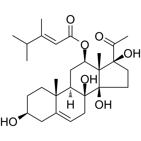
| SMILES | CC(C)/C(=C/C(=O)O[C@@H]1C[C@@H]2[C@]3(CC[C@@H](CC3=CC[C@]2([C@@]4([C@]1([C@@](CC4)(C(=O)C)O)C)O)O)O)C)/C |
| InChi Key | VWLXIXALPNYWFH-UXGQNDOZSA-N |
| InChi Code | InChI=1S/C28H42O7/c1-16(2)17(3)13-23(31)35-22-15-21-24(5)9-8-20(30)14-19(24)7-10-27(21,33)28(34)12-11-26(32,18(4)29)25(22,28)6/h7,13,16,20-22,30,32-34H,8-12,14-15H2,1-6H3/b17-13+/t20-,21+,22+,24-,25+,26+,27-,28+/m0/s1 |
| Chemical Name | [(3S,8S,9R,10R,12R,13S,14R,17S)-17-acetyl-3,8,14,17-tetrahydroxy-10,13-dimethyl-1,2,3,4,7,9,11,12,15,16-decahydrocyclopenta[a]phenanthren-12-yl] (E)-3,4-dimethylpent-2-enoate |
| HS Tariff Code | 2934.99.9001 |
| Storage |
Powder-20°C 3 years 4°C 2 years In solvent -80°C 6 months -20°C 1 month Note: This product requires protection from light (avoid light exposure) during transportation and storage. |
| Shipping Condition | Room temperature (This product is stable at ambient temperature for a few days during ordinary shipping and time spent in Customs) |
Biological Activity
| Targets |
The action targets of Caudatin are the VEGF-VEGFR2-AKT/FAK signaling axis; IC50 for inhibiting HUVEC proliferation = 23.6 ± 2.1 μM[1] |
| ln Vitro |
1. HUVEC proliferation inhibition: Caudatin (10/20/40 μM) inhibited proliferation in concentration-time-dependent manner. At 72 h, inhibition rates were 28.3±3.1% (10 μM), 45.6±3.8% (20 μM), 68.2±4.5% (40 μM); IC50 = 23.6±2.1 μM (MTT) [1] 2. HUVEC migration suppression: 40 μM Caudatin reduced VEGF-induced migration by 72.4±5.8% (Transwell, crystal violet staining) [1] 3. HUVEC tube formation inhibition: 40 μM Caudatin decreased tube length by 65.7±4.9% and branch number by 68.3±5.2% (Matrigel assay) [1] 4. Signaling downregulation: 40 μM Caudatin reduced VEGF-induced p-VEGFR2 (78.5±6.1%), p-AKT (71.2±5.4%), p-FAK (69.3±5.7%); total proteins unchanged (Western blot) [1] |
| ln Vivo |
Nude mouse HCT116 xenograft model (groups: control, Caudatin 20/40 mg/kg, ip, q2d×21d): 1. Tumor growth inhibition: 40 mg/kg reduced volume by 56.7±4.5% and weight by 58.3±4.2% [1] 2. MVD reduction: 40 mg/kg decreased CD31+ MVD from 48.1±3.5 to 18.2±2.3 vessels/field (62.1±5.3% reduction) [1] 3. Signaling inhibition: 40 mg/kg reduced tumor p-VEGFR2 (68.5±5.8%), p-AKT (63.2±4.9%), p-FAK (61.7±5.1%) (Western blot) [1] |
| Cell Assay |
1. HUVEC culture: EGM-2 (10% FBS, growth factors), 37°C/5% CO₂, passages 3–8 [1] 2. MTT assay: 5×10³ cells/well (96-well), Caudatin + VEGF, 24/48/72 h; MTT (5 mg/mL) → DMSO, read 570 nm [1] 3. Transwell assay: 1×10⁵ cells/upper chamber (8 μm), VEGF + Caudatin; 24 h, crystal violet staining, count migrated cells [1] 4. Tube formation: Matrigel-coated 96-well, 2×10⁴ cells/well + Caudatin + VEGF; 6 h, quantify tube length/branches [1] 5. Western blot: Caudatin + VEGF for 1 h; RIPA lysis, BCA quantification, detect p-VEGFR2/VEGFR2/p-AKT/AKT/p-FAK/FAK/β-actin [1] |
| Animal Protocol |
1. Animals: BALB/c nude mice (4–6 weeks, 18–22 g), SPF environment (22±2°C, 12 h light/dark) [1] 2. Xenograft: 1×10⁷ HCT116 cells/mouse (sc, right flank); tumor volume = (length×width²)/2 [1] 3. Drug admin: Caudatin dissolved in DMSO + saline (DMSO <1%, 2/4 mg/mL); 20/40 mg/kg ip, q2d×21d; control = saline-DMSO [1] 4. Sample collection: Sacrifice, excise tumor (weigh/photograph); fix part in 4% paraformaldehyde (CD31 IHC), store rest at -80°C (Western blot) [1] |
| Toxicity/Toxicokinetics |
1. In vitro: Caudatin (up to 40 μM) kept NHFF viability >85% (MTT) [1] 2. In vivo: No body weight loss; normal serum ALT/AST/BUN/Cr; no liver/kidney/heart/lung/spleen damage (histopathology) [1] |
| References |
[1]. Antiangiogenic properties of caudatin in vitro and in vivo by suppression of VEGF‑VEGFR2‑AKT/FAK signal axis. Mol Med Rep. 2017 Dec;16(6):8937-8943. |
| Additional Infomation |
Caudatin has been reported in Araujia sericifera with data available. 1. Caudatin is a C21 steroidal glycoside from Cynanchum species (traditional use: anti-inflammation/antitumor) [1] 2. Mechanism: Suppresses VEGF-VEGFR2-AKT/FAK axis to inhibit endothelial cell functions (angiogenesis) [1] 3. Significance: Potential antiangiogenic/antitumor lead compound with good safety [1] |
Solubility Data
| Solubility (In Vitro) | DMSO : ~50 mg/mL (~101.91 mM) |
| Solubility (In Vivo) |
Solubility in Formulation 1: ≥ 5 mg/mL (10.19 mM) (saturation unknown) in 10% DMSO + 40% PEG300 + 5% Tween80 + 45% Saline (add these co-solvents sequentially from left to right, and one by one), clear solution. For example, if 1 mL of working solution is to be prepared, you can add 100 μL of 50.0 mg/mL clear DMSO stock solution to 400 μL PEG300 and mix evenly; then add 50 μL Tween-80 to the above solution and mix evenly; then add 450 μL normal saline to adjust the volume to 1 mL. Preparation of saline: Dissolve 0.9 g of sodium chloride in 100 mL ddH₂ O to obtain a clear solution. Solubility in Formulation 2: ≥ 5 mg/mL (10.19 mM) (saturation unknown) in 10% DMSO + 90% (20% SBE-β-CD in Saline) (add these co-solvents sequentially from left to right, and one by one), clear solution. For example, if 1 mL of working solution is to be prepared, you can add 100 μL of 50.0 mg/mL clear DMSO stock solution to 900 μL of 20% SBE-β-CD physiological saline solution and mix evenly. Preparation of 20% SBE-β-CD in Saline (4°C,1 week): Dissolve 2 g SBE-β-CD in 10 mL saline to obtain a clear solution. Solubility in Formulation 3: ≥ 5 mg/mL (10.19 mM) (saturation unknown) in 10% DMSO + 90% Corn Oil (add these co-solvents sequentially from left to right, and one by one), clear solution. For example, if 1 mL of working solution is to be prepared, you can add 100 μL of 50.0 mg/mL clear DMSO stock solution to 900 μL of corn oil and mix evenly. (Please use freshly prepared in vivo formulations for optimal results.) |
| Preparing Stock Solutions | 1 mg | 5 mg | 10 mg | |
| 1 mM | 2.0382 mL | 10.1910 mL | 20.3820 mL | |
| 5 mM | 0.4076 mL | 2.0382 mL | 4.0764 mL | |
| 10 mM | 0.2038 mL | 1.0191 mL | 2.0382 mL |