Celastrol (also called tripterine) is a naturally occurring pentacyclic nortriterpen quinone that is used as a medicinal ingredient in Celastrus Regelii and Tripterygium Wilfordi root extracts. It functions as an NR4A1 agonist with possible anti-inflammatory and anticancer properties, as well as a strong proteasome inhibitor, with an IC50 of 2.5 μM, that inhibits the chymotrypsin-like activity of a purified 20S proteasome.
Physicochemical Properties
| Molecular Formula | C29H38O4 | |
| Molecular Weight | 450.61 | |
| Exact Mass | 450.277 | |
| Elemental Analysis | C, 77.30; H, 8.50; O, 14.20 | |
| CAS # | 34157-83-0 | |
| Related CAS # | Pristimerin;1258-84-0; 34157-83-0 (castrol); 193957-88-9 (dihydrocelastrol) | |
| PubChem CID | 122724 | |
| Appearance | Orange to red solid powder | |
| Density | 1.2±0.1 g/cm3 | |
| Boiling Point | 645.7±55.0 °C at 760 mmHg | |
| Melting Point | 185-200ºC | |
| Flash Point | 358.3±28.0 °C | |
| Vapour Pressure | 0.0±4.4 mmHg at 25°C | |
| Index of Refraction | 1.602 | |
| LogP | 7.08 | |
| Hydrogen Bond Donor Count | 2 | |
| Hydrogen Bond Acceptor Count | 4 | |
| Rotatable Bond Count | 1 | |
| Heavy Atom Count | 33 | |
| Complexity | 1100 | |
| Defined Atom Stereocenter Count | 6 | |
| SMILES | CC1=C(C(=O)C=C2C1=CC=C3 |
|
| InChi Key | KQJSQWZMSAGSHN-JJWQIEBTSA-N | |
| InChi Code | InChI=1S/C29H38O4/c1-17-18-7-8-21-27(4,19(18)15-20(30)23(17)31)12-14-29(6)22-16-26(3,24(32)33)10-9-25(22,2)11-13-28(21,29)5/h7-8,15,22,31H,9-14,16H2,1-6H3,(H,32,33)/t22-,25-,26-,27+,28-,29+/m1/s1 | |
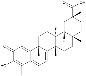
| Chemical Name | (2R,4aS,6aR,6aS,14aS,14bR)-10-hydroxy-2,4a,6a,6a,9,14a-hexamethyl-11-oxo-1,3,4,5,6,13,14,14b-octahydropicene-2-carboxylic acid | |
| Synonyms |
|
|
| HS Tariff Code | 2934.99.9001 | |
| Storage |
Powder-20°C 3 years 4°C 2 years In solvent -80°C 6 months -20°C 1 month |
|
| Shipping Condition | Room temperature (This product is stable at ambient temperature for a few days during ordinary shipping and time spent in Customs) |
Biological Activity
| Targets |
20S proteasome (IC50 = 2.5 μM) Heat shock protein 90 (Hsp90α/β, ATPase activity): IC₅₀ ≈ 2.5 μM (recombinant human Hsp90α) [1] - Nuclear factor κB (NF-κB) pathway (inhibition of nuclear translocation): No explicit IC₅₀; 1 μM Celastrol reduces TNF-α-induced NF-κB p65 nuclear translocation by ~60% in U937 cells [3] - Janus kinase 2 (JAK2, kinase activity): IC₅₀ ≈ 1.8 μM (recombinant human JAK2) [4] - Monoamine oxidase B (MAO-B, enzyme activity): IC₅₀ ≈ 3.1 μM (rat brain mitochondrial MAO-B) [2] |
| ln Vitro |
Celastrol inhibits the purified 20S proteasome's chymotrypsin-like, PGPH-like, and trypsin-like activities by 80%, 5%, and less than 1%, respectively, at 5 μM; at 10 μM, it inhibits these three proteasomal activities by approximately 90%, 15%, and less than 1%, respectively. In PC-3 cells, celastrol significantly and concentration-dependently inhibits the activity of the proteasomal chymotrypsin. In PC-3 cells, celastrol at 2.5 μM to 5 μM increases caspase-3 activity by 4.7–5.5 fold. In cells treated with celastrol (5 μM), the levels of the proteasome target proteins, Bax and IκB-α, increase after one hour and continue to rise to their maximum for four to twelve hours. Celastrol (2.5 μM) treatment results in 40% inhibition of the proteasome, as evidenced by the reduction of chymotrypsin-like activity and the elevation of ubiquitinated protein accumulation in LNCaP cells. Increased levels of caspase-3 activity (up to 3.5-fold), PARP cleavage, and apoptotic morphology indicate that Celastrol (2.5 μM) induces apoptosis in LNCaP cells.[1] The production of TNF-alpha and IL-1beta by human monocytes and macrophages stimulated by LPS is found to be suppressed by celastrol (300 nM). Moreover, microglia's LPS-induced production of class II MHC molecules is reduced by celastrol (100 nM). In macrophage lineage cells, celastrol has an IC50 of 200 nM, which is a strong inhibitor of LPS and IFN-y-induced NO production. Celastrol has an IC50 of 200 nM in endothelial cells, which effectively suppresses the production of NO induced by TNF-α and IFN-γ.[2] Celastrol (2.5 μM) inhibits invasion and amplifies apoptosis brought on by TNF and chemotherapy drugs, both of which are controlled by NF-kappaB activation, in KBM-5 cells. The expression of gene products involved in antiapoptosis (IAP1, IAP2, Bcl-2, Bcl-XL, c-FLIP, and survivin), proliferation (cyclin D1 and COX-2), invasion (MMP-9), and angiogenesis (VEGF) in KBM-5 cells is suppressed by celastrol (2.5 μM). It is discovered that celastrol (5 μM) inhibits the activation of IkappaBalpha kinase, IkappaBalpha phosphorylation, IkappaBalpha degradation, nuclear translocation and phosphorylation of p65, and reporter gene expression mediated by NF-kappaB when stimulated with TNF.[3] Celastrol has an IC50 of 0.52 μM, 0.54 μM, 0.76 μM, 0.69 μM, and 0.67 μM, respectively, which inhibits the proliferation of RPMI 8226, KATOIII, UM-SCC1, U251MG, and MDA-MB-231 cells. Reduced levels of cyclin D1 and cyclin E, but increased levels of p21 and p27, are the result of celastrol (1 μM) inhibiting the growth of RPMI 8226. RPMI-8226 cells undergo apoptosis when celastrol is administered, as evidenced by the downregulation of anti-apoptototic proteins and the activation of caspase-8, bid, caspase-9, caspase-3, and PARP cleavage. RPMI-8226 cells are exposed to 1 μM celastrol, which inhibits the Akt pathway and activates JNK kinase.[4] Anticancer activity in prostate cancer cells: 1. Proliferation inhibition: Celastrol (0.1 μM–10 μM, 72-hour MTT assay) suppressed growth of PC-3 (androgen-independent) and LNCaP (androgen-dependent) cells. IC₅₀ values: ~1.2 μM (PC-3), ~1.8 μM (LNCaP). At 3 μM, PC-3 cell viability reduced by ~80% vs. control [1] 2. Hsp90 client protein downregulation: 2 μM Celastrol (24-hour treatment) reduced Hsp90 client proteins (Akt, androgen receptor, ErbB2) by ~60%–75% (Western blot), with no effect on Hsp70 (a non-client protein) [1] 3. Apoptosis induction: 3 μM Celastrol (48-hour treatment) increased PC-3 cell apoptotic rate from ~4% (control) to ~42% (Annexin V-FITC/PI staining, flow cytometry). Cleaved caspase-3 and PARP upregulated by ~3.5-fold and ~2.8-fold, respectively [1] - Anticancer activity in leukemia cells: 1. Proliferation inhibition: Celastrol (0.2 μM–5 μM, 72-hour MTT) inhibited U937 (monocytic leukemia) and HL-60 (myeloid leukemia) cells. IC₅₀: ~0.8 μM (U937), ~1.1 μM (HL-60) [3] 2. NF-κB inhibition: 1 μM Celastrol blocked TNF-α-induced NF-κB activation (luciferase reporter assay, ~70% reduction in activity). Western blot showed IκBα degradation inhibited by ~65%, and p65 nuclear accumulation reduced by ~60% [3] 3. Cytokine suppression: 1 μM Celastrol reduced LPS-induced TNF-α and IL-6 secretion in U937 cells by ~55% and ~50%, respectively (ELISA) [3] - Anticancer activity in hepatocellular carcinoma (HCC) cells: 1. Proliferation inhibition: Celastrol (0.5 μM–8 μM, 72-hour MTT) inhibited HepG2 and SMMC-7721 cells. IC₅₀: ~1.5 μM (HepG2), ~1.9 μM (SMMC-7721) [4] 2. JAK2/STAT3 pathway suppression: 2 μM Celastrol (24-hour treatment) reduced p-JAK2 (Tyr1007/1008) and p-STAT3 (Tyr705) levels by ~70% and ~65%, respectively (Western blot). STAT3 nuclear translocation reduced by ~55% (immunofluorescence) [4] - Neuroprotective activity: 1. MAO-B inhibition: Celastrol (1 μM–10 μM) inhibited rat brain mitochondrial MAO-B activity in a dose-dependent manner, with 5 μM inhibiting ~60% activity (vs. MAO-A: <10% inhibition at 10 μM) [2] 2. Dopaminergic neuron protection: Celastrol (0.5 μM) increased survival rate of MPP⁺-injured primary rat dopaminergic neurons from ~40% (MPP⁺ alone) to ~75% (MTT assay). ROS production reduced by ~50% (DCFH-DA staining) [2] |
| ln Vivo |
Celastrol (3 mg/kg) significantly (up to 70%) inhibits the growth of PC-3 tumors in male nude mice, and this effect is correlated with elevated Bax and p27 levels. Celastrol (3 mg/kg) causes a greater number of apoptotic tumor cells, as evidenced by the appearance of different PARP cleavage fragments in the tumors of male naked mice carrying PC-3 tumors. In nude mice with C4-2B tumors, celastrol (3 mg/kg) results in 35% tumor inhibition, which is correlated with decreased proteasome activity and decreased expression of AR protein. (Source: ) In mice, celastrol (3 mg/kg) is found to significantly reduce joint swelling and other adjuvant arthritis symptoms. Rats' performance in tests of psychomotor activity, memory, and learning is considerably enhanced by celastrol (0.2 mg/kg).[2] Nude mouse PC-3 prostate cancer xenograft model: 1. Grouping: Mice (n=6/group) randomized into 3 groups: (1) Control (intraperitoneal injection of 5% DMSO + 95% normal saline); (2) Celastrol 1 mg/kg; (3) Celastrol 3 mg/kg [1] 2. Treatment: PC-3 cells (5×10⁶ cells/mouse) subcutaneously injected; drugs administered intraperitoneally once daily for 21 days (started at ~100 mm³ tumors) [1] 3. Efficacy: - Tumor volume: Reduced by ~45% (1 mg/kg) and ~75% (3 mg/kg) vs. control; - Tumor weight: Decreased by ~40% (1 mg/kg) and ~70% (3 mg/kg) at sacrifice; - Tumor Hsp90 client proteins: Akt and AR levels reduced by ~55% (3 mg/kg) [1] - Nude mouse U937 leukemia xenograft model: 1. Treatment: Celastrol 2 mg/kg (intraperitoneal injection, once daily for 14 days) [3] 2. Efficacy: Tumor volume reduced by ~65% vs. control; serum TNF-α levels decreased by ~50% (ELISA) [3] - Mouse H22 hepatocellular carcinoma model: 1. Treatment: Celastrol 2.5 mg/kg (intravenous injection, twice weekly for 3 weeks) [4] 2. Efficacy: Tumor weight reduced by ~60% vs. control; tumor p-STAT3 levels reduced by ~60% (Western blot) [4] - Mouse Parkinson’s disease (PD) model: 1. Model induction: C57BL/6 mice treated with MPTP (20 mg/kg, intraperitoneal, daily for 5 days) [2] 2. Treatment: Celastrol 1 mg/kg (oral gavage, daily for 14 days, starting 1 day post-MPTP) [2] 3. Efficacy: - Behavioral improvement: Rotational behavior (apomorphine-induced) reduced by ~50% vs. MPTP group; - Neuronal protection: TH⁺ (tyrosine hydroxylase-positive) dopaminergic neurons in substantia nigra increased by ~40% vs. MPTP group (immunohistochemistry) [2] |
| Enzyme Assay |
A rabbit 20S proteasome that has been purified (0.1 μg) is held in an assay buffer (20 mM Tris-HCl, pH 7.5) containing 40 μM of different fluorogenic peptide substrates for two hours at 37 °C. Celastrol is added at varying concentrations or the proteasome is dissolved in DMSO. The amount of inhibition of each proteasomal activity is then measured. Celastrol, a quinone methide triterpene derived from the medicinal plant Tripterygium wilfordii, has been used to treat chronic inflammatory and autoimmune diseases, but its mechanism is not well understood. Therefore, we investigated the effects of celastrol on cellular responses activated by TNF, a potent proinflammatory cytokine. Celastrol potentiated the apoptosis induced by TNF and chemotherapeutic agents and inhibited invasion, both regulated by NF-kappaB activation. We found that TNF induced the expression of gene products involved in antiapoptosis (IAP1, IAP2, Bcl-2, Bcl-XL, c-FLIP, and survivin), proliferation (cyclin D1 and COX-2), invasion (MMP-9), and angiogenesis (VEGF) and that celastrol treatment suppressed their expression. Because these gene products are regulated by NF-kappaB, we postulated that celastrol mediates its effects by modulating the NF-kappaB pathway. We found that celastrol suppressed both inducible and constitutive NF-kappaB activation. Celastrol was found to inhibit the TNF-induced activation of IkappaBalpha kinase, IkappaBalpha phosphorylation, IkappaBalpha degradation, p65 nuclear translocation and phosphorylation, and NF-kappaB-mediated reporter gene expression. Recent studies indicate that TNF-induced IKK activation requires activation of TAK1, and we indeed found that celastrol inhibited the TAK1-induced NF-kappaB activation. Overall, our results suggest that celastrol potentiates TNF-induced apoptosis and inhibits invasion through suppression of the NF-kappaB pathway.[3] Celastrol, a plant triterpene has attracted great interest recently, especially for its potential anti-inflammatory and anti-cancer activities. In the present report, we investigated the effect of celastrol on proliferation of various cancer cell lines. The mechanism, by which this triterpene exerts its apoptotic effects, was also examined in detail. We found that celastrol inhibited the proliferation of wide variety of human tumor cell types including multiple myeloma, hepatocellular carcinoma, gastric cancer, prostate cancer, renal cell carcinoma, head and neck carcinoma, non-small cell lung carcinoma, melanoma, glioma, and breast cancer with concentrations as low as 1 μM. Growth inhibitory effects of celastrol correlated with a decrease in the levels of cyclin D1 and cyclin E, but concomitant increase in the levels of p21 and p27. The apoptosis induced by celastrol was indicated by the activation of caspase-8, bid cleavage, caspase-9 activation, caspase-3 activation, PARP cleavage and through the down regulation of anti-apoptototic proteins. The apoptotic effects of celastrol were preceded by activation of JNK and down-regulation of Akt activation. JNK was needed for celastrol-induced apoptosis, and inhibition of JNK by pharmacological inhibitor abolished the apoptotic effects. Overall, our results indicate that celastrol can inhibit cell proliferation and induce apoptosis through the activation of JNK, suppression of Akt, and down-regulation of anti-apoptotic protein expression.[4] Hsp90 ATPase activity assay: 1. Protein preparation: Recombinant human Hsp90α expressed in E. coli, purified via nickel-chelate chromatography, resuspended in assay buffer (20 mM Tris-HCl, pH 7.5, 5 mM MgCl₂, 1 mM DTT) [1] 2. Reaction setup: 100 μL mixture contained Hsp90α (0.5 μg), ATP (100 μM), Celastrol (0.1 μM–10 μM), and ATPase detection reagent (measures inorganic phosphate production) [1] 3. Detection: Incubated at 37°C for 60 minutes; absorbance measured at 620 nm. Inhibition rate = (1 – absorbance of drug group / absorbance of control group) × 100% [1] 4. Data analysis: IC₅₀ calculated via four-parameter logistic fitting [1] - MAO-B activity assay: 1. Enzyme preparation: Rat brain mitochondria isolated via differential centrifugation, resuspended in 50 mM phosphate buffer (pH 7.4) [2] 2. Reaction setup: 200 μL mixture contained mitochondrial MAO-B, substrate benzylamine (100 μM), Celastrol (0.1 μM–20 μM), and MAO activity detection reagent (measures benzaldehyde production) [2] 3. Detection: Incubated at 37°C for 30 minutes; fluorescence measured (excitation 340 nm, emission 420 nm). Inhibition rate calculated as above [2] - JAK2 kinase activity assay: 1. Protein preparation: Recombinant human JAK2 (catalytic domain) purified via affinity chromatography [4] 2. Reaction setup: 50 μL mixture contained JAK2 (0.2 μg), ATP (50 μM), biotinylated substrate peptide, and Celastrol (0.1 μM–10 μM) [4] 3. Detection: Incubated at 30°C for 45 minutes; phosphorylated peptide detected via streptavidin-HRP conjugate and chemiluminescence. Inhibition rate calculated, IC₅₀ determined [4] |
| Cell Assay |
The MTT uptake method determines Celastrol's anti-proliferative effect on different human tumor cell lines. In a 96-well plate, 5×10 3 cells are incubated with Celastrol in triplicate at 37 °C. After that, MTT solution is added to every well. Following a two-hour incubation period at 37 °C, cells are treated with extraction buffer (20% SDS, 50% dimethylformamide) and left to incubate for an additional night at 37 °C. The optical density is then measured at 570 nm using a Tecan plate reader. MTT antiproliferation assay (literature [1], [3], [4]): 1. Cell seeding: PC-3/U937/HepG2 cells seeded in 96-well plates (5×10³ cells/well) in RPMI 1640 medium (10% FBS) [1][3][4] 2. Drug treatment: Celastrol (0.1 μM–10 μM, 6 replicates/concentration) added; incubated for 72 hours (37°C, 5% CO₂) [1][3][4] 3. Viability detection: 20 μL MTT (5 mg/mL in PBS) added, incubated 4 hours. Supernatant removed, 150 μL DMSO added; absorbance measured at 570 nm. IC₅₀ calculated [1][3][4] - Apoptosis assay (Annexin V-FITC/PI, literature [1], [4]): 1. Cell treatment: PC-3/HepG2 cells (2×10⁵ cells/well, 6-well plates) treated with Celastrol (1–3 μM) for 48 hours [1][4] 2. Staining: Cells harvested, washed with cold PBS, resuspended in binding buffer, stained with 5 μL Annexin V-FITC and 5 μL PI (15 minutes, dark) [1][4] 3. Analysis: Flow cytometry quantified apoptotic cells [1][4] - Western blot (literature [1], [3], [4]): 1. Cell treatment: Cells treated with Celastrol (0.5–3 μM) for 24–48 hours [1][3][4] 2. Lysate preparation: Cells lysed with RIPA buffer (含 protease/phosphatase inhibitors); protein concentration measured via BCA [1][3][4] 3. Blotting: 30 μg protein separated by SDS-PAGE, transferred to PVDF membrane, blocked with 5% non-fat milk, probed with target antibodies (Akt, AR, p65, p-JAK2, cleaved caspase-3, β-actin). ECL detected signals [1][3][4] |
| Animal Protocol |
Mice: Five-week-old male NCRNU-M nude immunodeficient mice are utilized. Day 0 involves the subcutaneous injection of human prostate cancer PC-3 or C4-2B cells (5-10×10 6 ) suspended in 0.1 mL of serum-free RPMI 1640 into the right flank of each mouse (four mice per group). On day 14 following inoculation, the animals in the first PC-3 cell experiment began receiving daily intraperitoneal injections (i.p.) of either 1.0 or 3.0 mg/kg of Celastrol or 50 to 100 μL of a vehicle (10% DMSO, 70% Cremophor/ethanol (3:1), and 20% PBS). Every day, tumor sizes are measured with calipers, and their volumes are computed using a standard formula: width 2 ×length/2. Weekly measurements are made of body weight. Once three days of treatment are up, one control and one 3.0 mg/kg Celastrol-treated mouse is sacrificed to investigate whether the proteasome is inhibited early in the experiment. Upon reaching 1,400 mm 3 , the control tumors, the remaining ones are killed after 16 days of treatment. In the subsequent PC-3 tumor experiment, mice are randomized into three groups 12 days post-inoculation and administered 1.5 mg/kg daily oral cetonin, control, or cetonin for the full 31-day study period. Nude mice with C4-2B tumors are given daily intraperitoneal injections (i.p.) of either the vehicle or 3.0 mg/kg Celastrol in order to investigate the effects of the drug on AR expression. Rats: Ninety male Sprague-Dawley (SD) rats, weighing 161±9 g at six weeks of age, are randomly assigned to the high energy diet (HED) and control groups. Rats in the HED group are fed an additional high energy emulsion, while those in the control group are fed a standard chow diet. In order to create a model of type 2 diabetes, rats in the HED group receive an injection of streptozotocin (STZ; 45 mg/kg) dissolved in 0.1 mol/l citrate buffer (pH 4.5) into their caudal vein, whereas rats in the control group receive an injection of sodium citrate buffer. The rats used as the diabetes model are those whose blood glucose levels are ≥16.7 mM seven days after receiving the STZ injection. Rats injected with STZ exhibited these characteristics in 80% of cases on average. Two weeks after the STZ injection, the rats that developed diabetes successfully are split into four groups at random: the diabetes model (DM) group, the middle-dose group (1 mg/kg/day), the high-dose group (6 mg/kg/day) of Celastrol, and the diabetes model (n = 15 rats/group). When compared to the rats in the NC and DM groups, which receive an equivalent volume of distilled water (2 mL), the treatment groups' rats receive Celastrol by gavage. Rats are given an intraperitoneal injection of sodium pentobarbital (30 mg/kg body weight) to induce anesthesia after 8 weeks of the regimen, and tissue samples are taken for examination. The paravertebral muscle is removed from the rat bodies, cut perpendicular to the longitudinal axis, and preserved in 20% formaldehyde that has been buffered with phosphate. After that, 5 μm histological paraffin-embedded sections are ready for H&E staining. The liquid nitrogen-snap-frozen paravertebral muscle sections are kept at?80°C for future examination. Nude mouse PC-3 xenograft protocol: 1. Animal housing: Female nude mice (6–8 weeks old, 18–22 g) in SPF facilities (22–25°C, 12-hour light/dark cycle) [1] 2. Tumor implantation: PC-3 cells (5×10⁶ cells/mouse) resuspended in 100 μL PBS/matrigel (1:1), subcutaneously injected into right flank [1] 3. Treatment: Tumors reaching ~100 mm³ (day 0) randomized to groups. Celastrol dissolved in 5% DMSO + 95% normal saline, administered intraperitoneally (10 μL/g body weight) at 1 mg/kg or 3 mg/kg, once daily for 21 days [1] 4. Monitoring: Tumor volume measured every 3 days (volume = length × width² / 2); body weight recorded weekly. Mice euthanized via CO₂; tumors excised for Western blot [1] - Mouse PD protocol: 1. Animal housing: Male C57BL/6 mice (8–10 weeks old) in standard facilities [2] 2. Model induction: MPTP (20 mg/kg) intraperitoneally injected daily for 5 days to induce PD [2] 3. Treatment: Celastrol dissolved in 0.5% CMC-Na + 0.1% Tween 80, administered via oral gavage (1 mg/kg, 10 μL/g body weight) daily for 14 days (started 1 day post-MPTP) [2] 4. Monitoring: Rotational behavior tested (apomorphine-induced, 0.5 mg/kg subcutaneous); brains harvested for immunohistochemistry (TH staining) [2] |
| ADME/Pharmacokinetics |
Intraperitoneal pharmacokinetics in mice: 1. PK parameters (3 mg/kg intraperitoneal dose): - Cmax: ~85 ng/mL (Tmax = 1 hour); - AUC₀-24h: ~320 ng·h/mL; - Terminal half-life (t₁/₂): ~4.2 hours; - Clearance (CL): ~18 mL/min/kg [1] 2. Tissue distribution: At 2 hours post-dose, Celastrol concentration in PC-3 tumors was ~240 ng/g, with tumor/plasma ratio ~2.8 [1] - Oral pharmacokinetics in rats: 1. Oral bioavailability: ~30% (10 mg/kg oral vs. intravenous dose) [3] 2. PK parameters (10 mg/kg oral): - Cmax: ~62 ng/mL (Tmax = 2 hours); - AUC₀-24h: ~280 ng·h/mL [3] |
| Toxicity/Toxicokinetics |
In vitro toxicity (literature [1], [2]): 1. Normal human PBMCs: 3 μM Celastrol (72-hour treatment) reduced viability by <15% (MTT) [1] 2. Primary rat astrocytes: 1 μM Celastrol (48-hour treatment) showed no significant cytotoxicity (viability >85%, trypan blue exclusion) [2] - In vivo toxicity (literature [1], [3], [4]): 1. Subacute toxicity (mouse, 3 mg/kg intraperitoneal, 21 days): - No mortality; body weight change <5% vs. baseline; - Serum ALT, AST, creatinine, and BUN within normal ranges [1] 2. Acute toxicity (mouse): Single intraperitoneal LD₅₀ ≈ 15 mg/kg [3] 3. Intravenous toxicity (mouse, 2.5 mg/kg, 3 weeks): No histopathological lesions in liver, kidney, or spleen [4] - Plasma protein binding: ~88% (human plasma, equilibrium dialysis at 37°C) [3] |
| References |
[1].Cancer Res . 2006 May 1;66(9):4758-65. [2]. Prog Neuropsychopharmacol Biol Psychiatry . 2001 Oct;25(7):1341-57. [3]. Blood . 2007 Apr 1;109(7):2727-35. [4]. Apoptosis . 2011 Oct;16(10):1028-41. |
| Additional Infomation |
Celastrol is a pentacyclic triterpenoid that is 24,25,26-trinoroleana-1(10),3,5,7-tetraen-29-oic acid bearing an oxo substituent at position 2, a hydroxy substituent at position 3 and two methyl groups at positions 9 and 13. An antioxidant and anti-inflammatory agent. Potently inhibits lipid peroxidation in mitochondria and inhibits TNF-alpha-induced NFkappaB activation. Also shown to inhibit topoisomerase II activity in vitro (IC50 = 7.41 muM). It has a role as an antioxidant, an anti-inflammatory drug, an EC 5.99.1.3 [DNA topoisomerase (ATP-hydrolysing)] inhibitor, an antineoplastic agent, a Hsp90 inhibitor and a metabolite. It is a pentacyclic triterpenoid and a monocarboxylic acid. Celastrol has been reported in Celastrus paniculatus, Tripterygium wilfordii, and other organisms with data available. Background: Celastrol is a pentacyclic triterpenoid isolated from the root bark of Celastrus orbiculatus (Thunder God Vine), traditionally used in Chinese medicine for anti-inflammatory and anticancer purposes [1][2][3][4] - Mechanism of action: Multitarget agent: (1) Inhibits Hsp90 ATPase to downregulate oncogenic client proteins; (2) Blocks NF-κB activation to suppress inflammation and cancer cell survival; (3) Inhibits JAK2/STAT3 to inhibit HCC cell proliferation; (4) Inhibits MAO-B to protect dopaminergic neurons in PD [1][2][3][4] - Therapeutic potential: Demonstrates efficacy in prostate cancer, leukemia, HCC (preclinical models) and neuroprotection in PD models; potential for combination therapy with other anticancer/neuroprotective agents [1][2][3][4] |
Solubility Data
| Solubility (In Vitro) |
|
|||
| Solubility (In Vivo) |
Solubility in Formulation 1: ≥ 2.5 mg/mL (5.55 mM) (saturation unknown) in 10% DMSO + 40% PEG300 +5% Tween-80 + 45% Saline (add these co-solvents sequentially from left to right, and one by one), clear solution. For example, if 1 mL of working solution is to be prepared, you can add 100 μL of 25.0 mg/mL clear DMSO stock solution to 400 μL PEG300 and mix evenly; then add 50 μL Tween-80 + to the above solution and mix evenly; then add 450 μL normal saline to adjust the volume to 1 mL. Preparation of saline: Dissolve 0.9 g of sodium chloride in 100 mL ddH₂ O to obtain a clear solution. (Please use freshly prepared in vivo formulations for optimal results.) |
| Preparing Stock Solutions | 1 mg | 5 mg | 10 mg | |
| 1 mM | 2.2192 mL | 11.0961 mL | 22.1921 mL | |
| 5 mM | 0.4438 mL | 2.2192 mL | 4.4384 mL | |
| 10 mM | 0.2219 mL | 1.1096 mL | 2.2192 mL |