Physicochemical Properties
| Molecular Formula | C12H6BR4O |
| Molecular Weight | 485.79 |
| Exact Mass | 481.715 |
| Elemental Analysis | C, 29.67; H, 1.24; Br, 65.79; O, 3.29 |
| CAS # | 5436-43-1 |
| PubChem CID | 95170 |
| Appearance | White to off-white solid powder |
| Density | 2.2±0.1 g/cm3 |
| Boiling Point | 395.5±42.0 °C at 760 mmHg |
| Melting Point | 83-84℃ (ethanol ) |
| Flash Point | 162.8±26.4 °C |
| Vapour Pressure | 0.0±0.9 mmHg at 25°C |
| Index of Refraction | 1.665 |
| LogP | 7.39 |
| Hydrogen Bond Donor Count | 0 |
| Hydrogen Bond Acceptor Count | 1 |
| Rotatable Bond Count | 2 |
| Heavy Atom Count | 17 |
| Complexity | 227 |
| Defined Atom Stereocenter Count | 0 |
| SMILES | C1=CC(=C(C=C1Br)Br)OC2=C(C=C(C=C2)Br)Br |
| InChi Key | XYBSIYMGXVUVGY-UHFFFAOYSA-N |
| InChi Code | InChI=1S/C12H6Br4O/c13-7-1-3-11(9(15)5-7)17-12-4-2-8(14)6-10(12)16/h1-6H |
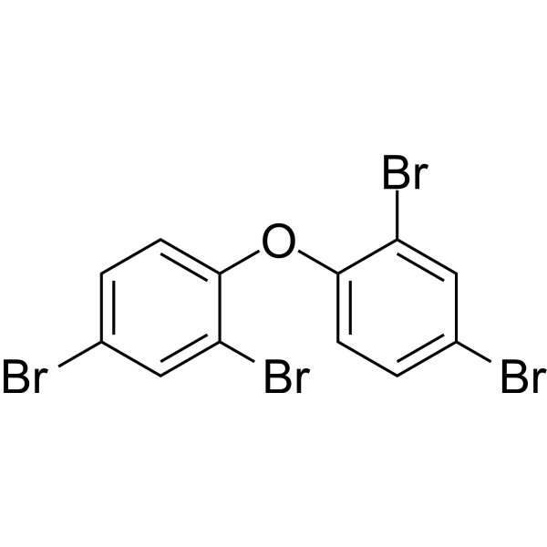
| Chemical Name | 2,4-dibromo-1-(2,4-dibromophenoxy)benzene |
| Synonyms | 5436-43-1; 2,2',4,4'-Tetrabromodiphenyl ether; BDE-47; PBDE 47; BDE 47; PBDE-47; Benzene, 1,1'-oxybis(2,4-dibromo-; 2,2,4,4-tetrabromodiphenyl ether; |
| HS Tariff Code | 2934.99.9001 |
| Storage |
Powder-20°C 3 years 4°C 2 years In solvent -80°C 6 months -20°C 1 month |
| Shipping Condition | Room temperature (This product is stable at ambient temperature for a few days during ordinary shipping and time spent in Customs) |
Biological Activity
| Targets | JNK signaling pathway; Mitochondrial oxidative phosphorylation (OXPHOS); Reactive oxygen species (ROS) [1] |
| ln Vitro |
Exposure to BDE - 47 led to a significant decrease in cell viability and a prominent increase in apoptosis in RAW264.7 cells. A decrease in mitochondrial membrane potential (MMP) and an increase in cytochrome c release and caspase cascade activation demonstrated that cell apoptosis induced by BDE - 47 occurred via the mitochondrial pathway. BDE - 47 also inhibited phagocytosis in RAW264.7 cells, changed the related immune factor index, and caused immune function damage. Moreover, there was a significant increase in the level of cellular reactive oxygen species (ROS), and the regulation of genes linked to oxidative stress was also demonstrated using transcriptome sequencing |
| ln Vivo |
- BDE - 47 exposure (70 mg/kg) significantly elevated the body weight and worsened hepatic steatosis along with increased inflammation in high - fat diet (HFD) - fed mice. BDE - 47 up - regulated triglyceride synthesis but suppressed lipid exportation and β - oxidation, aggravating the accumulation of hepatic lipid in HFD - fed mice. In addition, the increase of liver fibrosis, serum transaminase levels, as well as lipid peroxidation have been observed in mice co - treated with BDE - 47 and HFD - BDE - 47 stimulated the production of dopamine and 5 - hydroxytryptamine, but inhibited expression of nestin, gfap, gap43, and psd95 in 24 hpf embryos. BDE - 47 also inhibited neural crest - derived melanocyte differentiation and melanin syntheses process, evidenced by disrupted expression of wnt1, wnt3, sox10, mitfa, tyrp1a, tyrp1b, tryp2, and oca2 gene in 72 hpf embryos and decreased tyrosinase activities in embryos at 48 and 72 hpf. The transcriptional activities of myosin vaa, kif5ba, rab27a, mlpha, and cdc42 genes, which are associated with intracellular transport process, were also disturbed during zebrafish development, leading to fast spontaneous movement and melanin accumulation deficit in zebrafish embryos |
| Animal Protocol | Zebrafish embryos were exposed to BDE-47 at concentrations of 1, 5, 10, and 50 μg/L for 5 days. The compound was dissolved in embryo medium (vehicle control: 0.1% DMSO). Post-exposure, embryos/larvae were collected for oxidative stress markers (SOD, CAT, GSH, MDA, ROS), gene expression analysis (Sod1, *Ucp-2*, Gstp2, Nqo1), and comet assay to assess DNA damage [1]. |
| Toxicity/Toxicokinetics |
- BDE - 47 increased mortality, morphological damage, and altered population dynamics and fecundity of rotifer Brachionus plicatilis EPA IRIS Information 2,2',4,4'-Tetrabromodiphenyl ether (BDE-47) Toxicity Summary EPA IRIS Summary PDF (Update: Jun-30-2008 ) Critical Effect Systems Nervous Reference Dose (RfD), chronic 1 x 10 ^-4 mg/kg-day EPA Integrated Risk Information System (IRIS) RAIS Toxicity Values Oral Chronic Reference Dose (RfDoc) (mg/kg-day) 0.0001 Oral Chronic Reference Dose Reference IRIS Current |
| References |
[1]. BDE-47 induced apoptosis in zebrafish embryos through mitochondrial ROS-mediated JNK signaling. Chemosphere. 2020 Nov;258:127385. |
| Additional Infomation |
2,2,4,4-tetrabromodiphenyl ether (BDE-47) has received considerable attention because of its high detection level in biological samples and potential developmental toxicity. Here, using zebrafish (Danio rerio) as the experimental animal, we investigated developmental effects of BDE-47 and explored the potential mechanism. Zebrafish embryos at 4 h post-fertilization (hpf) were exposed to 0.312, 0.625 and 1.25 mg/L BDE-47 to 74-120 hpf. We found that BDE-47 instigated a dose-related developmental toxicity, evidenced by reduced embryonic survival and hatching rate, shortened body length and increased aberration rate. Meanwhile, higher doses of BDE-47 reduced mitochondrial membrane potential and ATP production but increased apoptosis in zebrafish embryos. Expression of genes involved in mitochondrial oxidative phosphorylation (OXPHOS) (ndufb8, sdha, uqcrc1, cox5ab and atp5fal) were negatively related to BDE-47 doses in zebrafish embryos. Moreover, exposure to BDE-47 at 0.625 or 1.25 mg/L impaired mitochondrial biogenesis and mitochondrial dynamics. Our data further showed that BDE- 47 exposure induced excessive reactive oxygen species (ROS) and oxidative stress, which was accompanied by the activation of c-Jun N-terminal Kinase (JNK). Antioxidant NAC and JNK inhibition could mitigate apoptosis in embryos and improve embryonic development in BDE-47-treated zebrafish, suggesting the involvement of ROS/JNK pathway in embryonic developmental changes induced by BDE-47. Altogether, our data suggest here that developmental toxicity of BDE-47 may be associated with mitochondrial ROS-mediated JNK signaling in zebrafish embryo.[1] Mechanism: BDE-47 targets mitochondria, inhibits OXPHOS, reduces mitochondrial membrane potential (MMP), and induces ROS. This activates JNK signaling, leading to caspase-dependent apoptosis in zebrafish embryos [1]. Developmental Toxicity: The study confirms BDE-47's role in disrupting embryonic development via mitochondrial dysfunction [1]. 2,2',4,4'-Tetrabromodiphenyl ether is an organobromine compound and an aromatic ether. |
Solubility Data
| Solubility (In Vitro) | DMSO : 200 mg/mL (411.70 mM; with sonication) |
| Solubility (In Vivo) |
Note: Listed below are some common formulations that may be used to formulate products with low water solubility (e.g. < 1 mg/mL), you may test these formulations using a minute amount of products to avoid loss of samples. Injection Formulations (e.g. IP/IV/IM/SC) Injection Formulation 1: DMSO : Tween 80: Saline = 10 : 5 : 85 (i.e. 100 μL DMSO stock solution → 50 μL Tween 80 → 850 μL Saline) *Preparation of saline: Dissolve 0.9 g of sodium chloride in 100 mL ddH ₂ O to obtain a clear solution. Injection Formulation 2: DMSO : PEG300 :Tween 80 : Saline = 10 : 40 : 5 : 45 (i.e. 100 μL DMSO → 400 μLPEG300 → 50 μL Tween 80 → 450 μL Saline) Injection Formulation 3: DMSO : Corn oil = 10 : 90 (i.e. 100 μL DMSO → 900 μL Corn oil) Example: Take the Injection Formulation 3 (DMSO : Corn oil = 10 : 90) as an example, if 1 mL of 2.5 mg/mL working solution is to be prepared, you can take 100 μL 25 mg/mL DMSO stock solution and add to 900 μL corn oil, mix well to obtain a clear or suspension solution (2.5 mg/mL, ready for use in animals). Injection Formulation 4: DMSO : 20% SBE-β-CD in saline = 10 : 90 [i.e. 100 μL DMSO → 900 μL (20% SBE-β-CD in saline)] *Preparation of 20% SBE-β-CD in Saline (4°C,1 week): Dissolve 2 g SBE-β-CD in 10 mL saline to obtain a clear solution. Injection Formulation 5: 2-Hydroxypropyl-β-cyclodextrin : Saline = 50 : 50 (i.e. 500 μL 2-Hydroxypropyl-β-cyclodextrin → 500 μL Saline) Injection Formulation 6: DMSO : PEG300 : castor oil : Saline = 5 : 10 : 20 : 65 (i.e. 50 μL DMSO → 100 μLPEG300 → 200 μL castor oil → 650 μL Saline) Injection Formulation 7: Ethanol : Cremophor : Saline = 10: 10 : 80 (i.e. 100 μL Ethanol → 100 μL Cremophor → 800 μL Saline) Injection Formulation 8: Dissolve in Cremophor/Ethanol (50 : 50), then diluted by Saline Injection Formulation 9: EtOH : Corn oil = 10 : 90 (i.e. 100 μL EtOH → 900 μL Corn oil) Injection Formulation 10: EtOH : PEG300:Tween 80 : Saline = 10 : 40 : 5 : 45 (i.e. 100 μL EtOH → 400 μLPEG300 → 50 μL Tween 80 → 450 μL Saline) Oral Formulations Oral Formulation 1: Suspend in 0.5% CMC Na (carboxymethylcellulose sodium) Oral Formulation 2: Suspend in 0.5% Carboxymethyl cellulose Example: Take the Oral Formulation 1 (Suspend in 0.5% CMC Na) as an example, if 100 mL of 2.5 mg/mL working solution is to be prepared, you can first prepare 0.5% CMC Na solution by measuring 0.5 g CMC Na and dissolve it in 100 mL ddH2O to obtain a clear solution; then add 250 mg of the product to 100 mL 0.5% CMC Na solution, to make the suspension solution (2.5 mg/mL, ready for use in animals). Oral Formulation 3: Dissolved in PEG400 Oral Formulation 4: Suspend in 0.2% Carboxymethyl cellulose Oral Formulation 5: Dissolve in 0.25% Tween 80 and 0.5% Carboxymethyl cellulose Oral Formulation 6: Mixing with food powders Note: Please be aware that the above formulations are for reference only. InvivoChem strongly recommends customers to read literature methods/protocols carefully before determining which formulation you should use for in vivo studies, as different compounds have different solubility properties and have to be formulated differently. (Please use freshly prepared in vivo formulations for optimal results.) |
| Preparing Stock Solutions | 1 mg | 5 mg | 10 mg | |
| 1 mM | 2.0585 mL | 10.2925 mL | 20.5850 mL | |
| 5 mM | 0.4117 mL | 2.0585 mL | 4.1170 mL | |
| 10 mM | 0.2059 mL | 1.0293 mL | 2.0585 mL |